
Abstract
An aged, free-ranging, female, radio-collared American black bear (Ursus americanus) died after an approximately 5 month long period of weight loss. Gross necropsy findings included severe diffuse pyogranulomatous bronchopneumonia, marked granulomatous lymphadenitis of tracheobronchial lymph nodes and multiple intra-abdominal lymph nodes, chronic focal jejunal ulceration, and widespread alopecia. Histopathologic examination revealed abundant fungal organisms morphologically compatible with Blastomyces sp. within pyogranulomatous inflammatory lesions in the lungs, multiple lymph nodes, liver, kidneys, jejunum, and right adrenal gland. In addition, the haircoat had a mild infestation of chewing lice (Trichodectes pinguis euarctidos), and large numbers of rhabditid nematodes consistent with Pelodera sp. were histologically observed within hair follicles.
Keywords
A free-ranging, female American black bear (Ursus americanus) was observed and radio-tracked by researchers from the Wildlife Research Institute near Ely (St. Louis County), Minnesota, starting in June of 2004. Body weight was periodically recorded at a field weight station and demonstrated typical seasonal pre-hibernation weight gain, with recordings varying from 70 to 182 kg. The last weight obtained at the field station in August of 2009 was 102 kg, which was considered normal for the time of year. However in late November, the bear was observed to be severely underweight and inappetent. Shortly thereafter, the bear was found deceased, partially submerged in a shallow pool of water. The carcass was recovered and submitted to the University of Minnesota Veterinary Diagnostic Laboratory (St. Paul, Minnesota) for diagnostic evaluation.
At presentation, the bear weighed 62 kg. The haircoat of the trunk, rump, and limbs was thinned and unkempt, and scattered within the denser haircoat of the brisket were numerous round, dorsoventrally flattened, white to brown, small (1–3 mm diameter) lice, identified as Trichodectes pinguis euarctidos.
Necropsy confirmed generalized marked skeletal muscle atrophy and visible but inadequate subcutaneous and visceral adipose stores. Bilaterally, the lungs were diffusely voluminous and discolored pale gray to tan (Fig. 1). On cut section, the pulmonary parenchyma was meaty, soft, and mottled tan to gray with a small amount of brown mucoid material within the larger airways (Fig. 2). The tracheobronchial, mesenteric, perirenal, iliac, inguinal, and colonic lymph nodes were enlarged up to approximately 3 times normal size and, on section, were multifocally to diffusely effaced and expanded by homogenous, tan–yellow, soft tissue with occasional white, firm foci. Other lesions included infrequent strands of fibrin within the peritoneal cavity, mild diffuse hepatomegaly, focal chronic ulceration of the proximal jejunum (15 mm × 15 mm × 7 mm), focal nodular expansion of the medulla of the right adrenal gland (5 mm × 5 mm × 5 mm), and multiple, long, thin, white filariid nematodes (≤70 mm long) coiled within the connective tissues of the intrathoracic tracheal serosa, and fascia of the left axillary region.
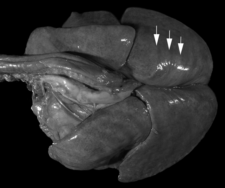
Lungs; American black bear (Ursus americanus). The lungs were diffusely enlarged as evidenced by rib impressions best visible in the right caudal lung lobe (arrows).
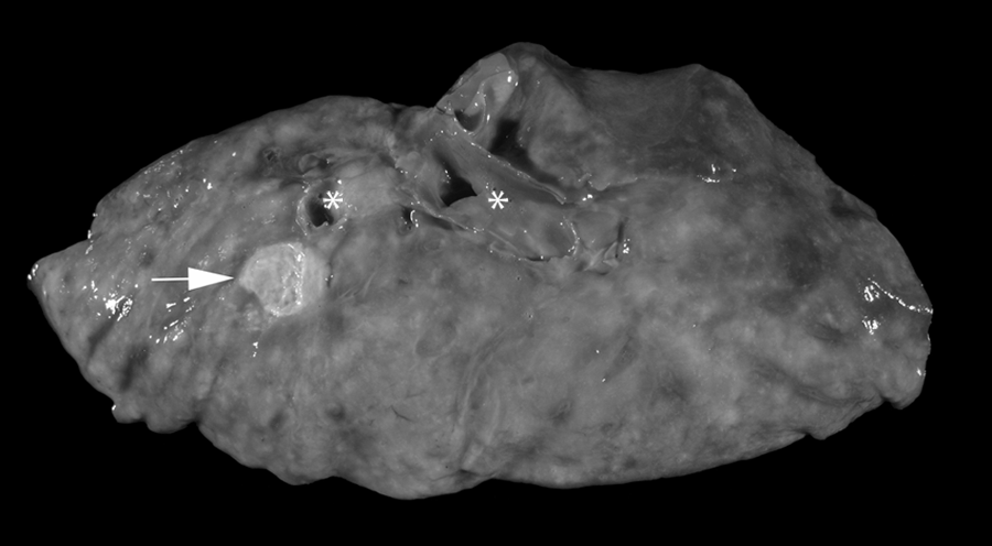
Cut section of the lungs; American black bear (Ursus americanus). The lungs were diffusely consolidated and brownish. Besides the diffuse consolidation, a focal, well-demarcated, beige pyogranuloma (arrow) is present next to a larger airway (asterisks).
Samples of all major organs, endocrine glands, eyes, brain, skeletal muscle, and haired skin were fixed in 10% neutral buffered formalin for histopathology. The main histologic lesion, involving the lungs and lymph nodes, consisted of marked pyogranulomatous inflammation with numerous multinucleated macrophages surrounding areas of eosinophilic cellular debris infiltrated by variable numbers of yeast cells. The lungs were diffusely affected. The round to oval, 10–15 μm in diameter yeast organisms were characterized by a 1–2 μm thick, refractile, double-contoured wall, a basophilic central nucleus surrounded by a thin rim of clear space, and occasional broad-based budding. The organisms were often observed within multinucleated macrophages (Fig. 3). Yeast cells were most frequent in the lungs and lymph nodes, with up to 30 organisms per 400× field, while lower numbers were observed within sites of granulomatous inflammation in the liver, kidneys, right adrenal gland, and jejunum at the site of ulceration. There was no evidence of viral inclusion bodies, and immunohistochemistry using a Canine distemper virus antigen–specific monoclonal antibody a yielded a negative result in lung tissue. These findings were consistent with the diagnosis of disseminated blastomycosis, with the presumptive primary pathogen being Blastomyces sp. given that no other predisposing factors were identified.
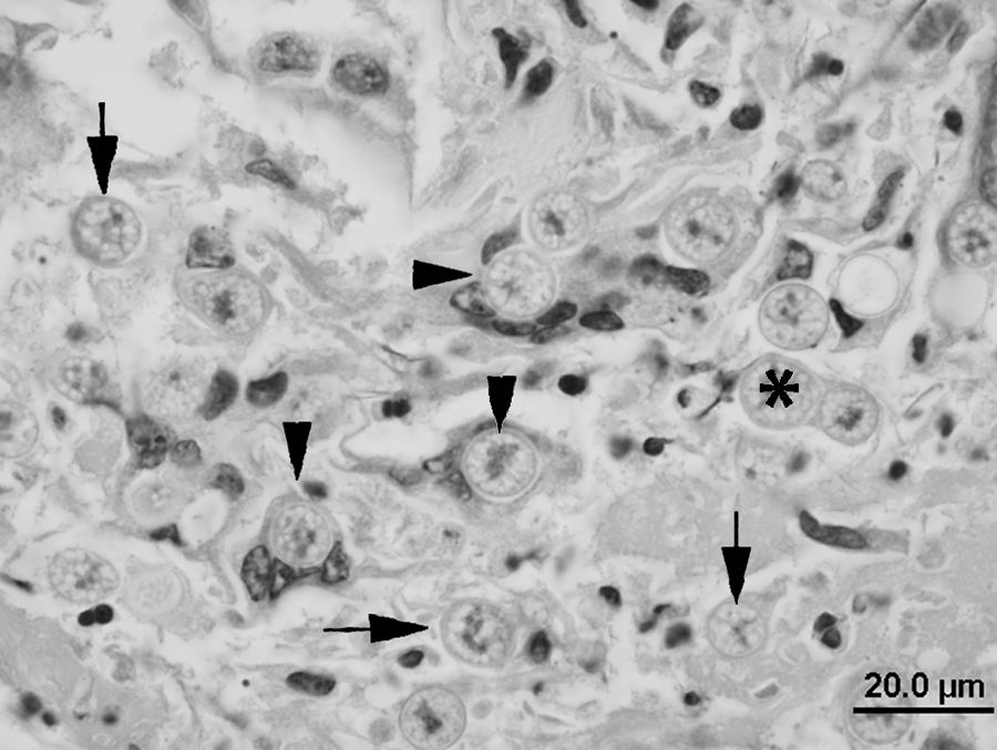
Photomicrograph of the lungs; American black bear (Ursus americanus). Numerous yeast cells were present in the alveolar lumina (arrows). The yeast cells were round to oval, 10–15 μm in diameter, and had a 1–2 μm thick, refractile, double-contoured wall, a basophilic central nucleus surrounded by a thin rim of clear space, and occasional broad-based budding (asterisk). The organisms were often observed within multinucleated macrophages (arrowheads). These features were consistent with Blastomyces dermatitidis. Bar = 20.0 µm.
In addition, sections of haired skin from the flanks and rump revealed diffuse moderate orthokeratotic hyperkeratosis, mild multifocal lymphoplasmacytic perifolliculitis and dermatitis, and numerous ectatic hair follicles filled with lamellar keratin and moderate to high numbers of larval nematodes, with up to 12 per follicle. The nematodes averaged 20 μm in diameter and were characterized by a thin cuticle with paired lateral alae, platymyarian musculature, and poorly discerned internal structures, consistent with a rhabditid species (Fig. 4). To verify the age of the bear, an upper first premolar was extracted and sent to Matson’s Laboratory LLC (Milltown, Montana) for age determination by cementum analysis, which determined the bear to have been 26.75 years old.
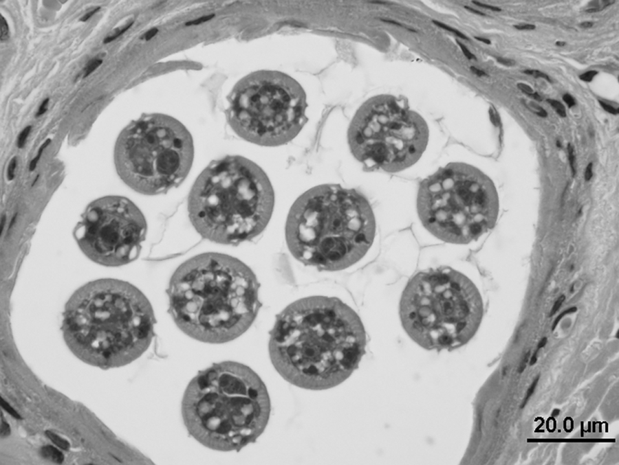
Photomicrograph of an ectatic hair follicle in the skin; American black bear (Ursus americanus). The hair follicle contained 12 nematodes in cross-section. The nematodes averaged 20 μm in diameter and were characterized by a thin cuticle with paired lateral alae, platymyarian musculature, and poorly discerned internal structures. These features were consistent with a rhabditid species. Bar = 20.0 µm.
Blastomycosis is a chronic pyogranulomatous disease caused by the dimorphic fungus Blastomyces dermatitidis. The identification of the fungus is frequently made based on morphological criteria using cytological or histological specimen as in the presented case. The yeasts of a Blastomyces sp. in tissues have rather distinctive features such as a size of approximately 8–20 μm, thick, double-contoured, refractile walls, and broad-based budding, but isolation of the organism is considered to be the gold standard. 19 More recently, polymerase chain reaction assays have become available for clinical samples because isolation is time consuming. 18 The morphologic features allow pathologists to distinguish the fungus from related fungi such as Paracoccidioides sp., Coccidioides sp., Cryptococcus sp., and Histoplasma sp. 16 Furthermore, Paracoccidioides sp. and Coccidioides sp. are not endemic in Minnesota or neighboring states and Canada.1,14
While best documented in dogs and human beings, blastomycosis has been reported in a wide variety of mammalian species including polar bears, wolves, exotic felids, marine mammals, a ferret, an Indian fruit bat, and a rhesus monkey (Clyde VL, Ramsay EC, Munson L: 1996, A review of blastomycosis in large zoo carnivores. In: Proceedings of the Annual Meeting of the American Association of Zoo Veterinarians, ed. Baer CK, pp. 554–556. Puerto Vallarta, Mexico, November 3–8).2–5,11,13,15,20–23 The fungal organism is associated with acidic sandy soils that are rich in decaying vegetation. Endemically infected areas include the Mississippi and Ohio River valleys and the Great Lakes states. 9 Geographic clustering of cases has been documented and, in Minnesota, blastomycosis is common in 3 adjacent northern counties (St. Louis, Itasca, and Beltrami; Minnesota Department of Health: 2003, Blastomycosis. Available at: http://www.health.state.mn.us/divs/idepc/diseases/blastomycosis/dcn603blasto.html. Accessed on July 30, 2012). Systematic surveillance for blastomycosis in Minnesota from 1999 to 2009 identified the probable county of exposure to B. dermatitidis for 258 human cases and 581 canine cases as St. Louis County, which is the county of residence of the bear at the time of its death. St. Louis County accounted for 31% of human cases and 23% of canine cases (http://www.health.state.mn.us/divs/idepc/diseases/blastomycosis/statistics.html). Interestingly, the only reported case of fatal blastomycosis in a wild wolf (Canis lupus) in Minnesota also occurred in St. Louis County. 22 It seems likely that the high exposure rate in this region contributed to the onset of disease in the bear described herein.
Infection is usually acquired through inhalation in human beings and in dogs, and a pulmonary route of infection is likely in the current case given the severity of the pyogranulomatous pneumonia and abundance of organisms in the lungs.2–4 In dogs, extrapulmonary dissemination of the organism via the vascular or lymphatic system most often affects the lymph nodes, skin, eyes, and bones, less frequently involving the brain, meninges, kidneys, liver, and spleen.3,4 In the present case, dissemination of the organism was evident in the visceral lymph nodes of the thoracic and abdominal cavities, liver, kidneys, intestine, and right adrenal gland. There was no evidence of dissemination to the skin, eyes, or central nervous system. Skeletal lesions were not apparent at necropsy, and there was no histopathological evidence of organisms in the femoral bone marrow; however, further evaluation of the bones was not conducted. In previous reports of nonfatal blastomycosis in 2 captive polar bears, the infection resulted predominately in respiratory disease, and dissemination to the central nervous system was not apparent (Clyde VL, et al.: 1996, A review of blastomycosis in large zoo carnivores). 13
The significance of the advanced age of the bear described in the current study to the onset of disease is not clear. While black bears can be long-lived, it is rare for an individual free-ranging animal to exceed 20–25 years of age. 10 Old age has not been identified as a risk factor for blastomycosis in human beings or dogs; however, older human patients tend to develop more severe pulmonary infection with a higher risk of mortality.3,8 It seems plausible that the greatest risk factor for this bear obtaining the disease was geographic exposure, but advanced age likely contributed to the overwhelming severity of the condition.
The appearance of the rhabditid nematodes within the hair follicles is consistent with Pelodera dermatitis, which has been previously reported in dogs, cattle, horses, sheep, and, in 2008, 1 case in an American black bear with concurrent sarcoptic mange.6,7 The saprophytic nematodes are common within decaying organic matter or superficial moist soil, and opportunistic parasitism occurs following direct contact of the skin with contaminated bedding or soil. 7 While the bear in the current case was unlikely to have been spending prolonged time in a den due to hibernation, fatigue and debilitation from the systemic mycotic disease may have increased the time the bear lay in contact with moist, infested bedding material. The other parasites identified at gross necropsy were less likely to have contributed to the bear’s declining condition and death. Trichodectes spp. are chewing lice that are reported to be common on free-ranging American black bears.12,17 When present in small numbers, there is often no apparent detrimental effect to the host. The filariid-like nematodes within the connective tissues are most consistent with Dirofilaria ursi, which are considered to be nonpathogenic in bears.12,17
This report of blastomycosis in a wild American black bear indicates that, although uncommon, systemic mycoses should be considered as differential diagnoses in bears with chronic infections or respiratory signs, particularly within endemic areas. In addition, the current report provides further support that Pelodera dermatitis should be considered a differential diagnosis for alopecia and dermatitis in black bears.
Footnotes
Acknowledgements
The authors are grateful to Ronda Aho and Jan Shivers, Minnesota Veterinary Diagnostic Laboratory, for their histology and immunohistochemistry expertise, and Dr. Bert Stromberg, Department of Veterinary Biosciences at the University of Minnesota, for identification of the parasites.
a.
Clone CDV-NP, VMRD Inc., Pullman, WA.
Declaration of conflicting interests
The author(s) declared no potential conflicts of interest with respect to the research, authorship, and/or publication of this article.
Funding
The author(s) declared that they received no financial support for their research and/or authorship of this article.