
Abstract
The objectives of the current study were to evaluate the use of DNA microarray for the rapid and direct detection of Mycobacterium tuberculosis and Mycobacterium bovis in bovine milk, blood, and pharyngeal swab samples, and to compare the use of DNA microarrays with current molecular detection techniques. The present study describes a microarray assay based on mtp40 and pncA gene sequences, which can be used to detect M. tuberculosis and M. bovis species. Each probe was spotted onto a silylated glass slide with an arrayer and used for hybridization with fluorescently labeled DNA derived from amplified DNA samples. The detection limit for mycobacterial DNA using this DNA microarray method was 50 fg (5 tubercle bacilli). Mycobacterium tuberculosis and/or M. bovis was detected in 7.1% (24/336) of the cattle specimens using the DNA microarray compared to 6.0% (20/336) using culture methods. Mixed infections were detected in 3 animals using the DNA microarray method, whereas the mixed infections were detected in 2 animals using either culture or polymerase chain reaction methods. The use of ancillary in vitro tests alongside the DNA microarray enhanced the detection of cattle infected with M. tuberculosis and/or M. bovis and reduced the number of false-positive animals that would be culled. More species may be easily added to this system, and supplementary probes can be added to increase the simultaneous detection power.
Keywords
Both Mycobacterium bovis and Mycobacterium tuberculosis can pose health hazards to animals and human beings. 6 Since infected animals are potentially capable of infecting human beings with tuberculosis (TB), the differential identification of M. tuberculosis and M. bovis is important. 1 Although M. bovis infections typically occur in cattle, infections have also been reported in human beings.7,13 Presently, there is limited data regarding the prevalence of M. tuberculosis and M. bovis in cattle and the possible zoonotic importance of bovine tuberculosis in Asia.
Currently, although laboratory diagnoses of Mycobacterium infections are made primarily using smear tests and culturing, the sensitivity of such tests is low and results may vary depending on the examiner. Culturing and biochemical tests can take 4–8 weeks and sometimes fail to provide precise identification. A diagnosis can also be made with immunological testing, which can take even longer. Direct staining and microscopic examination of clinical specimens can produce results more quickly, but this methodology lacks sensitivity and specificity. Tuberculin testing has traditionally been used to determine the prevalence of infections in animals and human beings. For instance, the primary antemortem diagnostic test for TB in cattle is the bovine tuberculin skin test, but various factors can reduce its specificity and sensitivity.7,8 Recently, several molecular methods have been developed that provide clear criteria for the identification of mycobacteria. The methods comprise a variety of polymerase chain reaction (PCR) assays, such as those based on DNA sequence variations in the direct repeat region of the M. tuberculosis complex strains (spoligotyping) or on single nucleotide polymorphisms in either the oxyR or the gyrB genes.2,4,5Additionally, human patients with compromised immune function due to chronic medical conditions may not show positive skin tests. Therefore, the development of effective, accurate, and rapid diagnostic methods for M. tuberculosis and M. bovis is necessary. For instance, the development of DNA microarray technologies can be applied in microbiology. 9 Recently, DNA microarrays have been widely and successfully employed to simultaneously monitor the expression of complete genomes and identify genes that are differentially expressed in distinct cell types, developmental stages, disease states, or in cells exposed to various reagents.11,14
The objective of the present study was to establish a DNA microarray method for detecting and identifying the main TB pathogens. The mtp40 and pncA genes were chosen as genetic markers to screen for specific probes, and primers were designed to amplify each of them. The PCR technique was combined with an oligonucleotide microarray to detect the M. bovis and M. tuberculosis amplicons.
The M. tuberculosis H37Rv, M. bovis AN5, and 14 nontuberculosis mycobacterial strains used in the present study were obtained from the Laboratory of Animal Quarantine, Guangdong Inspection and Quarantine Technology Center (China). To determine the specificity of the DNA microarray, 10 bacterial species were used that were readily available in the laboratory (i.e., Escherichia coli, Streptococcus agalactiae, Streptococcus dysgalactiae, Salmonella typhimurium, Sarcina lutea, Staphylococcus aureus, Pasteurella multocida, Streptococcus pneumoniae, Bacillus subtilis, and Legionella pneumophila).
A total of 536 samples were collected from 336 black and white dairy cattle housed in different farms in southern China. The intradermal tuberculin test was performed on 220 of the samples. The cattle were evaluated for clinical signs of TB, including lymphadenopathies, body weight loss, production loss, intermittent pyrexia, udder infection, and dry cough. Two or 3 samples were collected from each animal, namely, citrated blood, milk, or a pharyngeal swab. Each sample was divided into 2 aliquots. The aliquoted samples were transported within 8 hr of collection on wet ice to the laboratory. All samples were processed to isolate and culture mycobacteria in modified Lowenstein–Jensen media. Standard biochemical tests were used to identify the bacteria species level, including niacin production, nitrate reduction, catalase activity, Tween hydrolysis, arylsulphatase, and thiophene-2-carboxylic acid hydrazide sensitivity measurements. Twenty isolates derived from the 536 samples were identified by cultivation and standard biochemical tests (14 M. bovis isolates, 4 M. tuberculosis isolates, and 2 nontuberculosis mycobacteria isolates; see below).
The genomic DNA from the reference strains and blood samples were extracted using a genomic DNA kit. The milk samples were centrifuged at 12,000 × g for 20 min at 4°C. A sterile cotton swab was used to remove the fat layer, and the supernatant was discarded. The pellet was resuspended in phosphate buffered saline (PBS, pH 7.5) and processed. The pharyngeal swab was mixed with 1 ml of sterile PBS (pH 7.5) and centrifuged at 12,000 × g for 15 min. The pellet was resuspended in PBS (pH 7.5) and processed with a genomic DNA kit. PCR a was performed on the extracted DNA immediately, or the extracted DNA was stored at −80°C for subsequent experiments.
The complete sequences of the mtp40 and pncA genes were obtained from GenBank (http://www.ncbi.nlm.nih.gov). The primers were designed using Primer Premier 5.0 b and Oligo 6.0 c software. In the variable regions between the pair of primers, specific oligonucleotides that corresponded to the target species were selected as candidate probes. NH2- was added to each probe at the 3′ end. The reverse primers were labeled with fluorescein (cyanine [Cy]3) at the 5′ end. All of the oligonucleotide probes and primers are listed in Table 1. The nucleotide sequence data reported herein appear in the GenBank nucleotide sequence database under the following accession numbers: S69737.1 and BX248341.1, respectively. The probes were spottedd,e onto a silylated glass slide and used for hybridization. After the hybridization reaction, the chips were immediately scanned h at 532 nm (for Cy3), and the hybridization signal was analyzed with GenePix Pro 4.0 i software. The cutoff value equals the average negative signal intensity value plus 2 standard deviations. The positive signal intensity value was higher than the cutoff value.
Oligonucleotide probes and primers used in the current study.*
QC = quality control; pt = Mycobacterium tuberculosis probe; pm = Mycobacterium bovis probe; TB-F = M. tuberculosis forward primer; TB-R = M. tuberculosis reverse primer; MB-F = M. bovis forward primer; MB-F = M. bovis reverse primer.
The sensitivities of the DNA microarrays were evaluated using different concentrations of M. tuberculosis DNA at 100 pg, 10 pg, 1 pg, 100 fg, 50 fg, and 10 fg (1 bacilli). The hybridization results demonstrated that the selected probes could distinguish the single-stranded PCR products of TB-causing strains from other bacterial species. The species-specific probes detected M. tuberculosis and M. bovis accurately without cross-hybridization reactions, which allowed the probes to be used to in microarray experiments (Fig. 1). The hybridization sensitivity experiments indicated that the detection limit of the microarray is 50 fg, which was more sensitive than conventional PCR detection (Fig. 2). In order to appraise the specificity of the oligonucleotide probes, the 2 strains were amplified using optimized PCR conditions and then hybridized (shown in Fig. 2).
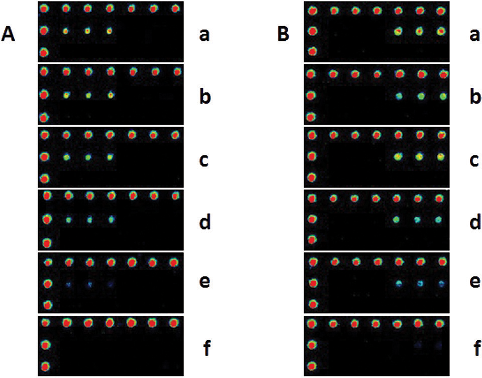
Microarray detection sensitivity. The sensitivity of the microarray was assessed with the following concentrations of Mycobacterium tuberculosis (
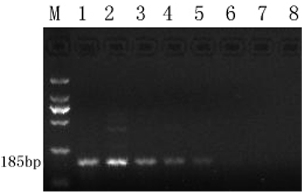
Conventional polymerase chain reaction sensitivity of Mycobacterium tuberculosis. Lane M: DL2000 DNA ladder marker; lanes 1–7: different concentrations of DNA: 10 ng, 1 ng, 100 pg, 10 pg, 1 pg, 100 fg, and 50 fg (5 bacilli), respectively; lane 8: distilled water.
To evaluate the DNA microarray, allelomorph PCRs f were performed as described previously. 3 The expected size of the amplicons in the allelomorph PCR assays was 185 bp. In all cases, positive amplification was obtained with the M. tuberculosis–specific primer pncAMT-2, but not with the M. bovis–specific primer pncAMB-2 (Fig. 3). The PCR results were compared with the DNA microarray and culture results (Table 2); the DNA microarray relevance ratio was 7.1% (24/336), which was higher than PCR and culture. Generally, culture followed by a panel of biochemical tests has been used for distinguish the mycobacteria; the disadvantage if this techniques is that most mycobacteria of clinical importance are slow growers and hence are difficult to isolate and cultivate. The time required for primary isolation ranges from 4 to 6 weeks in the case of solid culture media and 10–15 days by radiometric and other automated systems. A multiplex PCR–based assay detection and differentiation of M. tuberculosis and M. bovis has been reported previously, 10 with the sensitivities of the assays limited to detect 10–20 pg DNA of the tubercle bacilli. The DNA microarray assay described in the current study was developed to overcome these problems. The present study also has some limitations. Most Mycobacterium species cannot be identified with a single probe, but the microarray in the current study used single probes to distinguish M. tuberculosis and M. bovis from M. tuberculosis complex and nontuberculosis species. In future studies, a combination of probes will be used to identify individual species.
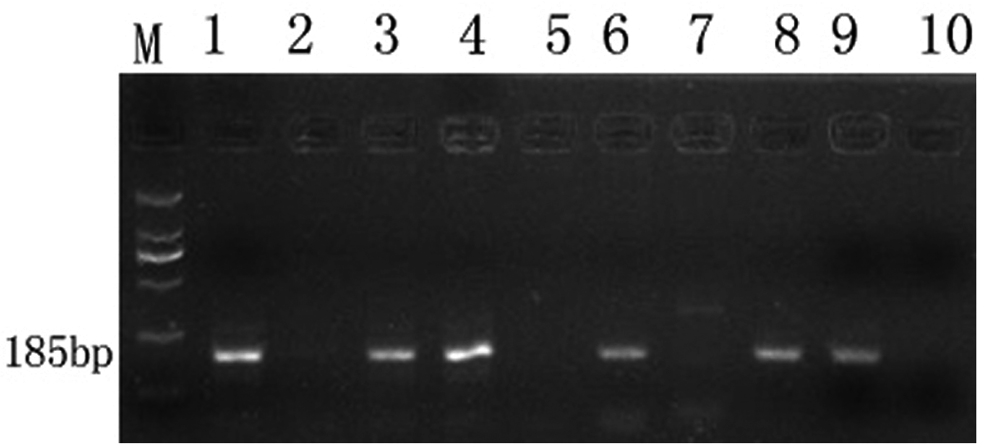
Allelomorph polymerase chain reaction (PCR) products of Mycobacterium tuberculosis and Mycobacterium bovis DNA (pncA gene). The products were electrophoresed on 1.5% agarose gels and stained with ethidium bromide. Lane M: DL2000 DNA ladder marker; lane 1: M. tuberculosis H37Rv amplified with the M. tuberculosis–specific primer; lane 2: M. tuberculosis H37Rv amplified with the M. bovis–specific primer; lanes 3–4: M. tuberculosis (clinical isolates); lanes 5, 10: negative controls with M. bovis–specific and M. tuberculosis–specific primers; lane 6: M. bovis AN5 amplified with the M. bovis–specific primer; lane 7: M. bovis AN5 amplified with the M. tuberculosis–specific primer; lanes 8–9: M. bovis (clinical isolates).
Detection of pathogenic mycobacteria in clinical samples.*
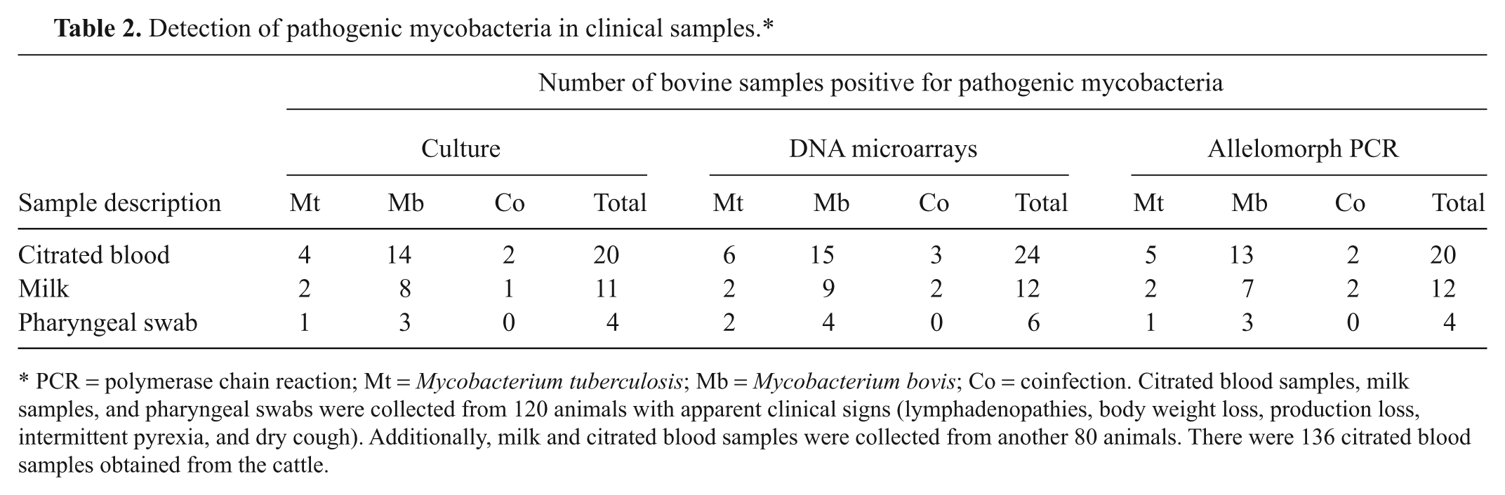
PCR = polymerase chain reaction; Mt = Mycobacterium tuberculosis; Mb = Mycobacterium bovis; Co = coinfection. Citrated blood samples, milk samples, and pharyngeal swabs were collected from 120 animals with apparent clinical signs (lymphadenopathies, body weight loss, production loss, intermittent pyrexia, and dry cough). Additionally, milk and citrated blood samples were collected from another 80 animals. There were 136 citrated blood samples obtained from the cattle.
Bovine tuberculosis can be transmitted through milk. Previous research has indicated that tuberculosis can be detected from the blood of cattle. Similar approaches have been reported to be useful in diagnosing mycobacterial infections in human beings. Among the samples screened by culturing, milk and blood showed the highest isolation rates. 12 Hence, pharyngeal swabs and other samples, such as rectal pinches, urine, and fecal samples were not analyzed.
In conclusion, a DNA microarray was developed that can efficiently and accurately detect M. tuberculosis and/or M. bovis pathogens from blood and milk. This method is a rapid (under 5 hr), accurate, and large-scale parallel handling method that can be applied to detection and identification of M. tuberculosis and M. bovis from clinical species. The DNA microarray described herein offers a higher sensitivity (50 fg) than conventional PCR. Additional species may be easily incorporated into this system, and supplementary probes can be added to increase the power of simultaneous detection.
Footnotes
Notes
The authors declared no potential conflicts of interest with respect to the research, authorship, and/or publication of this article.
This work was supported by a grant from the Guangdong Agriculture Key Scientific and Technological Issues (Grant No. 20081020100012) and Guangzhou Municipal Science and Technology Program (grant no. 2009Z1-E731) and Guangzhou Science and Technology Program 2010B060200040.