
Abstract
The Mythic 18 is a fully automated hematology bench-top analyzer using impedance technology for a complete blood cell count (CBC) and a 3-part white blood cell count (WBC) differential. The purpose of the current study was to evaluate the Mythic for assessment of agreement, precision, linearity, carry-over, stability, and usability under practice conditions. Ethylenediamine tetra-acetic acid–blood samples from 122 dogs, 140 cats, and 123 horses were analyzed with the Mythic and reference methods (Sysmex XT-2000iV, manual hematocrit, and microscopic WBC differentiation). Pearson’s coefficient of correlation, Passing–Bablok regression analysis, and Bland–Altman difference plots were performed to determine agreement. For precision, standard deviation and coefficients of variation were calculated. Linearity was determined according to Emancipator–Kroll. Red blood cell parameters showed excellent correlation and small biases, except for red cell distribution width and mean corpuscular hemoglobin concentration. Total WBC correlated excellently in canine and equine and very well in feline samples. In 23 feline specimens with platelet aggregates, the Mythic overestimated WBC. In all 3 species, absolute granulocyte counts correlated excellently. Equine lymphocyte counts showed good correlation whereas canine and feline lymphocyte counts correlated poorly. Feline platelets showed good correlation with a negative bias. The instrument showed good to excellent precision. The whole 3-part differential was found to be accurate in horses. In dogs and cats, absolute granulocyte counts were reliable. As with all impedance-based hematological instruments, evaluation of a blood smear is absolutely indicated to check for the presence of platelet aggregates, to verify WBC differentiation, and to identify possible abnormalities.
Introduction
Hematological results provide important information on the patient’s state of health, disease history, and response to treatment. 23 The invention of the Coulter cell counter and cell volume analyzer in 1956 highly reduced time-consuming manual work by automating the counting and sizing of cells. 9 Since then, several affordable, automated bench-top hematology analyzers have been developed for in-clinic use. 3 Most of these analyzers are primarily designed for human blood. When analyzing nonhuman hematology specimens, it is essential that the selected instrument is designed and validated for multispecies analysis. 20
The Mythic 18 a is an impedance-based hematology instrument originally designed for human application. To make the instrument suitable for veterinary application, settings for feline, canine, and equine blood samples have been developed in the Clinical laboratory of the Vetsuisse-Faculty of the University of Zurich (Zurich, Switzerland). The evaluation was conducted to assure the quality of the newly designed animal settings. Information on imprecision and inaccuracy of a hematological instrument are extremely valuable for the users. Each type of instrument should therefore be validated for each species before using results for clinical purpose.
The objective of the present study was to validate the Mythic 18 for use with blood samples from healthy and diseased cats, dogs, and horses. To this end agreement, precision, linearity, carry-over, and sample stability were determined. Biases were judged with respect to their clinical relevance.
Material and methods
Blood samples
Fresh ethylenediamine tetra-acetic acid (EDTA)-K3 blood samples from 122 dogs, 140 cats, and 123 horses from the Small Animal Clinic and the Clinic for Horses, at the Vetsuisse-Faculty, University of Zurich were analyzed on the Sysmex XT-2000iV b and reference methods, and usually with a time delay of 1.5 hr (until the routine work was finished) on the Mythic 18. All samples were collected by venipuncture regardless of sex, age, or breed and sent to the clinical laboratory in the framework of routine work to check the health status. Sample collection took place between May and December 2009. Complete sample analysis was performed within 6 hr after collection, most of them within 4 hr. The aforementioned blood samples were used to assess agreement and precision. To determine the range of linear measurement, 2 blood samples from cats, 2 from dogs, and 1 from a horse were used. Additionally, platelet-enriched plasma from a horse was used to assess linearity of the platelet count. Carry-over of blood from one sample to the following sample, thereby checking the effectiveness of cleaning of the instrument, was assessed for each species using 2 EDTA blood samples. To determine the effect of aging of samples, blood samples from 6 dogs and cats and 8 horses were used.
Instruments and methods used
Mythic 18
The Mythic 18 is a fully automated in-house hematology analyzer performing hematological analyses on EDTA anticoagulated blood. Nineteen species profiles can be created. For counting the cellular blood components, the Mythic 18 uses the impedance technique. A cyanide-free spectrophotometry method is used to measure hemoglobin by formation of oxyhemoglobin at 555 nm. Hematocrit is measured by volume integration. The sample volume is 10 µl. The instrument can determine 16 parameters in the normal mode and 18 in the research mode: white blood cell count (WBC) with absolute number and percentage of lymphocytes (LYM), monocytes (MONO), and granulocytes (GRAN), red blood cell count (RBC), hemoglobin concentration (HGB), hematocrit value (HCT), mean corpuscular volume (MCV), mean corpuscular hemoglobin (MCH), mean corpuscular hemoglobin concentration (MCHC), red cell distribution width (RDW), platelets (PLT), mean platelet volume (MPV), and for research, platelet crit (PCT) and platelet distribution width (PDW). For platelet counting, a floating threshold is used, whereas for RBC and WBC counts, the thresholds are predefined. Results are provided within 1 min on the liquid crystal display, printed out on a printer, and stored in the resident memory or in a USB key. Results were presented with flags; optionally, reference ranges can be reported. Additionally, the Mythic 18 shows histograms for WBC, RBC, and PLT. Prior to analysis, patient’s data can be entered manually or with a barcode reader. The instrument also displays message codes and histogram flags. However, message codes and flags have not been adapted yet to feline, canine, and equine blood samples. Therefore, conclusions about the usefulness of these message codes and flags cannot be drawn at this time.
Mythic 18 provides a 3-part WBC differential in samples with WBC within a range of 0.9 × 10³/µl to 150 × 103/µl. Quality control samples c are supplied as blood samples with 3 levels of RBC, WBC, and PLT levels. Results of each lot can be viewed on the display of the instrument in tables and Levey–Jennings graphs. The instrument uses 3 reagents d : a diluent, a lysis reagent, and a cleaning solution.
Sysmex XT-2000iV
The Sysmex XT-2000iV, equipped with software version 10b, was used as the reference instrument for total WBC, WBC differentiation, RBC, RBC indices, HGB, RDW, PLT count, and MPV. It is a fully automated hematology analyzer for animal blood providing 30 parameters. The impedance method with hydrodynamic focusing is used for RBC (RBC-I), HCT, and PLT (PLT-I). With these results, MCV, MCH, MCHC, RDW, MPV, and PDW are calculated. A flow cytometry device based on a sheath-flow and a semiconductor laser is used as an optical method for platelets (PLT-O) in cats, WBC, and WBC differentiation. Hemoglobin concentration is measured spectrophotometrically with a cyanide-free (sodium lauryl sulfate) method.
Manual methods
Manual HCT measurement was done with microhematocrit capillary tubes centrifuged at 13.000 × g for 5 min in a microhematocrit centrifuge. 10 Blood smears were stained using an automated staining instrument. e Microscopic differentiation of 2 modified Wright–stained blood smears, 100 WBC each, was conducted by 2 technicians with 10 years each of veterinary hematology experience. The results were used to calculate the absolute number of LYM, MONO, and GRAN counts by multiplying the percentage from the 200-cell count of each cell type with the total WBC from the Sysmex XT-2000iV.
Assessment of agreement
Analytical accuracy is defined by the International Council for Standardization in Hematology as a measure of agreement between the measured value of an analyte and its “true” value. 7 To determine accuracy, agreement between the results of the evaluated instrument and the results of a reference instrument were investigated. In the present study, assessment of agreement was determined by comparing the results of the Mythic 18 with those of the reference instrument, the manual HCT, and the microscopic differentiation. Sysmex XT-2000iV is widely used and accepted in veterinary clinical laboratories, and validation studies were conducted on the Sysmex XT-2000iV for cats 21 and cats, dogs, and horses. 13,14 For comparing results of granulocytes of the Mythic 18, results of the neutrophils, eosinophils, and basophils of the reference methods were added.
Precision and linearity
Within-series precision of the instrument was determined for each of the investigated species for low, normal, and high WBC values based on multiple analyses (more than 12 consecutive times). During the analysis, the sample was gently mixed. Afterwards, mean, standard deviation, and coefficient of variation as a measurement of the random error were calculated for all parameters. Precision from day to day was measured using commercially available quality control blood of low, intermediate, and high levels, which were analyzed once daily prior to analyzing patients samples over a 20-day period.
The linearity of the measurement range was assessed in all 4 species to determine the analytical range. Mythic 18 has a reportable range for WBC (−150 × 103/µl), RBC (–15 × 106/µl), HCT (−72%), and PLT (−4.000 × 10³/µl). The linearity of the measurement range was determined for WBC, RBC, HCT, HGB, and PLT by analyzing a series dilution of EDTA-K3 anticoagulated blood in triplicate. For cat and dog, 2 blood samples were used, 1 with high WBC to determine WBC linearity (5 ml) and 1 (cat, 12 ml; dog, 10 ml) for the remaining parameters. One equine sample (20 ml of ETDA-blood) was used for RBC, HGB, and HCT; additionally, platelet-enriched plasma of a horse was used to determine PLT linearity. The blood samples were centrifuged f at 390 × g for 10 min to receive results below and above the reference range. The plasma was then removed from the blood cells. Afterwards, concentrated blood cells were diluted with 0.9% saline solution in steps of 10%, to achieve a dilution series of 0–100% blood cell concentrate.
Carry-over
Carry-over was studied to assess if transfer of blood from a single sample will cause a falsely higher result in the following sample. For each species, 2 patient samples, 1 with high WBC, were analyzed twice followed by 3 replicates of diluents. 12
Cell aging
Cell aging studies were performed with blood samples from 6 cats, 6 dogs, and 8 samples from horses. They were analyzed at time point 1, 2, 4, 6, 8, 24, 32, and 48 hr after collection to calculate stability. The blood was stored at room temperature during the whole experiment. For each parameter, the difference in mean of results between each analysis and time point 1 hr was calculated. In cats, only RBC parameters were investigated.
Statistical analysis
All data were entered manually in a Microsoft Excel g spreadsheet. The Microsoft Excel add-in Analyse-it h was used for statistical analyses. For each parameter and each investigated species, Pearson coefficient of correlation (r), linear regression analysis according to Passing–Bablok provided intercept and slope with 95% confidence interval, 2 and Bland–Altman difference plot with biases and 95% limits of agreement 1 were calculated. Coefficient of correlation was considered excellent if r ≥ 0.95, very good if r = 0.90–0.94, good if r = 0.80–0.89, fair if r = 0.59–0.79, and poor if r < 0.59. 22 For precision analysis, standard deviation (SD) and coefficient of variation (CV) were calculated for each level, parameter, and species. The degree of linearity was determined with Analyse-it according to Emancipator–Kroll. 11
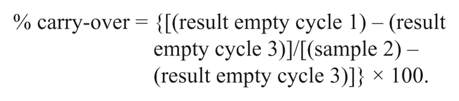
For WBC, RBC, HGB, and PLT, the percentage of carry-over was calculated using the following formula:
The stability of the blood samples was reviewed for statistically significant changes using the Friedman test and the Dunn multiple comparison post-test with GraphPad Prism. i Statistical significance was tested for the result of the first hour compared with the results of the following time points. Statistical significance was defined as P value <0.05.
Clinical relevance
For each sample, the data from the Mythic 18 and the reference methods were compared with hematology reference values that were established and currently used in the Clinical Laboratory of the Vetsuisse-Faculty University of Zurich (Table 1). The results were judged to be below or above the reference range, and the resulting interpretations from the Mythic 18 and the reference methods were compiled and compared to each other.
Reference values of hematological parameters for cats, dogs, and horses used in the current study (lower 5% reference limit and upper 95% reference limit).
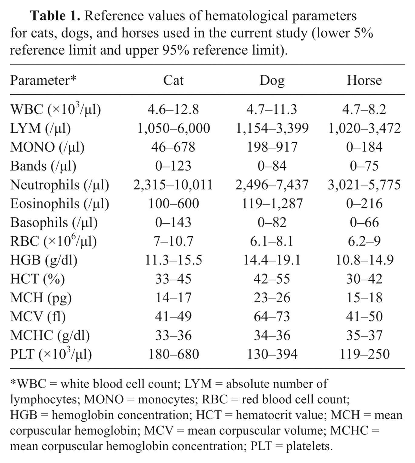
WBC = white blood cell count; LYM = absolute number of lymphocytes; MONO = monocytes; RBC = red blood cell count; HGB = hemoglobin concentration; HCT = hematocrit value; MCH = mean corpuscular hemoglobin; MCV = mean corpuscular volume; MCHC = mean corpuscular hemoglobin concentration; PLT = platelets.
Results
Assessment of agreement
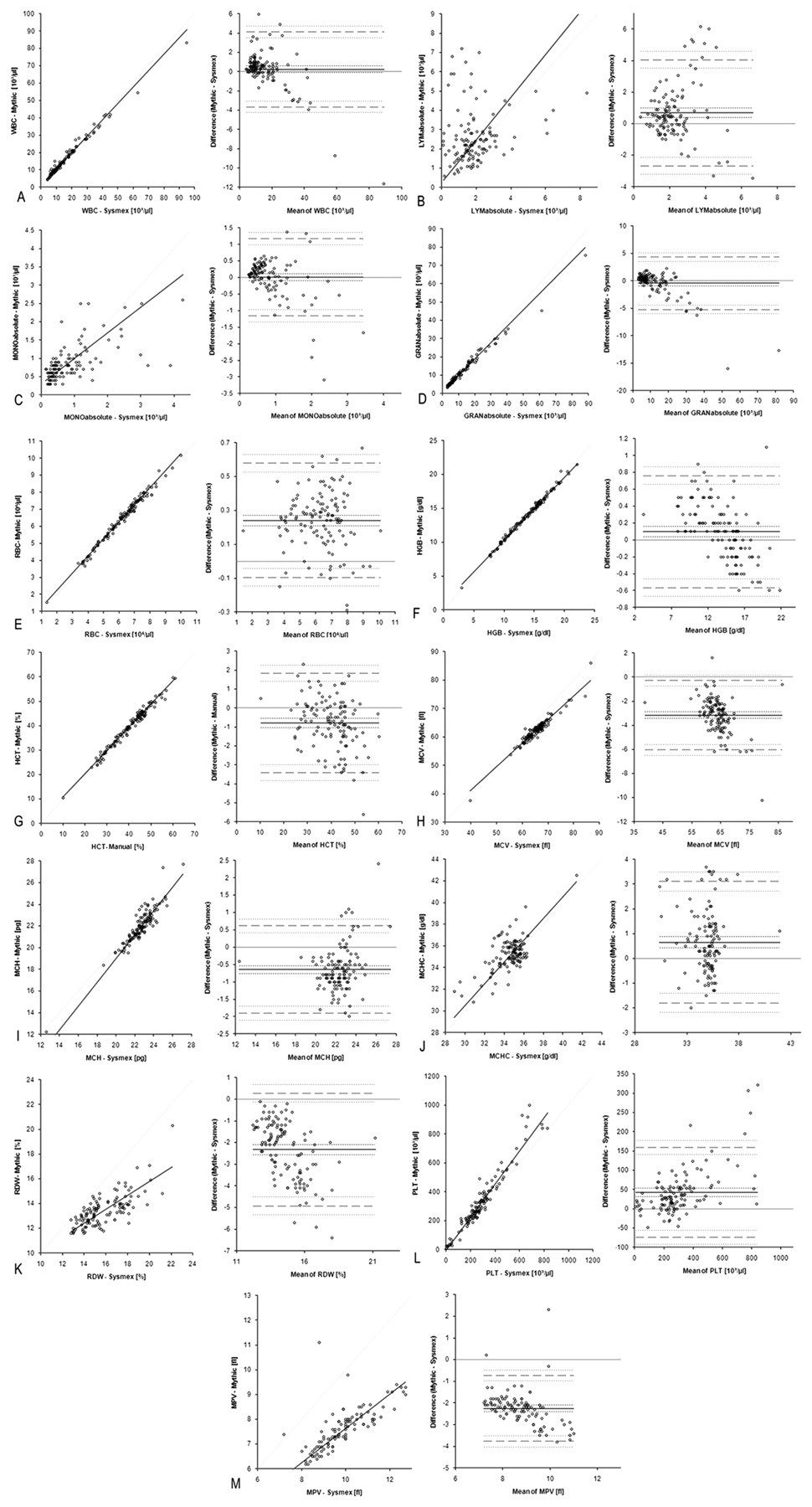
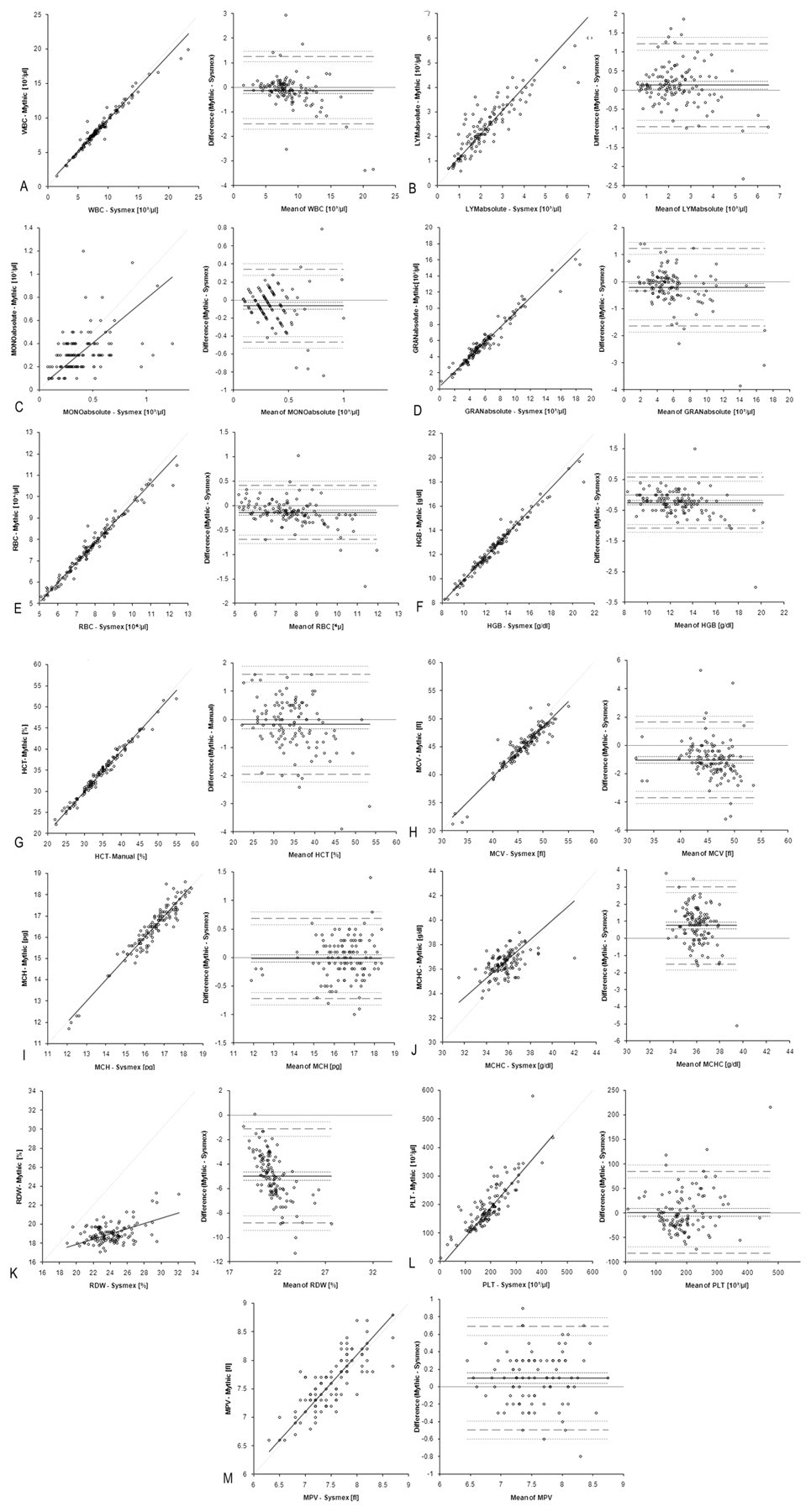
Pearson coefficient of correlation, intercept, and slope with 95% confidence intervals (CI) calculated by Passing–Bablok regression analysis, and biases with their 95% limits of agreement calculated by Bland–Altman difference plot are presented in Tables 2 and 3. Table 2 shows results for WBC, RBC, and PLT, and Table 3 presents results of the WBC differentiation compared with results from the Sysmex XT-2000iV and results of the manual WBC differentiation. Furthermore, linear regression analysis by Passing–Bablok and Bland–Altman difference plots are presented (Fig. 1 [cat], Fig. 2 [dog], and Fig., 3 [horse]). The results of the Mythic 18 for RBC, HGB concentration, HCT, and WBC (except for the cat) showed excellent correlation with the results provided by the reference instrument Sysmex XT-2000iV and manual HCT. The WBC results from the Mythic 18 were compared with the optical WBC results of the Sysmex XT-2000iV. Systematic errors with very small biases were observed in all 3 species for WBC. The RBC showed an excellent result for dogs with a small bias due to a constant systemic error. For HGB levels, a small proportional systemic error was seen in all investigated species. The MCV values showed excellent correlation in cat and dog and very good correlation in horses, with a systemic error and negative biases for horses and dogs. The feline MCH showed an excellent correlation with a small negative bias due to a constant systematic error, whereas the dog showed a proportional systemic error with a negative bias. For MCHC and PLT counts, a proportional systemic error was seen in all 3 species. In dogs, the Mythic 18 overestimated high PLT counts compared to the reference instrument. The MPV results for feline samples were not available, because the Sysmex XT-2000iV determines PLT counts optically via flow cytometry. The 3-part WBC differential showed for GRAN counts (absolute numbers) the best correlation and the smallest bias in all 3 species. The LYM counts showed a strong positive bias in cats and dogs with wide 95% limits of agreement. In horses, correlation was found to be good with a small bias. Results for MONO counts showed only fair correlation in canine samples and poor correlation in feline and equine samples. Some results of the WBC differential of the Sysmex XT-2000iV were excluded from statistical analysis due to the inability of the Sysmex XT-2000iV to differentiate WBC: 1 equine, 2 canine, and 4 feline blood samples. In the equine sample, both canine samples, and 3 of the 4 feline samples, the Sysmex XT-2000iV misclassified a left shift. In the remaining feline sample, normoblasts (52 normoblasts per 100 WBC) were seen in the blood smear while the Sysmex XT-2000iV classified them falsely to LYM.
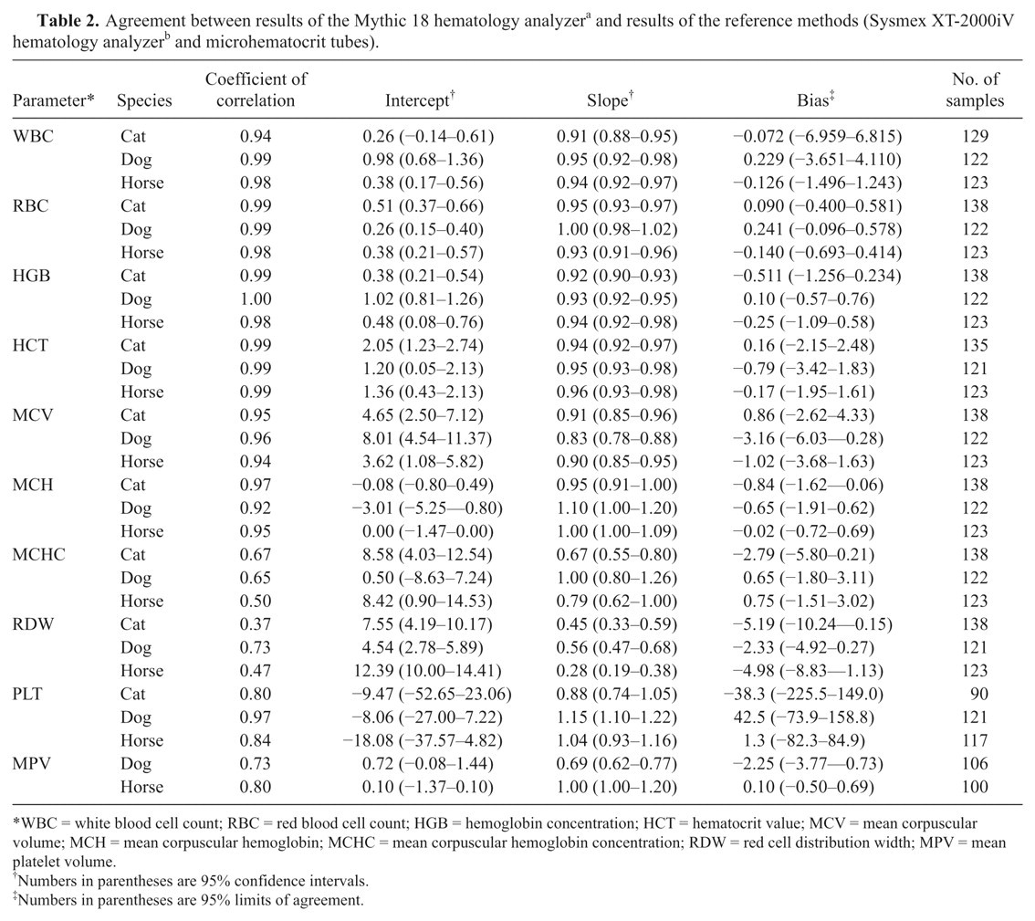
WBC = white blood cell count; RBC = red blood cell count; HGB = hemoglobin concentration; HCT = hematocrit value; MCV = mean corpuscular volume; MCH = mean corpuscular hemoglobin; MCHC = mean corpuscular hemoglobin concentration; RDW = red cell distribution width; MPV = mean platelet volume.
Numbers in parentheses are 95% confidence intervals.
Numbers in parentheses are 95% limits of agreement.
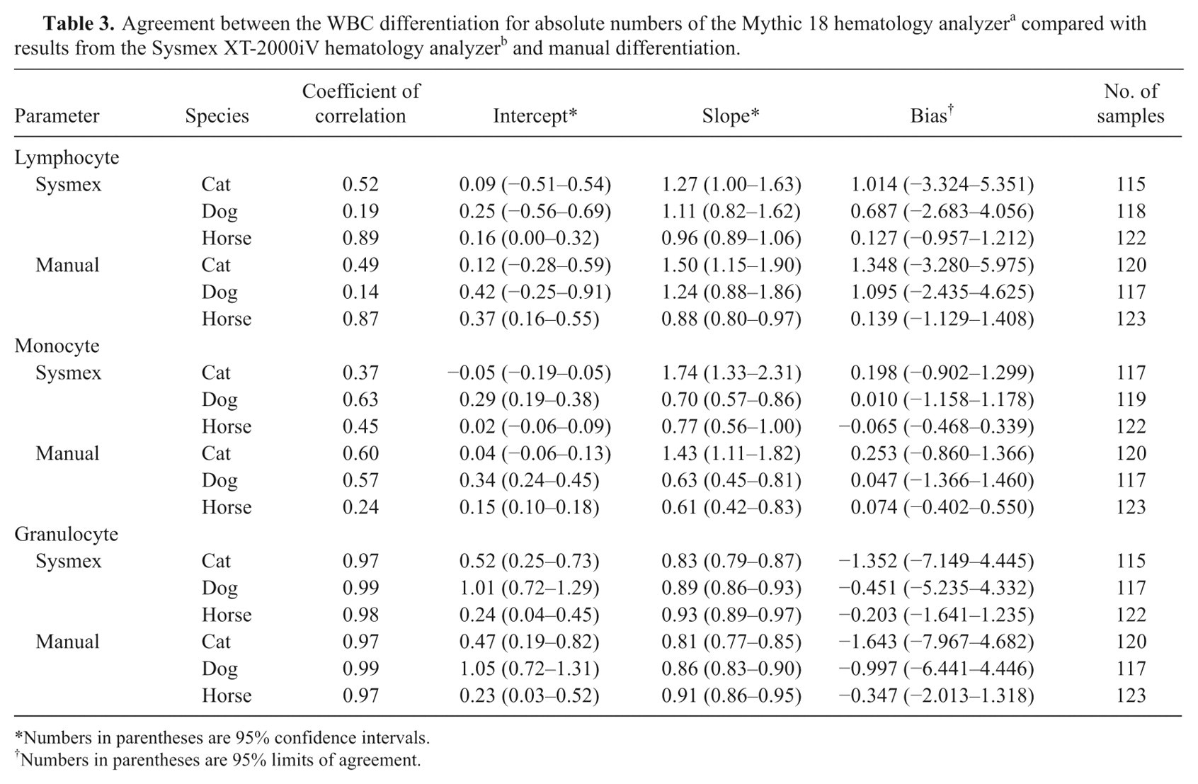
Numbers in parentheses are 95% confidence intervals.
Numbers in parentheses are 95% limits of agreement.
Precision and linearity
Mean values and CVs from the precision study are presented in Table 4 (precision in series) and Table 5 (precision from day to day). Results of the linearity study are presented in Table 6. Linearity plots are shown in Figures 4 (cat), 5 (dog), and 6 (horse). For all tested parameters, the instrument demonstrated good linearity. The tested ranges of linearity were within the ranges provided by the manufacturer for human blood except for canine WBC.
Precision in series: mean values and coefficients of variation for blood samples from cats, dogs, and horses with low, normal, and high values for total white blood cell count.*
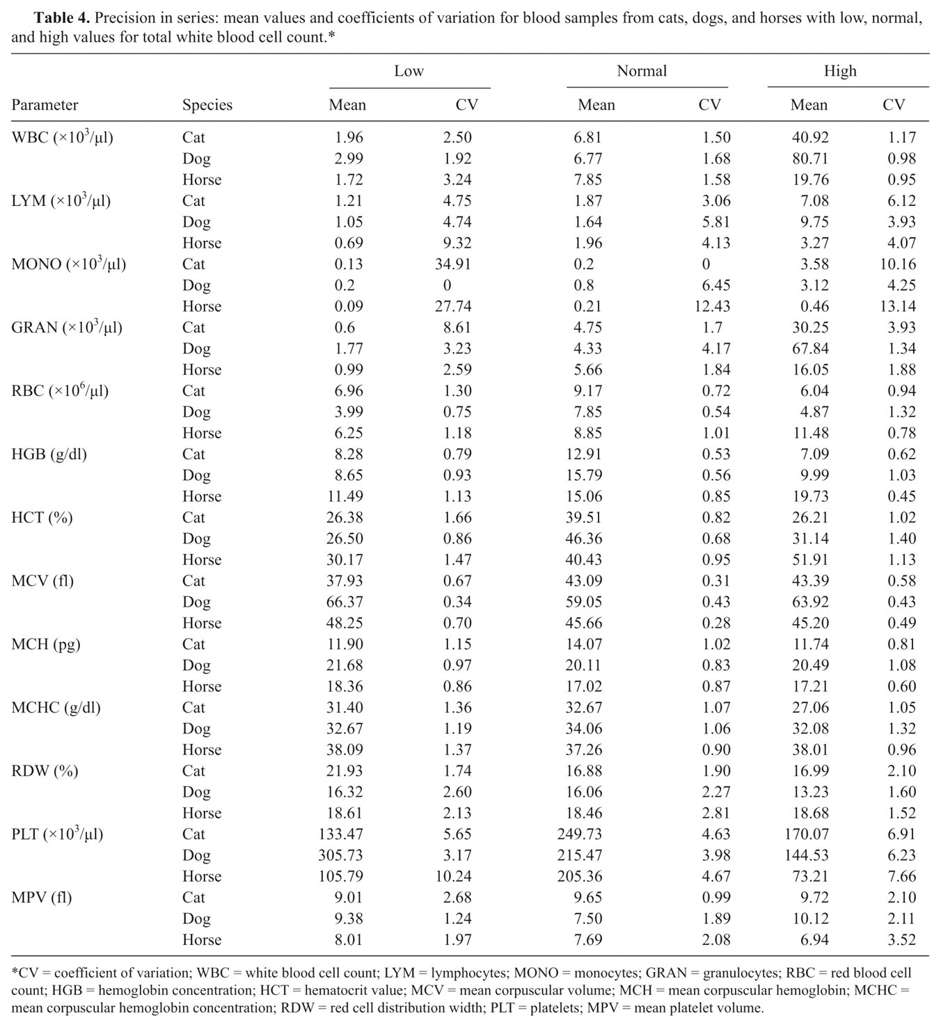
CV = coefficient of variation; WBC = white blood cell count; LYM = lymphocytes; MONO = monocytes; GRAN = granulocytes; RBC = red blood cell count; HGB = hemoglobin concentration; HCT = hematocrit value; MCV = mean corpuscular volume; MCH = mean corpuscular hemoglobin; MCHC = mean corpuscular hemoglobin concentration; RDW = red cell distribution width; PLT = platelets; MPV = mean platelet volume.
Precision from day to day: mean value and coefficient of variation for control blood samples.*
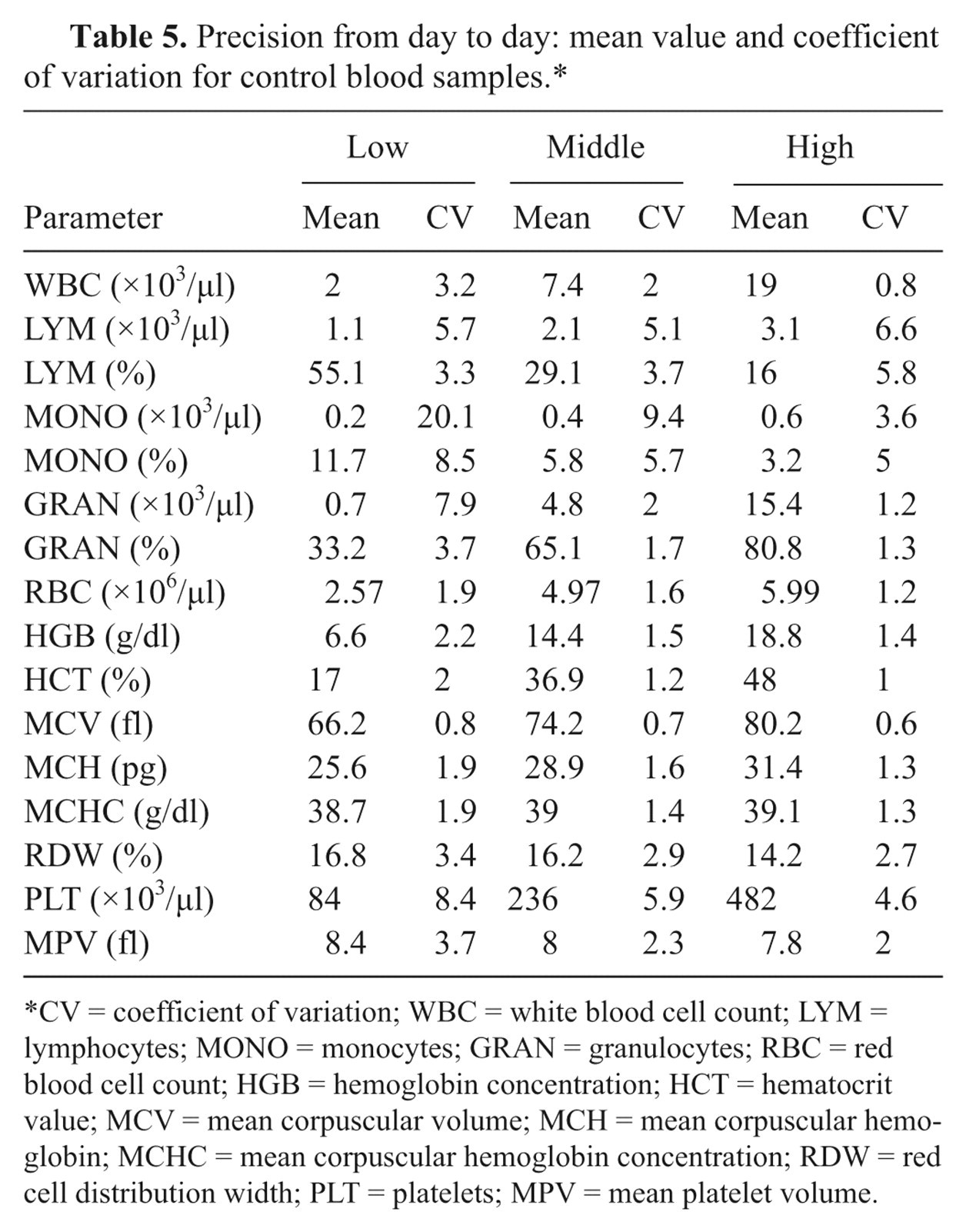
CV = coefficient of variation; WBC = white blood cell count; LYM = lymphocytes; MONO = monocytes; GRAN = granulocytes; RBC = red blood cell count; HGB = hemoglobin concentration; HCT = hematocrit value; MCV = mean corpuscular volume; MCH = mean corpuscular hemoglobin; MCHC = mean corpuscular hemoglobin concentration; RDW = red cell distribution width; PLT = platelets; MPV = mean platelet volume.
Range of linearity for canine and feline white blood cell count, red blood cell count parameter, and equine platelets in cats, dogs, and horses and for human blood samples.
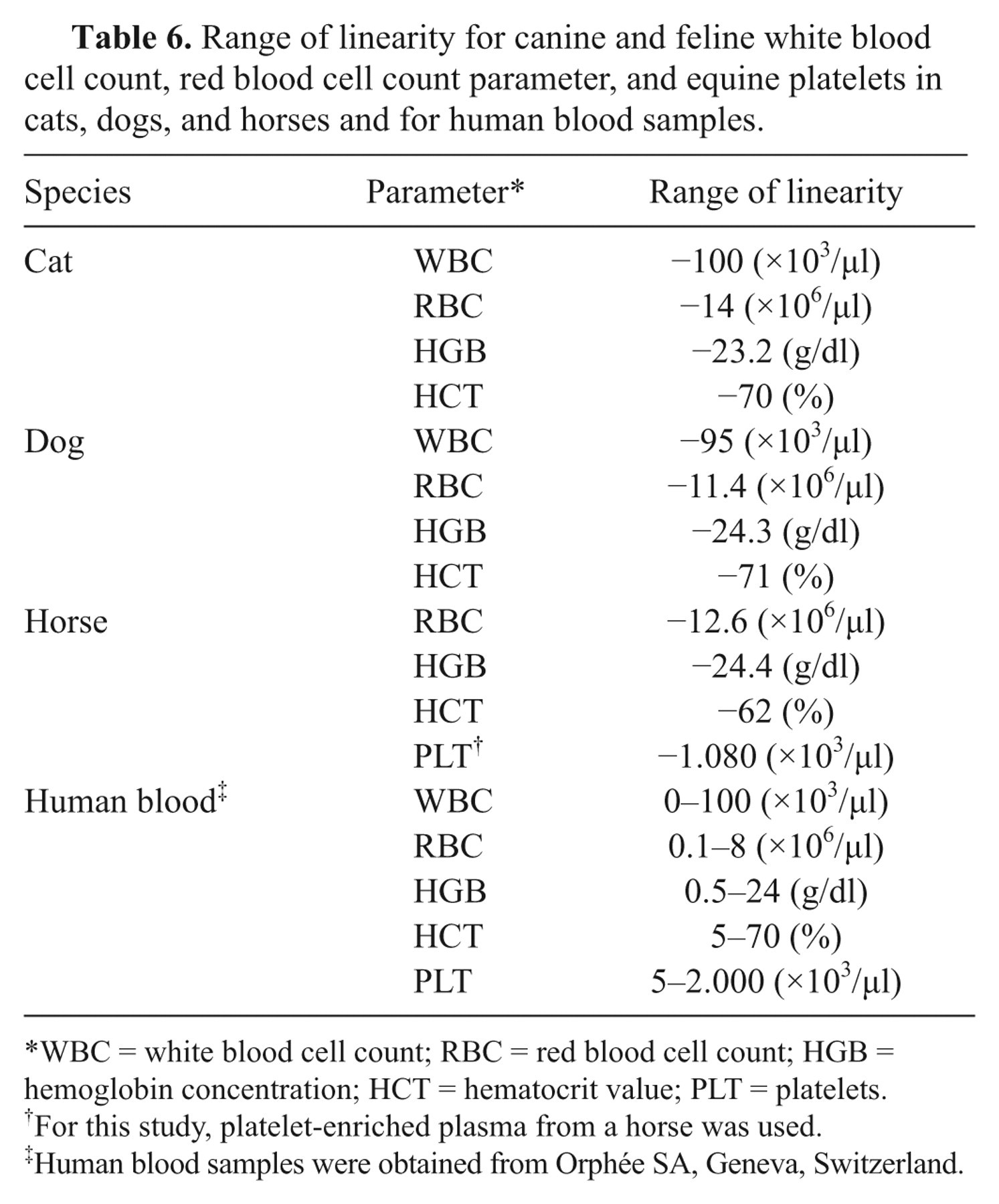
WBC = white blood cell count; RBC = red blood cell count; HGB = hemoglobin concentration; HCT = hematocrit value; PLT = platelets.
For this study, platelet-enriched plasma from a horse was used.
Human blood samples were obtained from Orphée SA, Geneva, Switzerland.
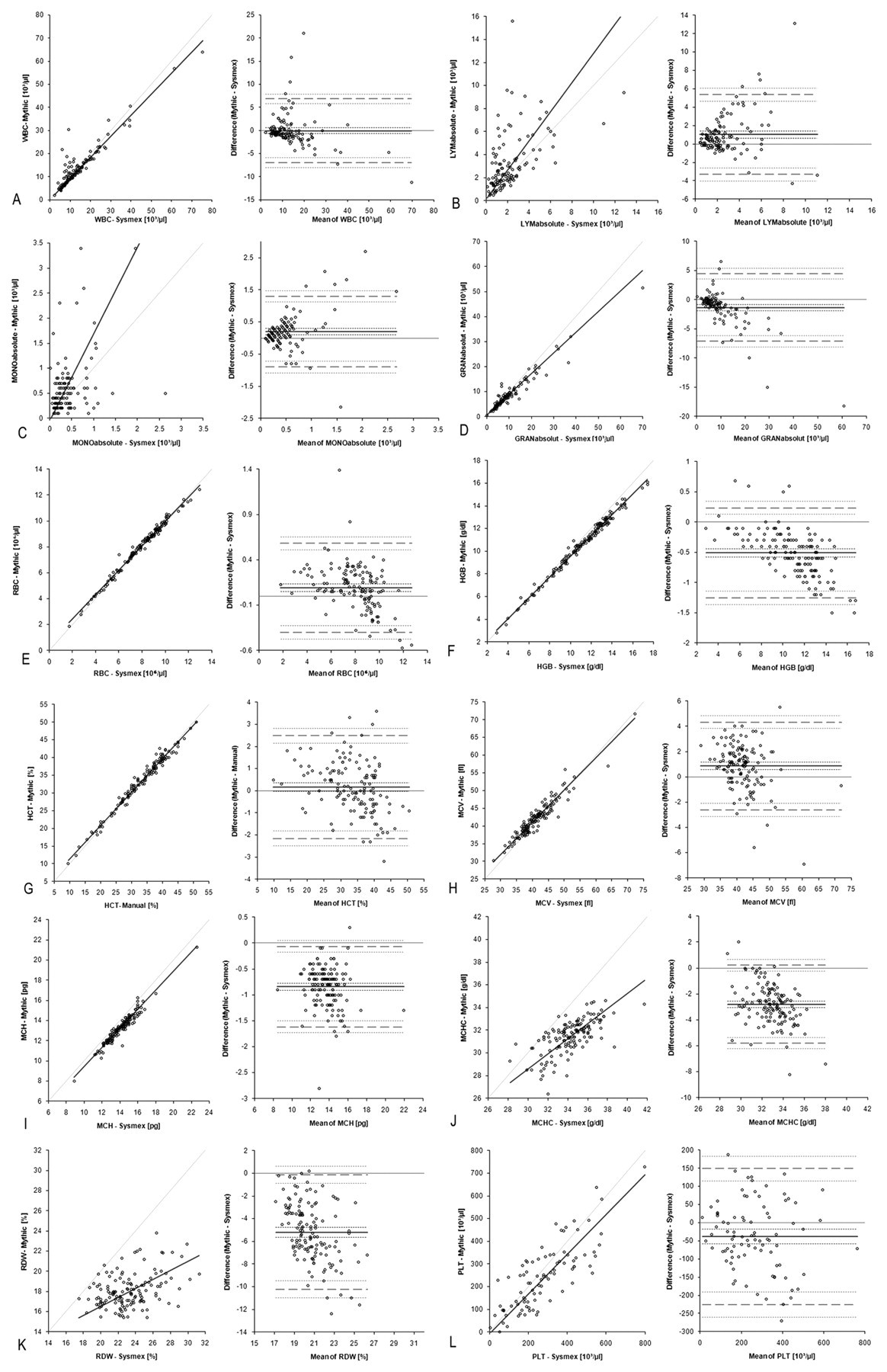
Bland–Altman analyses respectively Passing–Bablok regression for feline accuracy results. Comparison of the Mythic 18 hematology analyzer
a
with the Sysmex XT-2000iV hematology analyzer
b
resp. manual hematocrit. For feline white blood cell count (WBC;
Bland–Altman analyses respectively Passing–Bablok regression for canine accuracy results. Comparison of the Mythic 18 hematology analyzer
a
with the Sysmex XT-2000iV hematology analyzer
b
resp. manual hematocrit. For canine white blood cell count (WBC;
Bland–Altman analyses respectively Passing–Bablok regression for equine accuracy results. Comparison of the Mythic 18 hematology analyzer
a
with the Sysmex XT-2000iV hematology analyzer
b
resp. manual hematocrit. For equine white blood cell count (WBC;
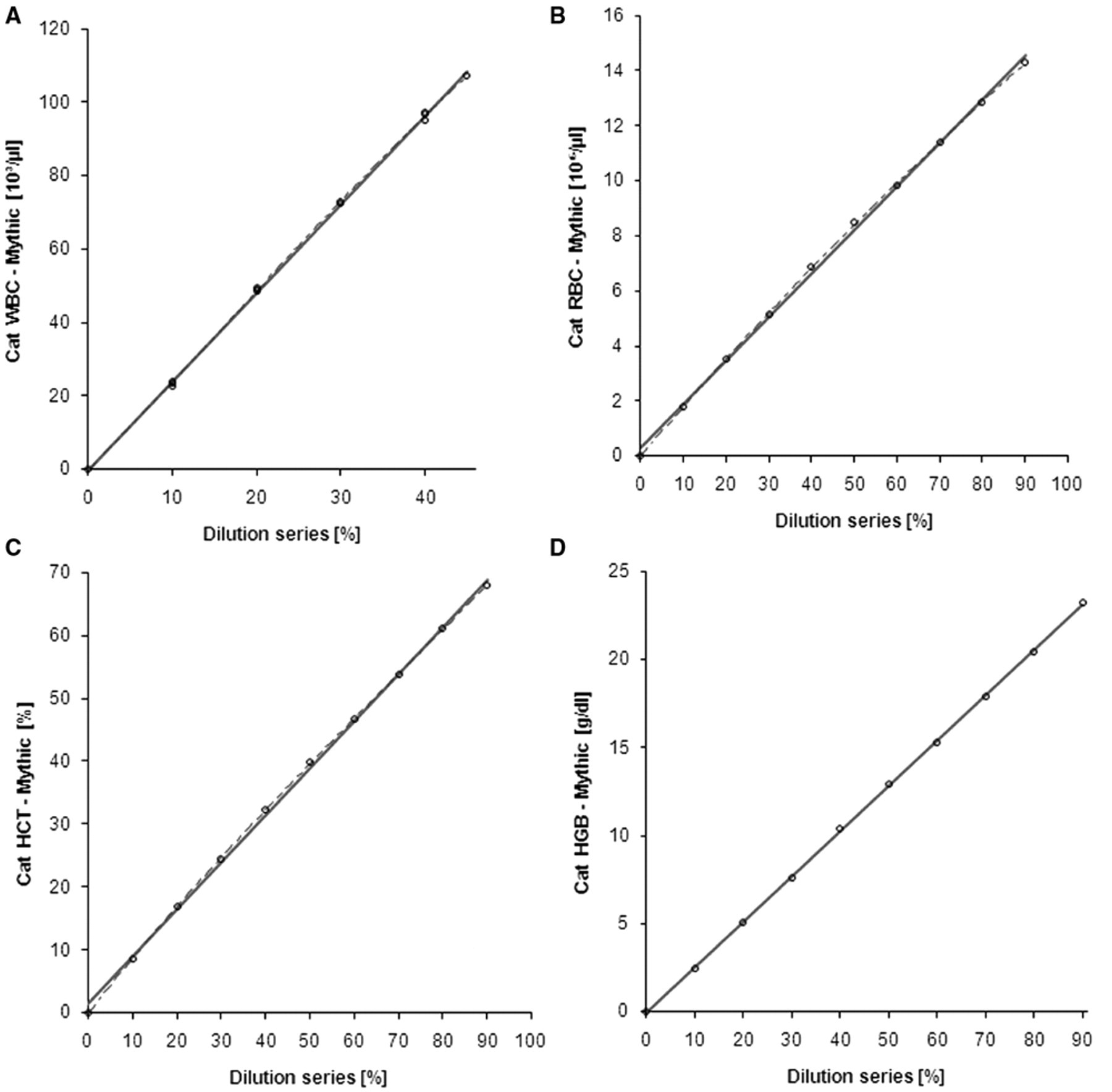
Linearity plot for feline white blood cell count (WBC;
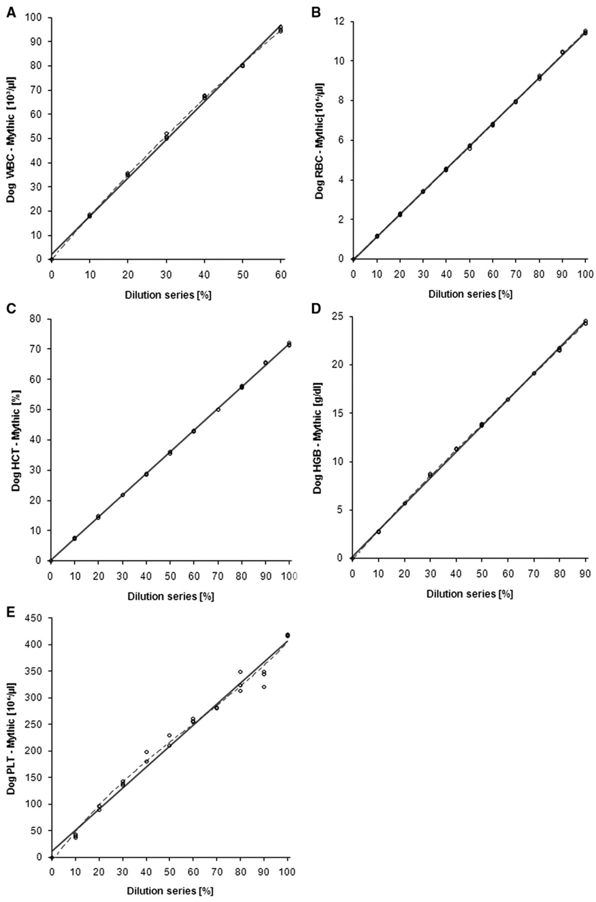
Linearity plot for canine white blood cell count (WBC;
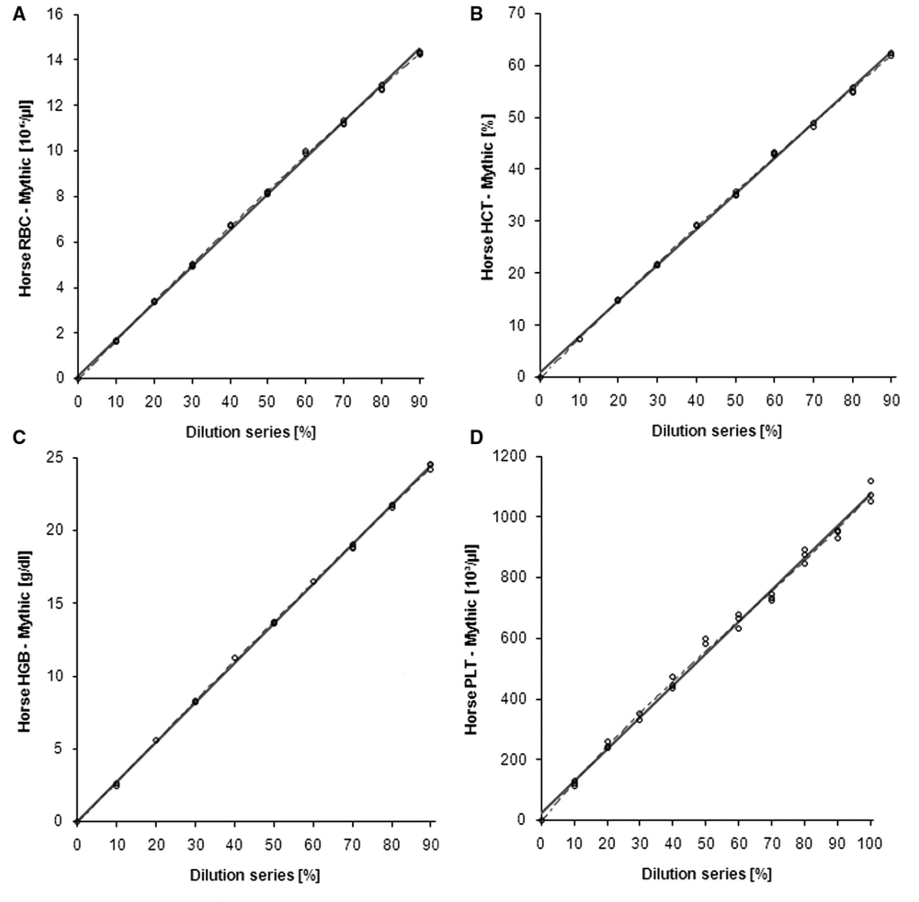
Linearity plot for equine red blood cell count (RBC;
Carry-over
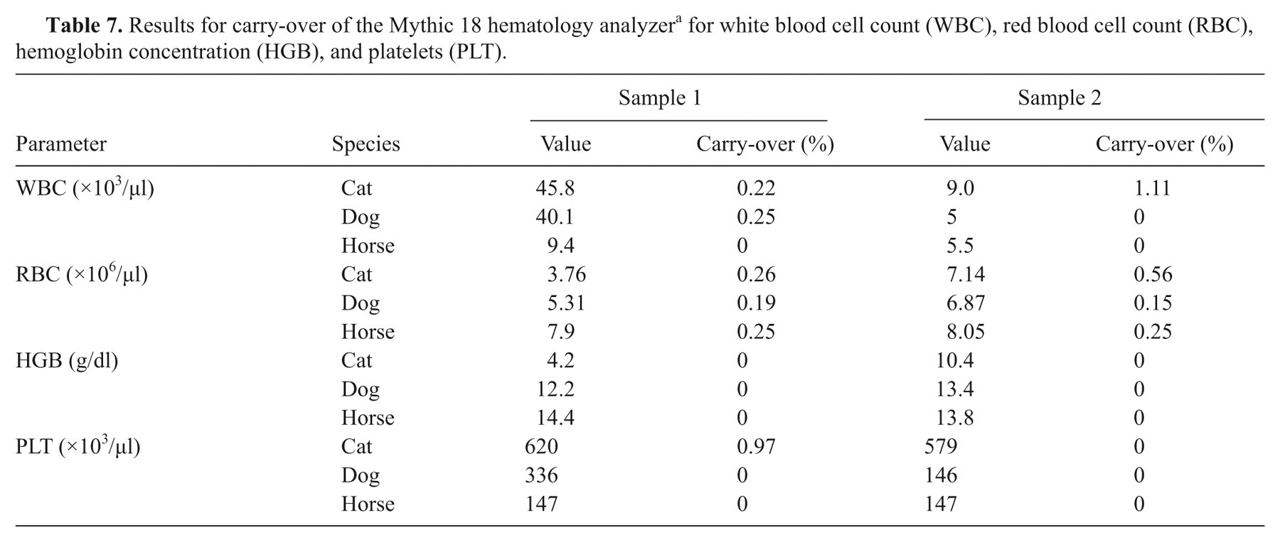
Table 7 presents the results of the carry-over experiment (“value” in Table 7 represents the result of the second sample analysis). The results of the second and third diluent analyses were always 0. All results for carry-over lie in the range provided by the manufacturer (<1%), except 1 sample for feline WBC.
Results for carry-over of the Mythic 18 hematology analyzer a for white blood cell count (WBC), red blood cell count (RBC), hemoglobin concentration (HGB), and platelets (PLT).
Cell aging
Table 8 shows the results of the cell aging study. The first significant changes appeared after 6 hr for HGB in the feline samples, but not at the remaining time points. In canine blood samples, a significant change in RBC was detected after 24 hr, but not at the remaining time points. Feline MCHC showed significant changes (i.e., a decrease) at time points 10, 24, 32, and 48 hr; in horses, such changes were also found at time points 32 hr and 48 hr. After 24 hr, equine WBC values started to decrease significantly, and the LYM-GRAN ratio moved in favor of LYM count. Canine and feline samples presented significant changes (i.e., an increase) for MCV values after 24, 32, and 48 hr. At time point 48 hr, feline HCT values, canine MCH values, and canine MONO% started to increase statistically significant.
Statistically significant changes in blood cell parameters in a cell aging study over 48 hr.
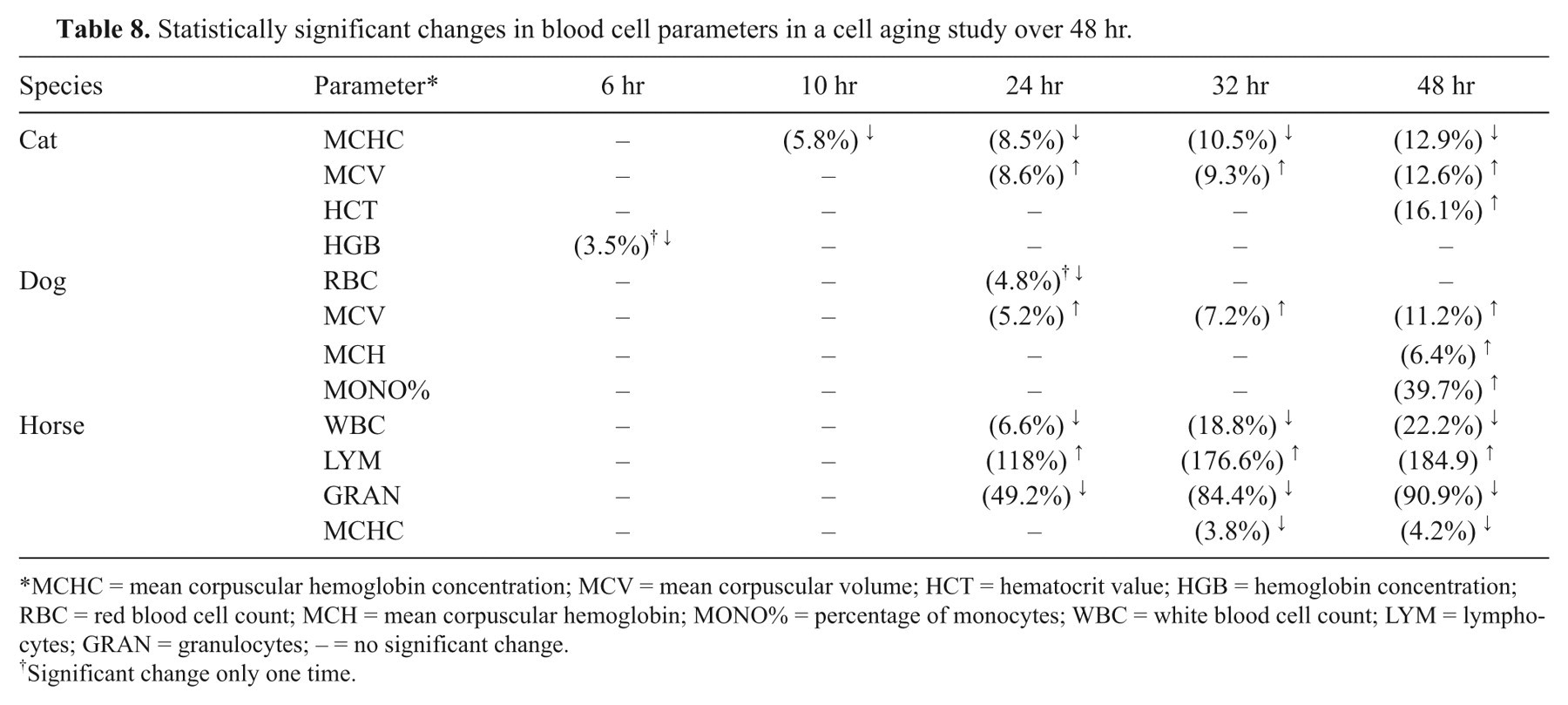
MCHC = mean corpuscular hemoglobin concentration; MCV = mean corpuscular volume; HCT = hematocrit value; HGB = hemoglobin concentration; RBC = red blood cell count; MCH = mean corpuscular hemoglobin; MONO% = percentage of monocytes; WBC = white blood cell count; LYM = lymphocytes; GRAN = granulocytes; – = no significant change.
Significant change only one time.
Clinical relevance
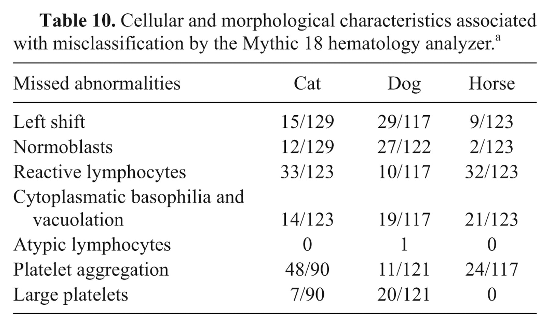
Some of the results deviate from those determined by the reference methods. In Table 9, the number of results that deviate and their clinical relevance are compiled. Table 10 presents the abnormalities seen in the blood smear during manual WBC differentiation.
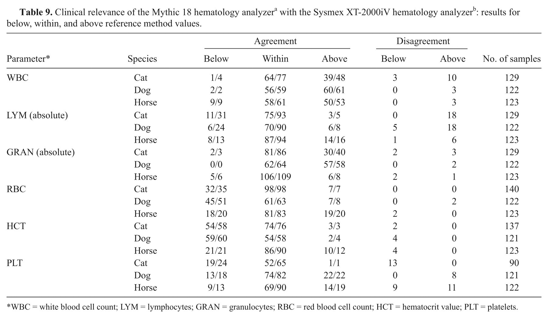
WBC = white blood cell count; LYM = lymphocytes; GRAN = granulocytes; RBC = red blood cell count; HCT = hematocrit value; PLT = platelets.
Cellular and morphological characteristics associated with misclassification by the Mythic 18 hematology analyzer. a
Discussion
The current study is based on the comparison of the 3-part WBC differential of the Mythic 18 with both manual and electronic WBC differentiation. Manual chamber counting of WBC, RBC, and PLT are well known as gold standard techniques. However, these methods show high imprecision due to the limited quantity of counted cells, artifacts, and classification of the cells. 8,10,14 Therefore, electronic blood cell counting methods have mainly replaced the former gold standard techniques. Microscopic WBC differentiation of a blood smear is still mandatory to confirm WBC abnormalities and to rule out the presence of platelet clumps, RBC parasites, and blood cell precursors. Nevertheless, this manual technique is prone to high imprecision, especially for those cells that are underrepresented in the blood. 21
Results for RBC parameter of the Mythic 18 showed very good to excellent agreement with the Sysmex XT-2000iV results, except for MCHC and RDW. Canine MCV values of the Mythic 18 showed a small negative bias compared to the Sysmex XT-2000iV, which could lead to different clinical conclusion in some cases. Small changes of the HCT correction factor in the canine settings could improve MCV agreement between the Sysmex XT-2000iV and the Mythic 18. Otherwise, adjustment of the reference limits for canine MCV would be indicated. Difference in the osmolarity of the diluents between the Sysmex XT-2000iV (250 mosm/kg) and the Mythic 18 (332 mosm/kg) can possibly cause a negative bias in canine MCV values. The hypotonic diluent of the Sysmex XT-2000iV causes swelling of the RBC whereas the relatively isotonic diluent of the Mythic 18 produces comparatively lower MCV values. 4 The agreement for MCHC values is less satisfactory in all 3 evaluated species. Low correlation for this parameter has been reported in previous studies 18,21,23 and can be mainly explained by the narrow concentration range of this parameter.
Generally, the Mythic 18 underestimated high total WBC in all 3 species on average by a few percentage points. In the feline samples, total WBC correlation is very good. However, in 23 out of 129 feline samples, the WBC were on average more than 1,000 WBC/µl higher than those of the reference method in which the WBC are determined by an optical flow cytometry principle 9 (Fig. 1A). This can readily be explained by the fact that feline PLT have a high tendency to aggregate. When 2 samples with the extremely high overestimation of more than 15.000 WBC/µl were removed from the statistic as outliers, the coefficient of correlation improved to 0.97 and the bias decreased from –0.043 to –0.364. In all 23 samples, platelet aggregates could be identified in the blood smear. It is a well-known phenomenon in cats that platelet clumps or large platelets can cause falsely increased WBC and decreased PLT counts in impedance-based hematological instruments. 10,16
In horses, cats, and dogs, GRAN are predominant in the blood (Table 1), therefore imprecision for this leukocyte subtype is low, and the Mythic 18 showed excellent agreement with both the Sysmex XT-2000iV and the manual WBC differentiation. 17 Agreement in canine and feline LYM counts in the Mythic 18 was not satisfactory. This finding has already been demonstrated for both species in the VetScan HMT, 6 and for canine samples in the CA530-Vet 17 and the Heska CBC (Becker M: 2007, A comparative study of seven in-house and two laboratory hematology instruments. Dissertation. Justus-Liebig University, Giessen, Germany). As LYM count in horses showed good agreement, the Mythic 18 can be judged as reliable for counting LYM in horses.
Compared to previous studies of impedance-based hematology instruments, the Mythic 18 showed better agreement with the reference methods for PLT counts for cats, dogs, and horses (Becker M: 2007, A comparative study of seven in-house and two laboratory hematology instruments). 5 The slight overestimation of canine PLT compared with results of the Sysmex XT-2000iV could be explained by the fact that the Mythic 18 has no sheath flow device. The good results for feline PLT counts were remarkable, particularly as samples with platelet aggregates were included in the calculation. Impedance-based hematology instruments have problems in counting feline PLT accurately. 16 The PLT and RBC sizes in cats often overlap, 24 and impedance-based instruments differentiate cells based on their volume. In the present study, more than 53% of the feline samples showed platelet aggregation.
Results for the linearity study showed that the Mythic 18 underestimated high WBC values and high HCT values. This is not of severe clinical relevance, as these values were far above the upper reference limit. In the majority of samples analyzed, results of the Mythic 18 would have led to the same clinical interpretation as the results obtained by the reference methods.
Misidentification of platelet aggregates as leucocytes was the most likely cause of lack of detection of leucopenia in 3 feline samples, and identification of erroneous leucocytosis in 10 feline samples. Twenty feline cases with lymphocytopenia were missed with the Mythic 18 due to the presence of platelet aggregates or large platelets. In all cases where the Mythic 18 revealed a lymphocytopenic cell count result, the results were accurate. This does not exclude that occasionally a lymphocytopenia may not be detected if more samples had been tested. For the high degree of misclassification in the LYM count of the dog, no obvious explanation can be provided.
The misidentified feline sample with granulopenia showed only a slight difference (7%), and would not have led to a different clinical conclusion. In the 10 cases where the Mythic 18 missed granulocytosis, the cases showed lower total WBC in the Mythic 18 compared to the Sysmex XT-2000iV. In 2 out of the 10 cases, the clinical interpretation would have been different. Platelet aggregates had led to overestimated WBC in 3 feline samples, as described above, which therefore led to false results of granulocytosis. False-positive granulocytosis and granulocytopenia in equine samples was mainly due to differences in total WBC between the Mythic 18 and the reference instrument.
For RBC, differences in canine samples were all below 10%. For equine HCT values, 4 false-positive samples assuming anemia occurred. The differences were less than 2%, which is attributed to the imprecision of the HCT reading in the capillary tube.
In 19 out of 90 cat samples, the Mythic 18 and the Sysmex XT-2000iV showed thrombocytopenia. Five feline samples with thrombocytopenia were detected only by the Sysmex XT-2000iV. Four out of these 5 feline blood samples demonstrated moderate to severe platelet aggregation in the blood smear. Additionally, in 13 feline samples, the Mythic 18 falsely showed a thrombocytopenia. Platelet aggregation was only found in 1 of these 13 cases, and giant platelets were found in 3 cases. In the remaining cases, no explanation for the detection of false-positive thrombocytopenia can be offered. It has been demonstrated that EDTA anticoagulated blood is prone to build platelet aggregates in cats. 15 High CVs in the equine precision study as well as the narrow range of reference limits may contribute to the high rate of misclassification in equine PLT counts.
In the present study, important abnormalities would have been missed when relying only on the electronic WBC differential of the Mythic 18 (Table 10). Two canine samples showed more than 4 nucleated red blood cells, while 1 feline sample showed 52 nucleated red blood cells. In these samples, WBC results were falsely increased and would have led to different clinical conclusions. One canine sample presented atypical LYM due to an immune-mediated disease. Left shifts and especially degenerative left shifts would have been missed in a remarkable number of blood samples in the canine and feline samples. Foamy cytoplasm of segmented neutrophils has been observed in all 3 species by manual microscopy. This is an important morphological indicator for severe inflammatory disease and toxicity. The presence of reactive LYM is a useful hint to antigenic stimulation in the patient. 19 These pathological findings give important information to the clinician and help to improve patient care.
The Mythic 18 was found to perform very well for RBC parameters and total WBC in all investigated species. In cats, it is important to ensure that no platelet aggregates are presented, otherwise WBC and PLT values should be determined by manual methods. The GRAN and LYM counts are accurate in horses. In dogs and cats, absolute GRAN counts are reliable. As with all impedance-based hematological instruments, a microscopic blood smear evaluation is needed to identify platelet aggregates, normoblasts, left shift, cell precursors, and blood parasites and to verify WBC differentiation. Flags for pathological values and reference limits need to be created by the manufacturer of the instrument.
Footnotes
Acknowledgements
The authors thank the technicians of the Clinical Laboratory, Vetsuisse Faculty, University of Zurich, for excellent laboratory assistance, especially Juliette Wälchli. The authors are grateful to Philippe Daire for his excellent support.
a.
Orphée SA, Geneva, Switzerland.
b.
Sysmex Corp., Kobe, Japan.
c.
Myt-3D, lots B059, B089, B119, Orphée SA, Geneva, Switzerland.
d.
Mythic 18 Vet M-Pack, Orphée SA, Geneva, Switzerland.
e.
HemaTek, Siemens, Switzerland.
f.
Rotina 35 R, Hettrich AG, Switzerland.
g.
Microsoft Excel® 2007, Microsoft Corp., Redmond, WA.
h.
Analyse-it Software Ltd., Leeds, United Kingdom.
i.
Version 3.00 for Windows, GraphPad Software, San Diego, CA.
The authors declared that they had no conflicts of interest with respect to their authorship or the publication of this article.
This study was funded in part by Orphee SA (Geneva, Switzerland). The authors declared that they received financial support for this project (salary for doctoral candidate AKW). A contract guaranteed independent publication of the results.