
Abstract
Bullfrog tadpoles (Rana catesbeiana) from a wastewater treatment facility were identified with severe lesions consisting of large, up to 1-cm in diameter, mineralized nodules protruding from the tail or gular region. Sectioning of formalin-fixed specimens revealed more extensive mineralization involving the vertebrae or muscles of the head and tail. Nodules examined microscopically were not associated with parasitic or infectious agents. Large nodules consisted of mineralized aggregates surrounded by a margin of granulomatous inflammation. Individual connective-tissue fibers and muscle cells were mineralized at some foci. The nodules consisted entirely of calcium phosphate, and the lesions appeared to be novel. Total serumcalcium concentrations of tadpoles and calcium concentrations in water samples did not differ significantly with increasing distance from the discharge site. Affected tadpoles had elevated cholecalciferol (25-OH-vitamin D3) levels. Effluent from this wastewater treatment facility is divided into 3 streams, each passing through a separate series of wetlands allowing for replicated evaluation of tadpoles with increasing distance from the proximate inputs of treated wastewater. The prevalence of lesions was correlated with proximity of cells to the initial wastewater discharge site, and 28.5% of bullfrog larvae in the first cells had lesions. None were affected in the fifth cells. Southern leopard frog larvae (Rana sphenocephala), the only other species affected, had a much lower prevalence of lesions (<1%) than bullfrog tadpoles and were only affected in the first cells. To date, the primary cause of elevated cholecalciferol is undetermined, but it appears to be remediated by passage of water through the wetlands.
Constructed wetlands are used in the tertiary treatment of water discharged from wastewater treatment facilities. 15 The site referenced in the current study is a municipal wastewater treatment facility with typical primary and secondary wastewater treatment systems. By the time water is discharged from the secondary system, many contaminants have been reduced to low level, and the water is deemed safe for release into a receiving stream. However, as a measure to reduce contaminant levels, the treated effluent is pumped into 3 parallel series of constructed wetlands. Each series of wetlands consists of 6-8 individual cells, which are shallow bodies of water that receive effluent in succession. Bacteria in the wetlands provide additional degradation of some chemicals not removed or degraded by the secondary treatment, and wetland plants absorb excess nitrogen and phosphorus. 15
The production of wildlife habitat is often touted as an incidental benefit of such constructed wetlands, and researchers at The University of Georgia (Ruiz, Fisk, Davis, and Maerz; Athens, Georgia) designed a study to investigate whether constructed wastewater-treatment wetlands were suitable habitat for amphibians. 7 A survey was initiated to assess the relative abundance and performance of tadpoles along the replicate lines of wetlands. Tadpoles of several species including bullfrogs (Rana catesbeiana), southern leopard frogs (Rana sphenocephala), green frogs (Rana clamitans), and green tree frogs (Hyla cinerea) were present, but bullfrog larvae were most abundant. Approximately 30% of the bullfrog larvae in the first wetland of each series had grossly visible nodules protruding from the tail. Initially, a subset of 12 second-year bullfrog larvae was collected for diagnostic investigation based on the presence of externally visible lesions. All were captured from the 3 initial wetlands, where the majority of the affected tadpoles were identified. The tadpoles were collected alive, euthanized by an overdose of 0.1% buffered tricaine methanesulfonate (MS-222), a and were immediately placed in 10% neutral buffered formalin.
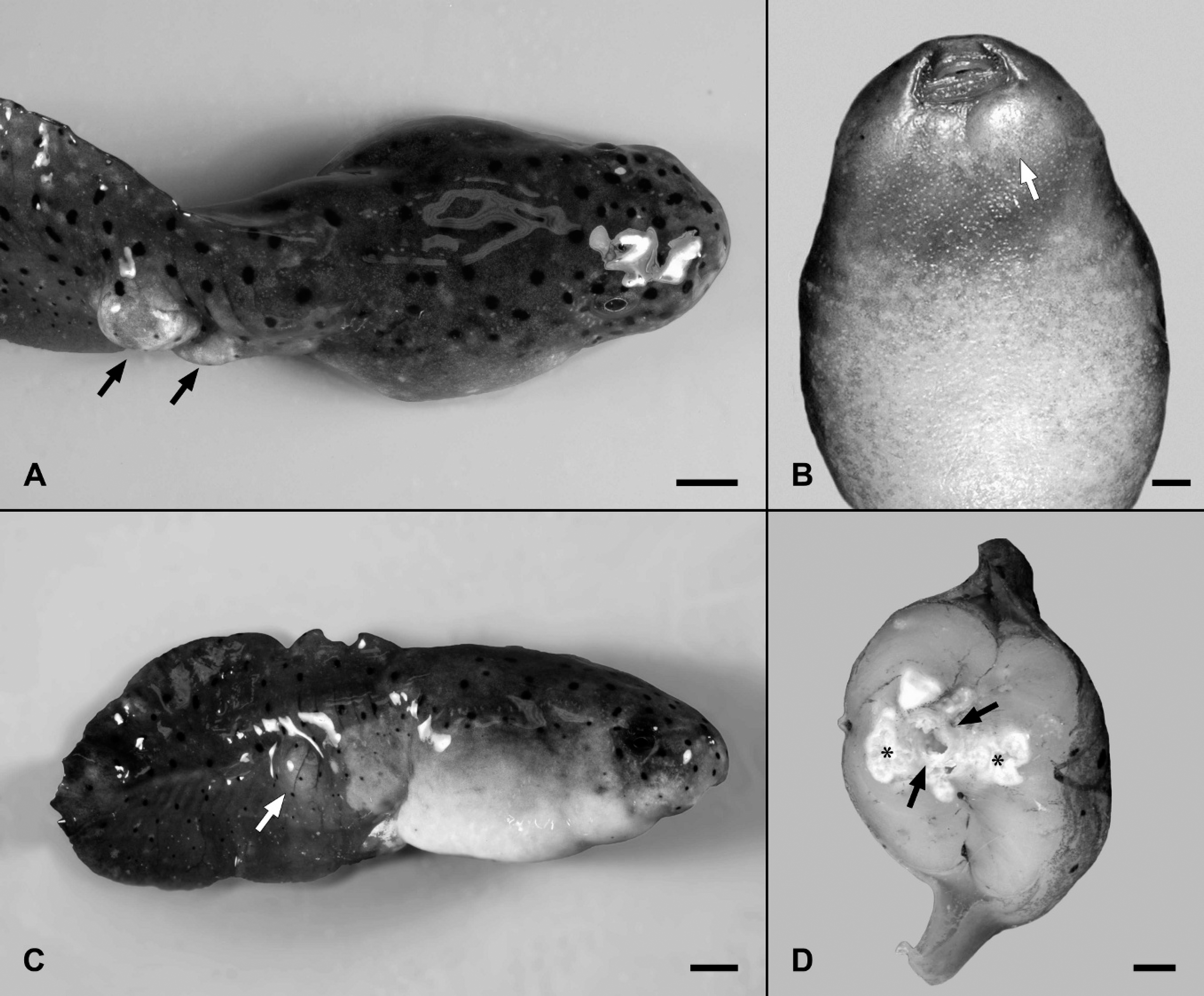
A, second-year bullfrog larvae with grossly visible nodules that elevate the skin and protrude laterally from the tail (arrows). Bar = 0.5 cm.
The 12 affected larvae were submitted to the Southeastern Cooperative Wildlife Disease Study, College of Veterinary Medicine, The University of Georgia. Ten of the tadpoles had firm, tan or light-yellow nodules that elevated the skin of the tail up to 2 cm (Fig. 1A). Four of these also had nodules deep in the gular region that slightly elevated the skin of the throat (Fig. 1B). Seven of the tadpoles had shortened tails, ruffled fin margins, and/or caudal scoliosis with stiffening of the affected portion of the tail (Fig. 1C). Transverse sections of all frog larvae revealed additional mineralized nodules not visible from external surfaces. The nodules were located deep in the skeletal muscle and sometimes extended to deformed caudal vertebrae (Fig. 1D). On cut surface, most nodules were variably hard and gritty, but some were softer and pasty. Rarely, they consisted of fluid-filled cystic structures with a margin of white to tan, pasty material. Formalin-fixed samples were embedded in paraffin, and 5-μm thick sections were stained with hematoxylin and eosin (HE).
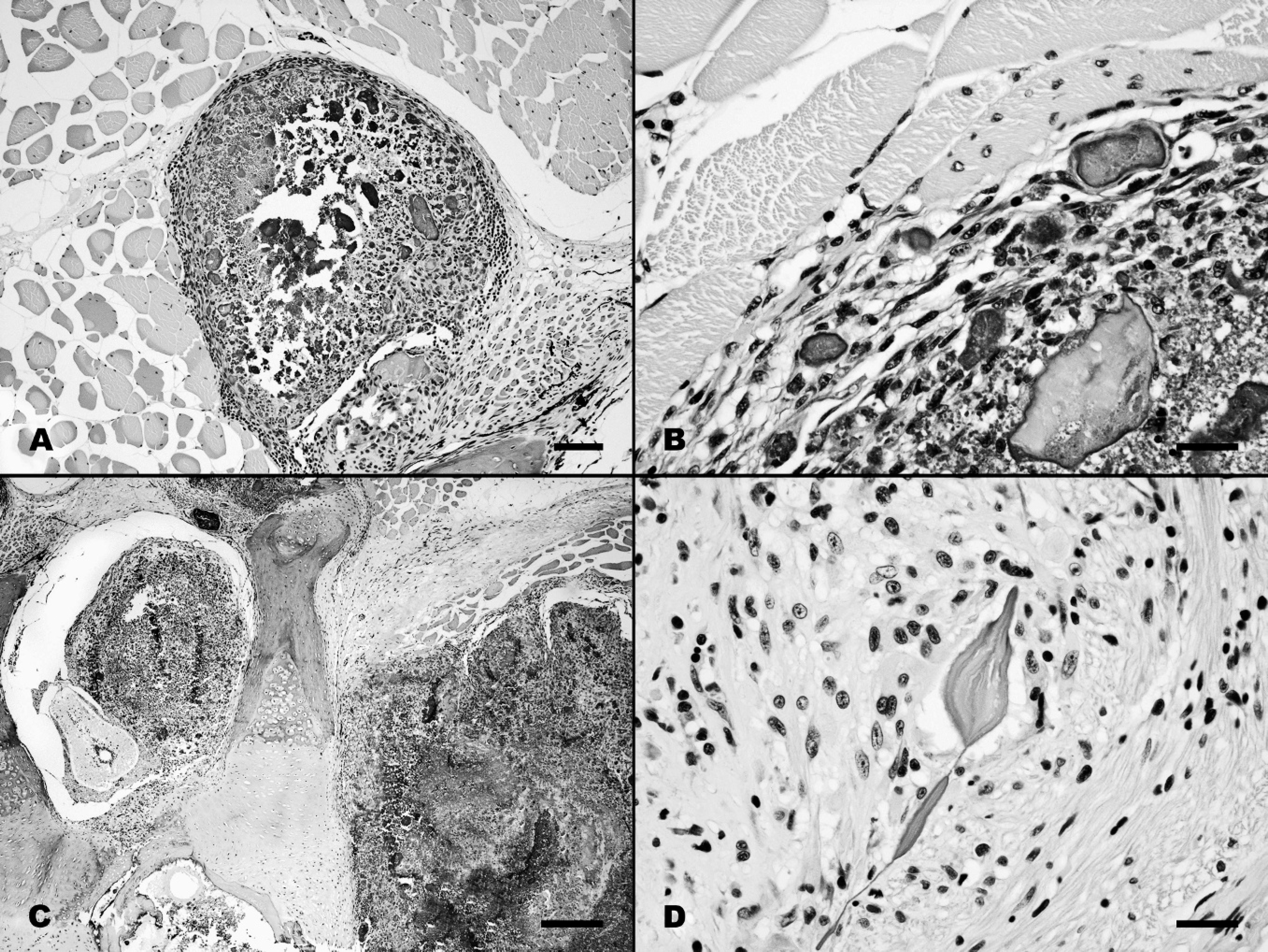
A, the large nodules consist of a central mass of calcified tissue surrounded by a foreign body inflammatory reaction with macrophages, giant cells, and concentric fibrosis. Hematoxylin and eosin (HE) stain. Bar = 100 μm.
Microscopically, the nodules consisted of variably sized and shaped mineralized fragments, surrounded by macrophages, giant cells, and fibroblasts with immature fibrous connective tissue (Figs. 2A, B). These mineralized foci were scattered in deep tissues of the tail causing focally extensive destruction of the muscles. Some foci effaced the cartilage of the caudal vertebrae and occasionally extended into the vertebral canal (Fig. 2C). The mineralized nodules in the gular region were microscopically identical to those in the tail. Some small isolated foci were identified in which individual myofibers or small connective tissue bundles were mineralized (Fig. 2D). A small number of macrophages and giant cells surrounded some of the smaller lesions, and there was a continuum between these and the larger mineralized aggregates with more extensive inflammation and tissue damage. The cystic lesions consisted of fluid-filled spaces within aggregates of mineralized debris; the mineralized material and associated inflammation and tissue damage appeared identical to those of the more solid nodules.
Serial sections were made through tissues containing the large mineralized aggregates to search for parasites or infectious agents that might have resulted in tissue damage with dystrophic mineralization. Representative sections were stained with HE, Gomori methenamine silver (GMS) stain, and a Giemsa stain. Histopathology confirmed that 6 of the 12 tadpoles were infected with Batrochytrium dendrobatidis. Microscopically, fungal elements were isolated to the keratinized denticles, and the denticles were variably eroded. Ten tadpoles had encysted metacercaria in various tissues, but these were associated with minimal inflammation. Ichthyophonus-like organisms were identified in the tissues of 1 tadpole. The parasites and fungi in these tadpoles were not associated with any mineralization and were determined to represent incidental findings.
Additional tadpoles with gross lesions were collected for additional diagnostic assays. An electrolyte panel, including analysis for sodium, potassium chloride, bicarbonate, anion gap, calcium, and phosphorus, was performed on serum from 5 bullfrog tadpoles with grossly visible nodules as described. All 5 of the affected tadpoles were collected from wetland C1, which receives direct inputs of treated wastewater. Additionally, 5 clinically normal bullfrog tadpoles, of the same developmental stage, were collected from a separate reference site 68.9 miles northeast of the constructed wetlands complex. 3 These apparently normal tadpoles were evaluated by the same electrolyte panel. No significant differences were evident between reference tadpoles and wastewater wetland tadpoles that had gross lesions. Water calcium concentrations were evaluated for the first and fifth cells in each series, but they did not differ appreciably. The average calcium concentration from the first and fifth cells was 1.4 mg/dl and 1.3 mg/dl, respectively.
Samples of the mineralized material from 2 tadpoles were collected and dried. The resultant material consisted of light-tan calculi from <1 mm in diameter to 4.9 mm × 4.1 mm × 2.8 mm, with a rough surface. Examination of the material at the Gerald V. Ling Urinary Stone Analysis Laboratory, University of California (Davis, California) revealed that material from both tadpoles consisted of 100% apatite, which is a salt of calcium phosphate [Ca5 (PO4)3 (OH)].
Six bullfrog tadpoles from wetland C1 were pooled, macerated, and analyzed by a commercially available radioimmunoassay for concentrations of 25-OH-vitamin D3 (cholecalciferol). b Six reference tadpoles of the same developmental stage were collected from the previously mentioned reference site and pooled for analysis in the same manner. The concentration in the pooled tadpole tissues from C1 was 6 nmol/l, whereas there were no detectable limits in reference tadpoles. There was no detectable cholecalciferol in water samples from C1, even though samples were concentrated by dehydration.
A previous study reported that 28.5% of the bullfrog tadpoles in first cells show externally visible lesions associated with mineralization but the frequency drops to 0.6% among third cells, and that mineralized tissues were never identified among fifth cells. 12 This is strong evidence that the cause of the mineralization is associated with proximate exposure to treated wastewater, and that the cause is remediated with transit of water through wetlands.
In addition to bullfrogs, southern leopard frog tadpoles were identified with the characteristic nodules but at a much lower prevalence. Only 2 southern leopard frog larvae, of the 358 collected from the initial wetlands, had lesions. None of the other species of frog larvae collected were ever identified with the characteristic nodules. However, the distributions of these species were nonrandom, and the first wetlands in each series contained few individuals of species other than bullfrogs. 12 This nonrandom pattern of distribution confounds the authors' ability to compare relative patterns of mineralization among the representative species. The domination of the initial wetlands by bullfrog larvae could have put them in a habitat more at risk for development of lesions, but the low prevalence among southern leopard frog larvae in the initial cells suggests the bullfrog larvae are more susceptible.
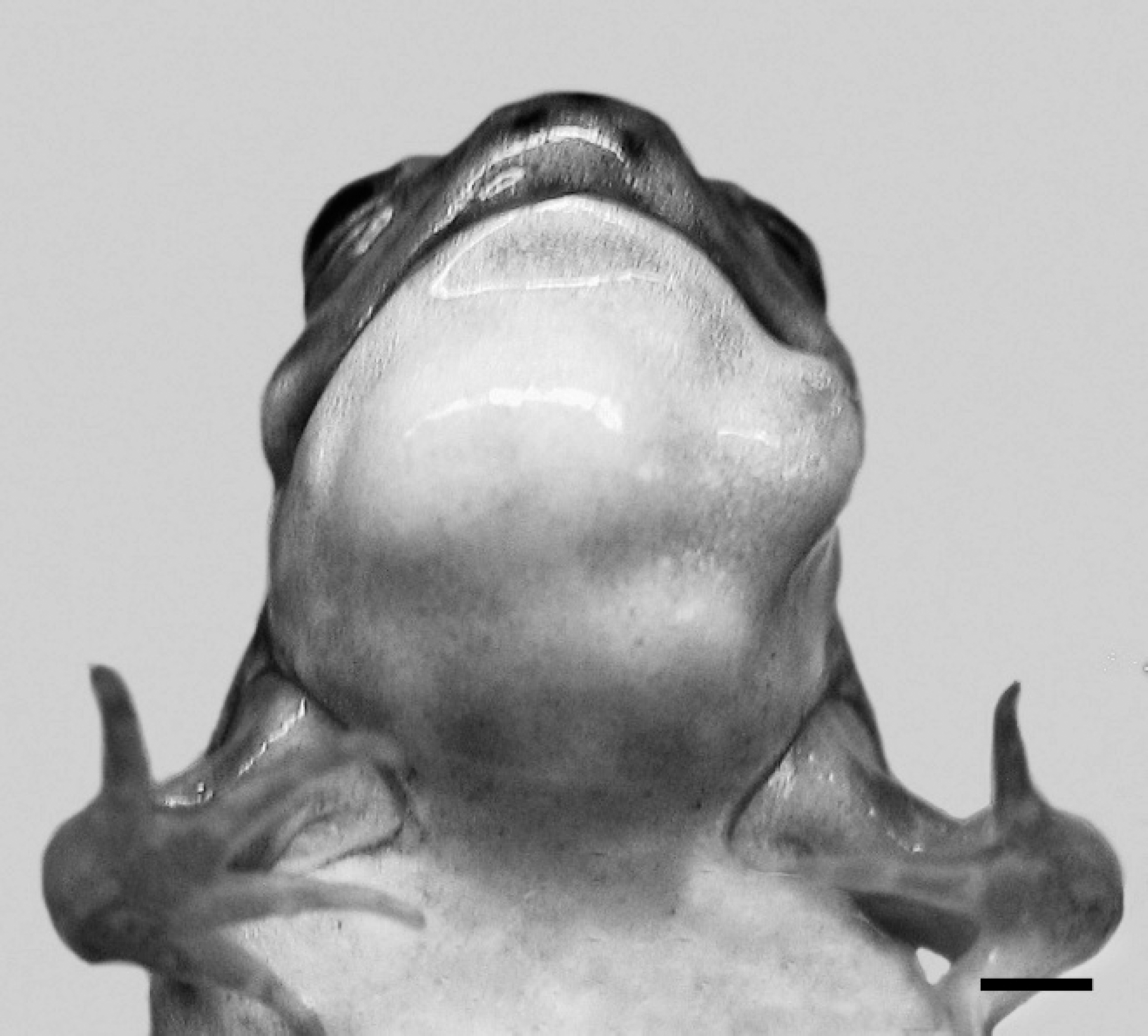
A recently metamorphosed frog that has failed to resorb the mineralized nodules that distort the soft tissues of the gular region. Bar = 0.25 cm.
Several bullfrog tadpoles with large nodules were permitted to complete metamorphosis in the laboratory to determine if the nodules would interfere with their maturation. In each instance, as the tails were resorbed, the caudal nodules were extruded. However, gular nodules remained, and the frogs died soon after metamorphosis (Fig. 3). Histopathology revealed that the skin overlying the nodules became ulcerated and resulted in severe localized bacterial infections with pleomorphic bacteria. Fresh samples were not available for bacterial culture and identification.
The ultimate cause of the mineralization in the frog larvae remains to be determined, but local destruction of tissues by parasitic or infectious agents with subsequent dystrophic mineralization could be excluded. The continuum between mild lesions, consisting of individual mineralized myofibers or fibrous connective tissue bundles, and the larger lesions, suggests they begin as isolated mineralized foci and expanded with increasing deposition of calcium salts around the original nidus. The mineralization results in a foreign body response with severe inflammation that ultimately causes progressive tissue destruction and further dystrophic calcification. Other diseases have been described with nodular aggregates of mineralized debris consisting of dystrophic mineralization occurring secondary to systemic mineral imbalances. Tumoral calcinosis of humans, for example, is a hereditary disease of dysfunctional phosphate metabolism with lesions that histologically bear a striking resemblance to the tadpole nodules. 10
The source of the elevated cholecalciferol could not be confirmed. The initial assumption was that it was an anthropogenic contaminant. However, fish, turtles, and adult bullfrogs examined from the site did not appear to be affected. If the environment was contaminated with high levels of cholecalciferol, it could be argued that other species and life stages of frogs should also have lesions indicative of toxicosis. Cholecalciferol was not found in the water. However, because it is a fat-soluble compound, this may not be an accurate measure of environmental availability. It is possible that it is sequestered in the environment by interaction with other molecules in the plant material, detritus, or sediments. If cholecalciferol is a primary contaminant, bullfrog tadpoles might be more susceptible than other species or life stages because of dietary differences, localization to specific microenvironments, or enhanced susceptibility because of their stage of development.
One potential but unproven source of cholecalciferol at the wetland site is production by plants. The plants most commonly associated with calcinosis, such as Cestrum diurnum and Solanum malacoxylon, produce 1,25-dihydroxycholecalciferol, the most metabolically active congener of vitamin D. 2,8,9 However, cholecalciferol intoxication can also result from ingestion of plant tissues, as is the case with the disease caused by Trisetum flavescens. 11 Plant-induced calcinosis among livestock is well documented, but only terrestrial plants have been described with vitamin D-like activity. 9 It remains to be seen if similar production occurs among aquatic species. However, the plants present at this site are well known, have not been associated with calcinosis or the production of calcinogenic compounds, and are present in both affected and unaffected cells.
The alternative to an unknown exogenous source of cholecalciferol is that exposure to treated wastewater is triggering the inappropriate endogenous production of cholecalciferol or slowing its degradation. 5,6 Potential causes are complex, but the sensitivity of amphibians to waterborne endocrine disruptors is well documented. 1 Endocrine-disrupting chemicals have been implicated in the reduced production of gonadotropin-releasing hormone in bullfrogs and green frogs with limb malformations. 13 Triclosan, a bactericidal compound present in many products, disrupts expression of the thyroid hormone-associated gene and can subsequently alter the postembryonic development of frogs through impaired thyroid hormone activity. 14 The herbicide atrazine, at low doses applied to tadpoles, induced hermaphroditism in frogs, possibly by inducing aromatase and promoting conversion of testosterone to estrogen. 4 There appear to be no published accounts of amphibian endocrine disruption associated with elevated cholecalciferol or the lesions observed in the present case. However, it would be naive to deny that there is much to learn regarding the range of compounds potentially having endocrine activity and the breadth of the endocrine systems that may be affected.
Regardless of the cause of the soft-tissue mineralization in the tadpoles in the current study, its effect is remediated quickly as treated wastewater transits through the constructed wetlands. 12 Lesions were almost exclusively identified in wetlands receiving direct inputs of treated wastewater, were reduced to <1% of tadpoles after transit through 2 wetland cells, and were no longer detectable by the fifth wetlands. The authors caution that a contaminant has not been demonstrated as the causative agent, but that it appears to be likely. Industrial approaches cannot remediate all potential contaminants in wastewater, but wetlands can be an effective means for removing a broad suite of unidentified contaminants. However, the findings of the current study and those originally reported in 2007 12 suggest that the discharge of treated wastewater into wetlands could degrade portions of wildlife habitats by exposing them to endocrine-disrupting compounds. 12
Although the ultimate cause of calcinosis in these tadpoles remains to be confirmed, it has been demonstrated that the mineralization is not associated with tissue damage caused by a viral, bacterial, or parasitic agent. The disease involves elevated tissue levels of cholecalciferol, but it remains to be determined if this is a primary contaminant or if it occurs through secondary effects on the endocrine axis in these developing frog larvae.
Acknowledgements
This work was supported primarily by Cooperative Agreement No. 2001-96130032-CA, Veterinary Services, APHIS, USDA; Cooperative Agreement 01ERAG0013, United States Geological Survey, Biological Resources Division, USDI; and sponsorship of SCWDS by the fish and wildlife agencies of Alabama, Arkansas, Florida, Georgia, Kentucky, Kansas, Louisiana, Maryland, Mississippi, Missouri, North Carolina, Ohio, Puerto Rico, South Carolina, Tennessee, Virginia, and West Virginia. Support from the states to SCWDS was provided in part by the Federal Aid to Wildlife Restoration Act (50 Stat. 917). The authors would like to thank Lonnie Philpot and the Clayton County Water Authority for continuing to provide access to the field site. The authors would also like to thank David Higginbotham for assistance with the fieldwork.
Footnotes
a.
Sigma-Aldrich, St. Louis, MO.
b.
DiaSorin Inc., Stillwater, MN.