
Abstract
A 10-day-old female alpaca (Vicugna pacos) cria with a history of urinary straining and dribbling was presented for evaluation. The animal had markedly elevated blood fibrinogen (800 mg/dl), mildly elevated phosphorus (9.3 mg/dl), and minimally elevated blood urea nitrogen (38 mg/dl) concentrations. The total protein (5.0 g/dl) concentration was mildly decreased. These findings were suggestive of mild renal disease. An abdominal ultrasound revealed bilateral hydronephrosis and hydroureter, and no urinary bladder was identified. Gross postmortem examination revealed urinary bladder agenesis and bilateral hydronephrosis and hydroureter, with both ureters opening into a sinus in the caudal vagina. Histologic examination of the kidneys showed necrosuppurative pyelonephritis with pelvic dilation, and both ureters had mild lymphoplasmacytic and histiocytic inflammation.
Congenital urinary tract anomalies are uncommon but can be part of a spectrum of lesions ranging from those that might be of no clinical significance (such as ureteric duplication), to those that produce renal failure or death (such as bilateral renal agenesis). 2,6,11 More severe anomalies in humans typically occur in conjunction with malformations of other organ systems, especially anorectal and spinal abnormalities. Cases of urinary bladder agenesis by itself are extremely rarely documented in domestic animals and humans. 3,8,12
A 10-day-old female alpaca (Vicugna pacos) cria was presented to the Large Animal Hospital at Tufts Cummings School of Veterinary Medicine (North Grafton, Massachusetts) for evaluation. Since birth, the animal had a history of urinary straining and dribbling and had been unable to produce a normal urine stream. Additional reported problems were lameness of the right front limb and a previous single episode of diarrhea. Upon physical examination, the hair of the ventral abdomen and perineal region was wet, and oral and conjunctival mucous membranes were slightly reddened. Lameness of the right forelimb was also observed. The forelimb was slightly deviated laterally at the fetlock but the animal was able to ambulate. Clinical pathology findings included a markedly elevated fibrinogen (800 mg/dl; reference interval: 100–400 mg/dl), mildly elevated phosphorus (9.3 mg/dl; reference interval: 3.3–8.9 mg/dl), and minimally elevated blood urea nitrogen (38 mg/dl; reference interval: 13–28 mg/dl) concentrations. Total protein (5.0 g/dl; reference interval: 5.5–7.6 g/dl) concentration was mildly decreased. These finding were suggestive of a mild renal disease. An abdominal ultrasound revealed marked bilateral hydronephrosis and hydroureter, and no urinary bladder was identified. Because of the poor prognosis, the animal was euthanized, and a routine postmortem examination was performed.
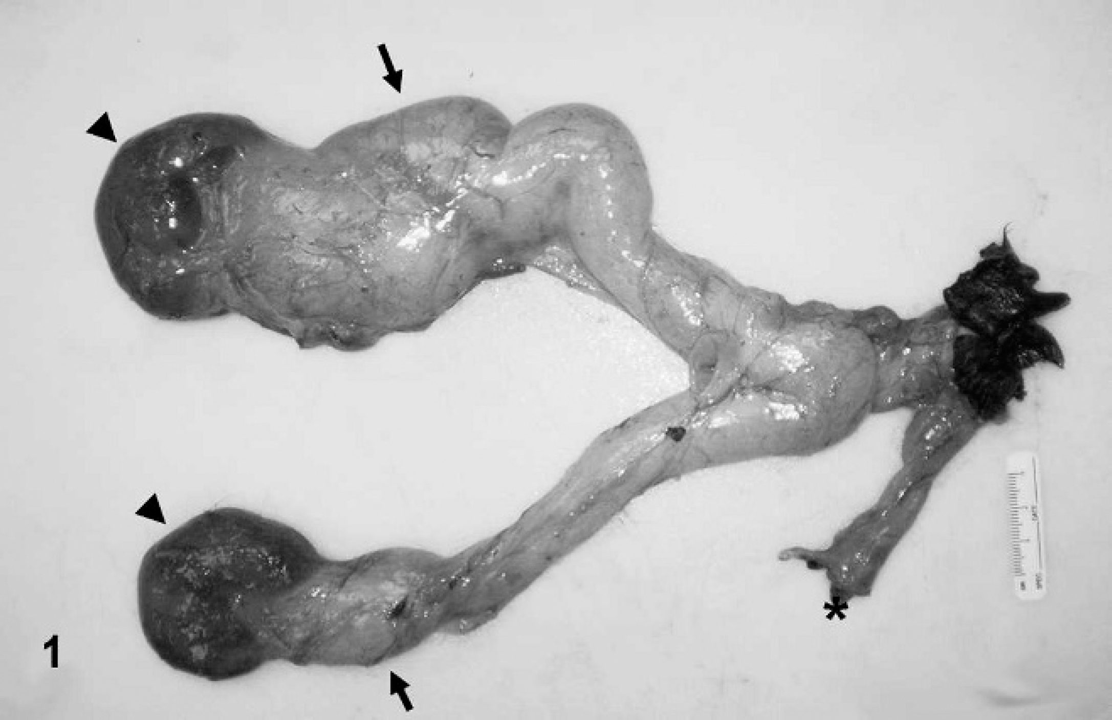
Ureters and kidneys; alpaca cria (Vicugna pacos). Both kidneys (arrowheads) are moderately enlarged. Both ureters (arrows) are markedly asymmetrically distended. A small portion of the colon is present (*). No urinary bladder is seen.
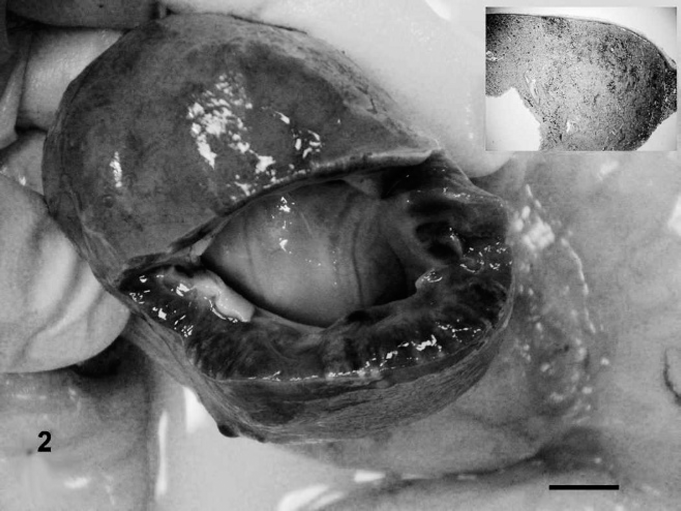
Right kidney; alpaca cria (Vicugna pacos). Cross section of the cortex and medulla of the right kidney with prominent hydronephrosis. Bar = 2 cm. Inset: Low-magnification photomicrograph of a histologic section of the kidney showing marked cortical thinning from hydronephrosis. Hematoxylin and eosin. Bar = 500 μm.
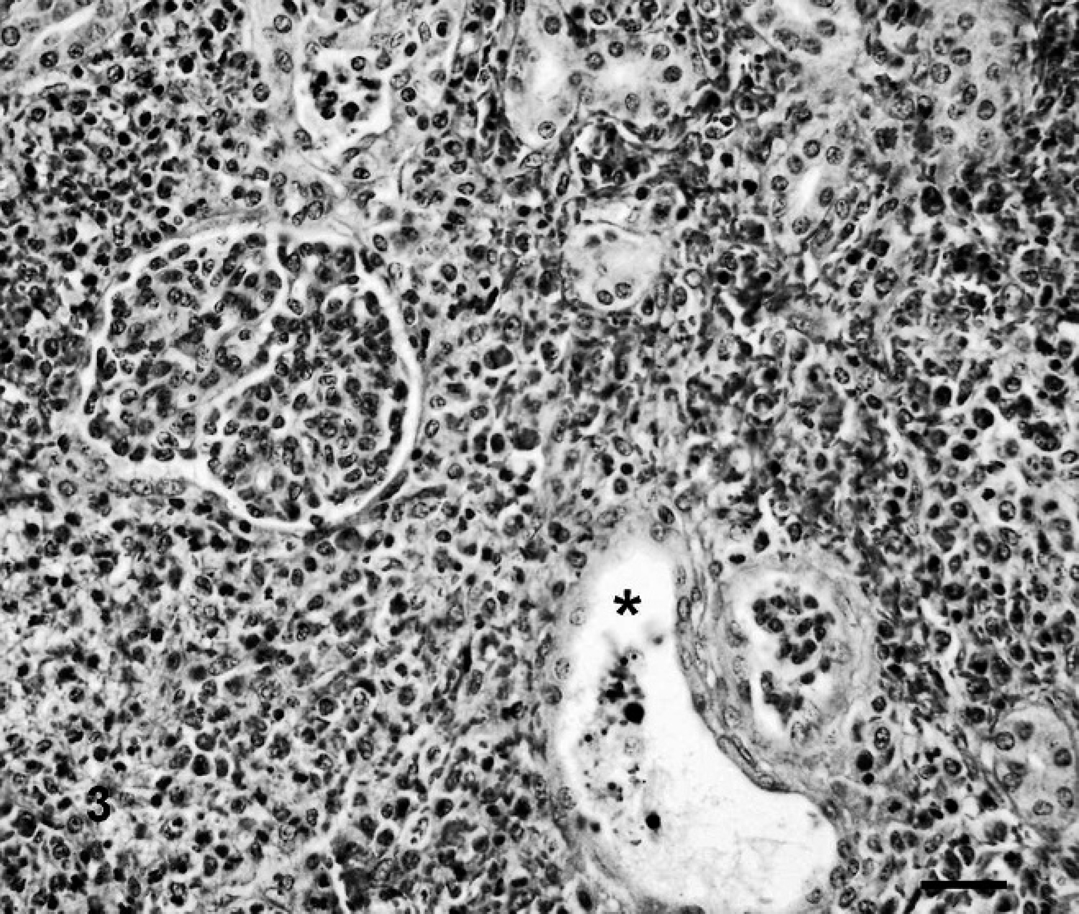
Kidney; alpaca cria (Vicugna pacos). Hemorrhage and fibrin as well as neutrophils and macrophages expand the cortical interstitium. Necrotic debris is present within a tubule (*). Hematoxylin and eosin. Bar = 100 μm.
On gross postmortem examination, no urinary bladder was found. There was marked bilateral hydroureters (the right ureter was 2.5 cm in diameter, whereas the left measured 5 cm), and distally, both ureters opened into a sinus (a 2-mm-diameter opening surrounded by fibrous tissue) in the caudal vagina, 2.5 cm cranial to the vulva (Fig. 1). Bilateral hydronephrosis was also present, with multiple, round, pale tan foci of discoloration throughout the renal cortices (Fig. 2). Additionally, the liver was pale brown and enlarged, and there was mild, diffuse, consolidation of both lungs. On longitudinal sectioning of the right forelimb, no additional gross osseous, articular, or soft tissue abnormalities were observed.
Tissues collected at postmortem examination were fixed in 10% neutral buffered formalin, routinely processed, and embedded in paraffin according to accepted histologic technique. Sections 5 μm thick were stained with hematoxylin and eosin (HE) for microscopic examination.
Examination of the HE-stained sections of kidney showed severe multifocal necrosuppurative pyelonephritis with marked pelvic dilation (consistent with ascending pyelonephritis; Fig. 3). Both ureters showed mild sub-mucosal edema with mild lymphoplasmacytic and histiocytic inflammation. Histologic changes in the liver were characterized by multifocal panlobular coagulation necrosis of a few contiguous lobules, often associated with mild hemorrhage; fibrin; and mild neutrophilic, histiocytic, and lymphocytic infiltrates. These changes were most likely due to septic embolic showers. Histologic changes in the lung were mild and characterized by slightly increased numbers of alveolar macrophages and occasional megakaryocytes within the alveolar septa. No histologic lesions were seen in other tissues.
Urinary tract congenital abnormalities, although uncommon, have been reported in many species, including dogs, 1 cats, 13 cattle, 10 and humans. 2,3,4,8,12 Sporadic case reports of congenital abnormalities affecting the urinary system of crias, including bilateral renal agenesis, have also been documented in the veterinary literature. 6,9,11,14
In general, the causes of congenital defects can be environmental, genetic, or a combination of both. Genetic defects are caused by an abnormal chromosome that contain a mutated gene. 9 Genetic defects are considered inheritable; however, noninheritable genetic defects can occur as well. Environmental factors, such as toxins, viruses, and nutritional imbalance, can also cause non-inheritable genetic defects. 12 Prenatal (6–9 gestational days) exposure to doxorubicin in rats also has been proven to cause several anomalies affecting the skeletal system, the gastrointestinal tract (imperforate anus, esophageal atresia, and tracheo-esophageal fistula), and the genitourinary tract with absence of the urinary bladder. 7
The urogenital tract in mammals originates from the intermediate mesoderm. Several steps characterize the formation of the urogenital tract with the formation of the pronephros (which develops in the neck region and has a transient existence), mesonephros (from the medial aspects of which develops the genital system), and metanephros (from which the adult kidney originates). 5,11
The urinary bladder originates from the urogenital sinus (ventral portion of the cloaca during embryogenesis), which is divided into 3 portions: a cranial (vesicular), middle (pelvic), and caudal (phallic) 5 part. The vesicular part is continuous with the allantois and will form the body of the urinary bladder. 1 The pelvic part will form the pelvic urethra (entire urethra in the female), and the phallic part will generate the penile urethra. 5 The trigone of the urinary bladder is derived from the wall of the mesonephric ducts. 1 As the body of the urinary bladder starts to expand, the walls of the distal ends of the mesonephric ducts are incorporated into the developing urinary bladder. 11
Scientific studies regarding the etiology of congenital diseases in alpacas have not been previously published. In the current case, a developmental defect of the vesicular part of the urogenital sinus, as well as the mesonephric ducts, is suspected because no drug treatment or toxin exposure was reported in the dam during pregnancy to explain the developmental abnormalities observed. Funding was not available to pursue further the etiology of this rare anomaly. Agenesis of the urinary bladder in mammals is an infrequent condition, and to the authors' knowledge, this is the first case reported in an alpaca.