
Abstract
Seven 5-week-old broad-breasted white commercial meat turkeys were submitted to the California Animal Health and Food Safety laboratory in Turlock with a history of respiratory illness. The primary diagnostic findings were mycotic pododermatitis and mycotic pneumonia. The unique feature of this case was the colonization of footpad epidermis and subcutis by fungal hyphae in commercial turkey species. No fungal cultures were undertaken at the time of the necropsy; therefore, paraffin-embedded tissue sections of lung and footpads were used to extract, amplify, and sequence mycotic DNA. A mixed population of fungi was identified in both lung and footpads by polymerase chain reaction amplification of part of the large subunit ribosomal RNA gene using broad-range fungal primers and DNA sequencing. In footpads, sequences matching Cryptococcus saitoi and Cladosporium and Cudoniella species were identified. It is believed that these fungi were opportunistic pathogens originating from the litter. The fungi identified from lungs were Aspergillus species, most closely matching Aspergillus flavus and Arxiozyma telluris (most likely a contaminant). Mycotic pododermatitis in avian species is considered a rare pathologic finding, and few documented reports are available. The on-farm prevalence of footpad lesions was estimated at 3%, and there was no associated increase in the incidence of lameness or weight depression in affected birds. Microscopically, a granulomatous inflammatory reaction associated with fungal hyphae was observed in lung parenchyma. Disruption of keratinized epidermis, encrustations, and acute inflammation were also noted in footpads invaded with fungal hyphae.
The overall incidence of reported avian mycotic dermatitis is relatively low. 15 Pathogenic dermatophytes belonging to the genera Trichophyton and Microsporum are most frequently isolated, but Aspergillus spp., Candida spp., Mucor spp., Rhizopus spp., and other species have also been isolated from the skin and feather follicles of clinically affected birds. 15 Candida albicans has been isolated from footpad lesions in Japanese quail (Coturnix cortunix japonica) between 5 and 30 weeks of age. 16 Avian mycotic dermatitis has also been documented in avian species, such as pigeons, psittacines, wild turkeys, bullfinches, canaries, budgerigars, wild passerine birds, and ostriches, 3,5,15 but mycotic pododermatitis in commercial turkeys has not been reported to the best of our knowledge.
Most cases of documented pododermatitis appear to be related to contact irritation associated with poor litter conditions and the use of coarse litter. Contact dermatitis is also sometimes associated with lesions on the posterior hock, thigh, and sternum. 5 Modern turkey production has seen a decrease in the incidence of contact pododermatitis cases due to improvements in litter management, as well as through the use of nipple drinkers. A direct correlation exists between contact dermatitis and increased stocking density, increased flock age, wet litter, use of particular feeds, male flocks, and winter conditions. 5,13 Dermatitis in turkey poults has been linked to feeding rations deficient in certain vitamins, such as riboflavin, 11 biotin, 14 and pantothenic acid, 10 as well as 40% or more soybean meal. 9 Footpad dermatitis associated with nutritional deficiencies can be considered an unlikely etiology in modern operations.
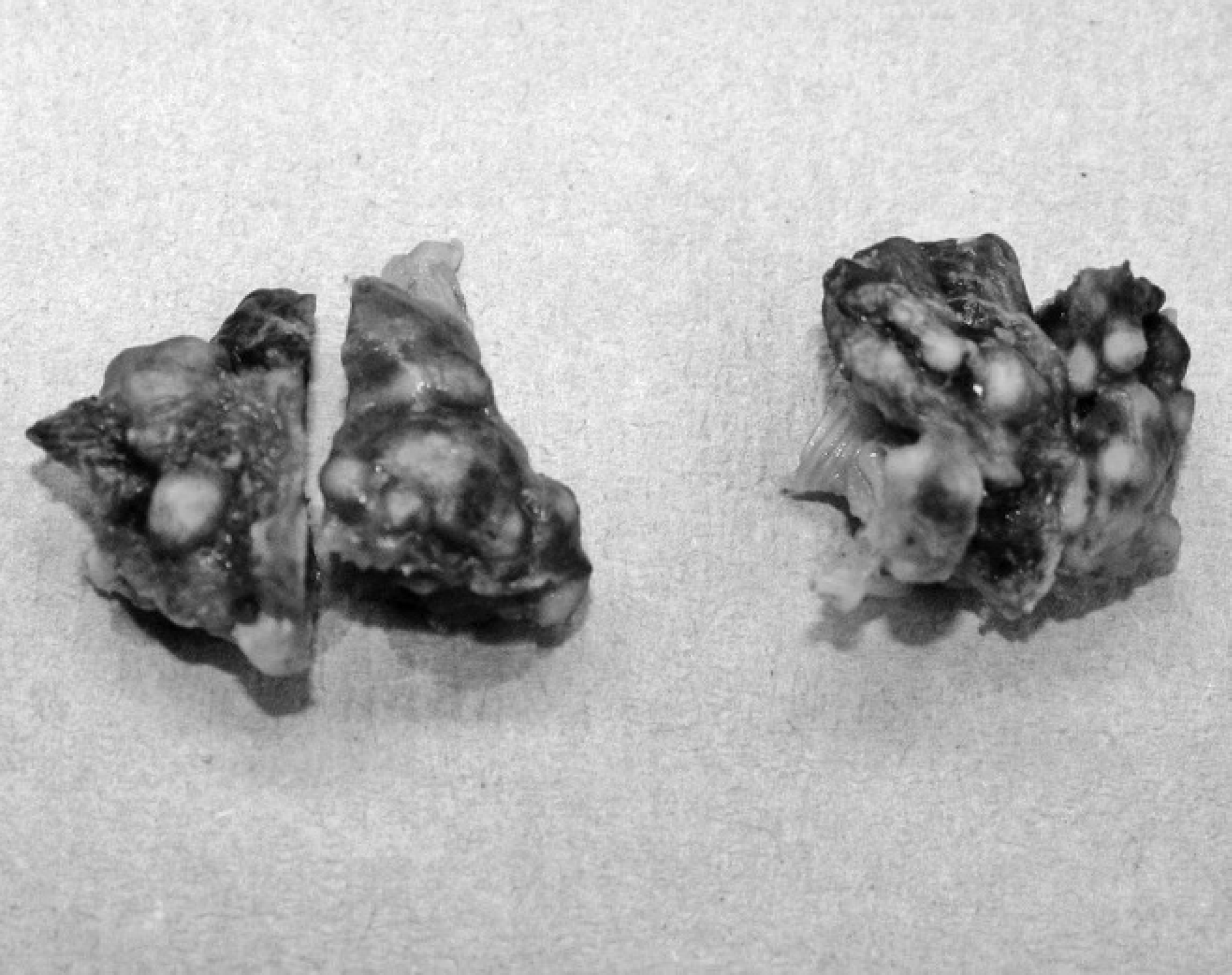
Cut sections showing an extensive infiltration of pale, circular nodules throughout the lung parenchyma (formalin-fixed specimens).
The current report was considered an interesting pathologic presentation of pododermatitis in commercial turkeys. Seven live 5-week-old commercial meat turkeys were submitted to the California Animal Health and Food Safety laboratory in Turlock by a northern California turkey ranch. Poults were submitted due to an increase in the incidence of mildly swollen infraorbital sinuses, foamy air sacs, and snicking, but there was no significant increase in mortality. The litter (wood shavings) was reported to be dry and in good condition.
At necropsy, there was an observable irregularity in the size of the poults. Most poults had a moderate hyperker-atosis of metatarsal pads, with raised, thickened scabs on the epithelial surface of 5 of 7 birds. White, firm nodules, averaging 2 mm in diameter, were distributed extensively throughout the lung parenchyma (5/7; Fig. 1). Additional gross lesions included mild bilateral conjunctivitis (5/7), mild thoracic and abdominal airsacculitis (6/7), pericarditis (3/7), and perihepatitis (1/7). Most poults exhibited signs of enteritis characterized by dilated, thin-walled small intestinal and cecal segments (5/7).
For histologic investigation, tissue sections from all major organs were collected and fixed in 10% neutral buffered formalin, routinely processed, embedded in paraffin wax, and sectioned at 3 μm. All tissue sections were stained using hematoxylin and eosin. 12 In addition, periodic acid-Schiff stains 17 were carried out on the footpad and lung sections. Hematoxylin and eosin-stained lung sections revealed an extensive infiltration of granulomas, as well as a lymphocytic inflammation distributed diffusely throughout the lung parenchyma of most sections. Necrotic debris and fungal hyphal elements were observed in multiple granulomas. Fungal morphology in the lung consisted of hyaline hyphae of irregular widths and occasional branching at right angles; rare septations were present. No yeast-like structures were observed. Multinu-cleated giant cells were observed scattered throughout the granulomas.
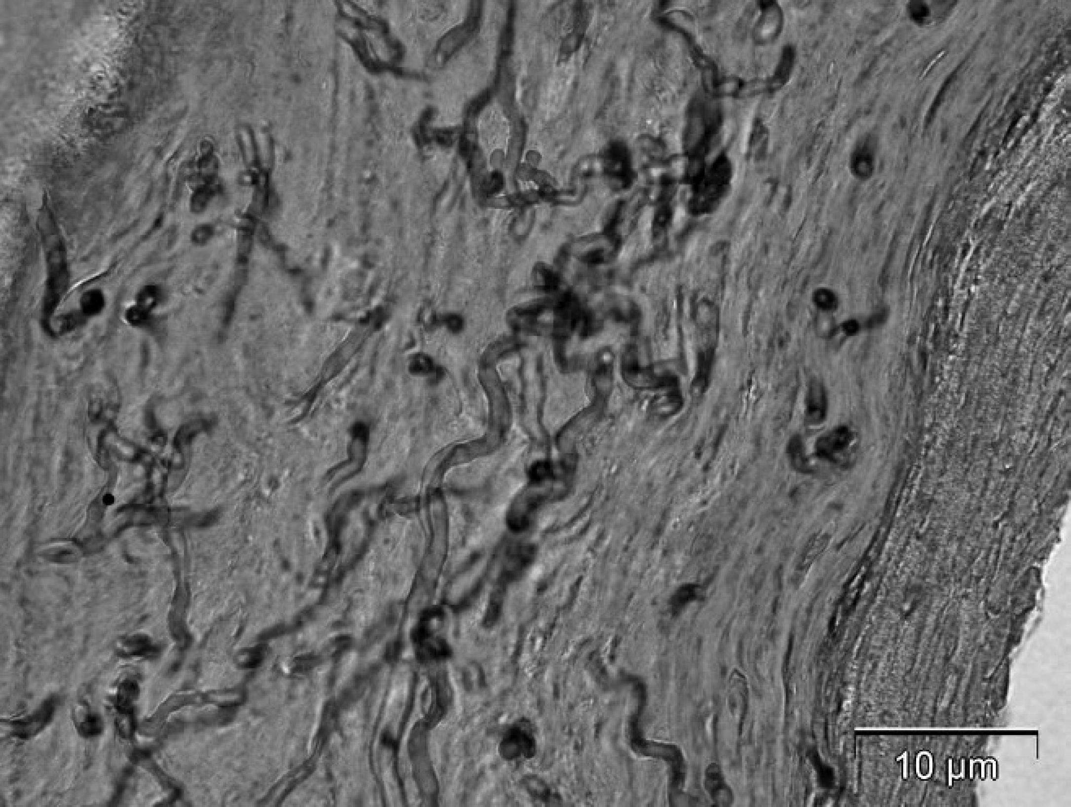
Periodic acid-Schiff-stained section of affected foot pad showing hyaline fungal hyphae within the dermis. Oval structures suggestive of yeast (arrow). Bar = 10 μm.
Hyperkeratosis and pododermatitis of multiple sections of footpads were also observed microscopically. Affected footpads had epidermal surface erosions, ulcerations, and fibrinopurulent crusts of degenerating keratin with dense areas of fungal mycelia within the crusts and also extending deep into the viable epidermis. Bacterial colonies and plant debris were also associated with the surface footpad scabs. The hyphae in the footpad were also hyaline but appeared thinner than those in the lung, and no consistent branching was seen. There was acute inflammation in the epidermis and superficial dermis characterized by hyperemia, edema, rounding of epithelial cells, increased fibroblasts, and heterophil infiltration.
Fungal hyphae were observed in the keratinized layer of the footpad, extending into the deeper viable layers of dermis in some areas. They were also rarely septate and appeared to be associated with oval structures suggestive of yeast, especially in the more superficial areas (Fig. 2). Some fungal hyphae were in close association with a dermal blood vessel in one periodic acid-Schiff section. No fungal hyphae were observed in the connective tissue of the footpad. Periodic acid-Schiff staining of lung sections also confirmed the presence of extensive mycelia scattered throughout multiple granulomas, especially around the periphery of lesions (Fig. 3).
Additional microscopic findings included mild crop mycoses characterized by budding yeast-like fungi and pseudomycelia in the superficial epithelial cell layer of multiple crop sections. Mild to moderate, primarily lymphocytic inflammation was noted in sections of the heart, small intestines, trachea, and conjunctiva. Aerobic bacterial investigation revealed small numbers of Escherichia coli from most tracheas. Small numbers of E. coli were also isolated from 1 of 4 air sacs. A group C1 Salmonella species was isolated from one sample, and Salmonella serotype Schwarzengrund was isolated from the other intestinal sample. Cloacal and pharyngeal swabs from poults were negative for avian influenza using real-time reverse transcription polymerase chain reaction. No significant serologic findings were detected by serologic tests performed on sera samples from the 7 poults.
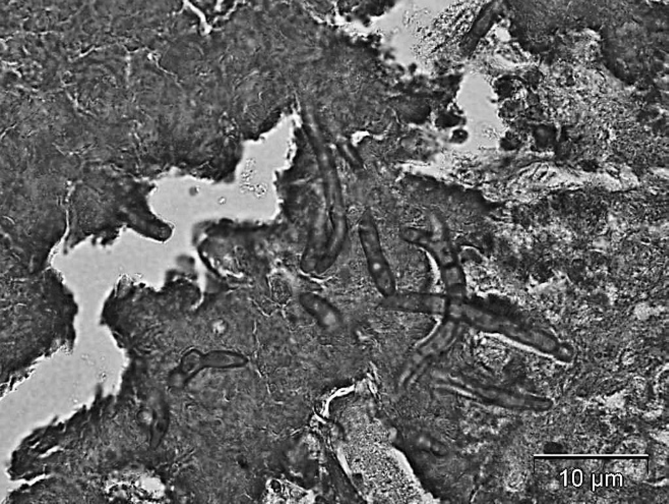
Periodic acid-Schiff-stained section of lung revealing the morphology of the hyaline fungal hyphae. Bar = 10 μm.
For molecular identification of fungi, DNA was extracted from paraffin-embedded tissue sections of footpad and lungs that exhibited mycotic hyphal lesions on histologic examination. Tissue sections were xylene extracted, 20 and DNA was obtained using a phenol-chloroform extraction method. 19 For detection of fungal DNA, approximately the first 600 bases from the 5′ end of the large subunit (LSU) rRNA gene were amplified using previously described fungal primers Ctb6 and TW13. 18 The resulting amplicon was sequenced by automated fluorescent cycle sequencing a in both directions using the same primers as in the initial amplification reaction. 18 Direct sequencing determined that more than one fungal genus was present in both lung and footpad samples. Hence, the amplified products from lung and footpad DNA were cloned, b and 3 clones from each sample were sequenced. A basic local alignment search tool search 1 of the consensus sequence results from the footpad lesion matched Cryptococcus saitoi (100% homology), Cladosporium, and Cudoniella species. Two of the clones from the lungs were an Aspergillus species, most closely matching Aspergillus flavus. The other clone matched Arxiozyma telluris, a type of yeast.
Because the 3 clones from the amplified fungal DNA from footpads yielded a different fungus for each clone, it was difficult to identify one primary fungus to associate with the lesion. It may also indicate that a mixed population of environmental fungi was able to invade and colonize the compromised footpad epidermis. One of the clones identified was Cladosporium species. This fungus is a dematiaceous (pigmented) mold, ubiquitous in air and organic material. Conidia are elliptical to cylindrical, are pale to dark brown, occur in branching chains, and have a distinct dark hilum. 4 The microscopic morphology of this mold is not analogous to that seen in histologic sections of the current case; thus, it was deduced that this was not the primary pathogen involved. DNA from one of the clones matched with 100% homology to the deposited sequences of C. saitoi. In spite of this high homology, a conservative approach to species identification based solely on the sequence homology was taken. This is primarily because other Cryptococcus species may also be closely related but there may be no deposited sequences for them. C. saitoi was formed from the segregation of Cryptococcus albidus into different species based on rRNA sequence data integrated with other physiologic and molecular characteristics. 7 Cryptococcus is a ubiquitous, basidiomycetous yeast. Microscopically, globose to ovoid yeast cells are formed and no true or pseudohypha are visible. 7 Histologic evidence of oval structures suggestive of a yeast, such as Cryptococcus, was seen in multiple periodic acid–Schiff-stained sections (Fig. 2). The third clone matched Cudoniella species, an external environmental fungus that most likely represents a contaminant within the footpad lesion. A specific identification of the irregularly septate, hyaline hyphal elements seen in the footpad sections could not be ascertained from molecular techniques. Although histopathology and molecular-based techniques were useful in the current case, no conclusive identification of a single primary fungus invading the footpads could be obtained.
The lung lesions were most likely a pulmonary aspergillosis, with A. flavus identified by genetic sequencing. Typically, Aspergillus mycelia are uniform in width, are regularly septated, and branch dichotomously at acute angles. 6 Interestingly, the hyaline hyphae seen in histopathologic sections of the lungs had irregular widths and appeared to branch occasionally at right angles. Aspergillus was not ruled out based on appearance in histologic sections, because Aspergillus species sometimes assume an atypical morphology in necrotic lesions. Globose varicosities and inconspicuous septa can occur in necrotic lesions; such hyphae can sometimes be mistaken for those of zygomycetes. 2 No yeast-like structures were identified by histopathology, and the A. telluris yeast that was identified in one of the clones from the lungs was considered an incidental finding and most likely represented a contaminant. Additional findings of inflammatory tissue reactions in multiple organs (heart, conjunctiva, tracheas), isolation of E. coli in tracheas and air sacs, crop mycosis, and intestinal salmonellosis may indicate that the birds were immunocompromised and susceptible to multiple opportunistic infections.
The plantar surface of the feet is usually well protected by thickened, cornified epithelium, but contact irritation can compromise the integrity of the keratinized epidermis. Because feet are removed during processing, footpad lesions do not usually result in downgrading of the carcass, but severe cases can result in an increased incidence of lameness and an associated depression in body weight. 13 Reports of hyperkeratosis, ulceration, and bacterial invasion of the footpad is not an uncommon finding in commercial turkeys. 5 Most of the cases reported are associated with poor litter conditions or dietary disturbances. Many fungi are ubiquitous, and the source of the footpad fungi was most likely from spores present in the litter. Overcrowding and poor litter management, particularly moisture build up, can predispose birds to abrasions and traumatic injury to footpads. 6 Erosive lesions can accentuate penetration, invasion, and colonization of the stratum epidermis and subcutis with fungi. The mixed populations of fungi isolated were most likely opportunistic environmental fungi that were able to gain entry in affected footpads. Because pododermatitis lesions are most often associated with bacterial invasion and proliferation, it is interesting to note that mycotic agents may also be able to invade and colonize the compromised plantar epidermis. No previous reports of plantar dermatitis in turkeys associated with fungal colonization were found in the veterinary literature. Hence, the current case was considered a novel presentation of pododermatitis in commercial turkeys. The foot lesions were considered an incidental finding in this case and did not appear to result in significant increases in morbidity and mortality in the flock. Management estimated that the prevalence of poults with excess crusting and darkened discoloration of the footpad was approximately 3%.
A lack of fungal culture for definitive speciation represents a limitation of the present report. Because the clinical presentation and gross pathologic lesions were nonspecific, mycosis was not suspected until histopathologic examination of tissue sections was performed. As a result, fungal identification relied on histology and isolation of fungal DNA from paraffin-embedded sections. In spite of the usefulness of fungal-specific DNA amplification and sequencing to detect fungal species, the gold standard for the identification and speciation of mycotic infection is fungal culture. 8
Footnotes
a.
Davis Sequencing Inc., Davis CA.
b.
Topo TA Cloning® Kit, Invitrogen Corp., Carlsbad, CA.