
Abstract
Concurrent tracheal hypoplasia and discrete subaortic stenosis are described in a 12-week-old Rottweiler puppy that presumably died of pulmonary edema. A brief literature review and comparison to previously published cases of tracheal hypoplasia in other breeds is presented along with a description of a subaortic septal ridge and comparison to the analogous condition in humans.
Pathological findings associated with tracheal hypoplasia in a Rottweiler puppy are presented. Concurrent dorsal and longitudinal concentric tracheal ring overlap is depicted, and a brief literature review regarding canine tracheal hypoplasia is presented. Additionally, a subaortic septal ridge is described that is speculated to represent an early step in the pathogenesis of subaortic stenosis, similar to the well-described syndrome in humans. 2,7
A 10.4-kg, 12-week-old, intact, male Rottweiler puppy with a body condition score of 1.5 out of 5 and respiratory distress was presented to the referring veterinarian. During initial attempts to stabilize the puppy, it died and was subsequently presented to the Louisiana State University School of Veterinary Medicine (Baton Rouge, LA) for necropsy. At necropsy, the heart had a subaortic area of septal wall thickening creating a 3.8-cm circumferential area of narrowing approximately 2–3 mm below the aortic valve. The aortic valve circumference was 4.2 cm, and the postvalvular aortic circumference was 4.6 cm. The aortic intimal lining was mildly roughened and dull. The right ventricle was dilated, and the right ventricular free-wall was 3 mm thick compared to a left ventricular free-wall thickness of 1.2 cm and an interventricular septal thickness of 1.3 cm. Microscopic examination of the heart demonstrated marked expansion of the subaortic interventricular septum with normal cardiomyocytes and mildly increased collagen deposition in the supravalvular aortic intimal lining (Fig. 1).
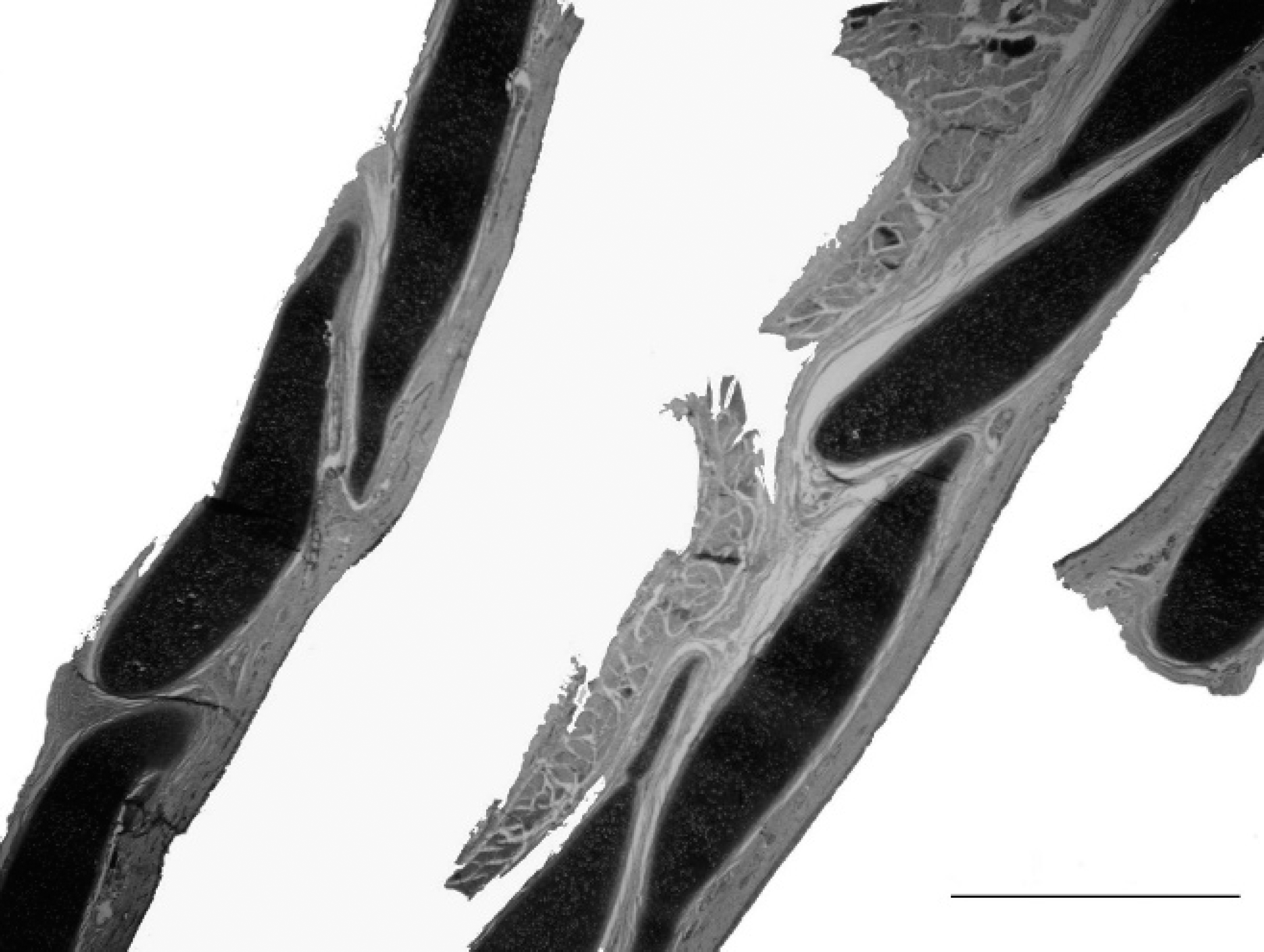
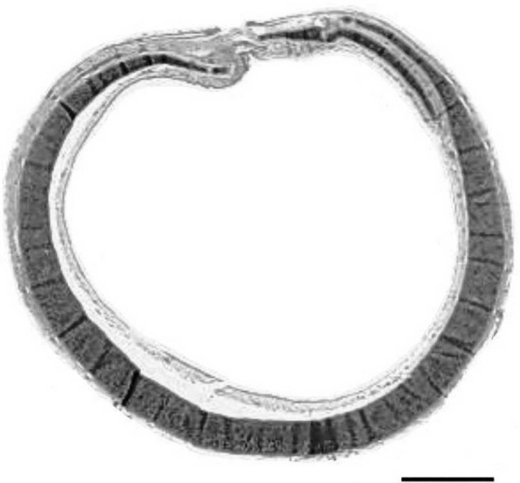
The trachea had a 5-mm-diameter lumen at the narrowest portion (and a maximal diameter of 6 mm) with overlap of the dorsal aspect of the tracheal rings causing plication of the narrow dorsal tracheal membrane, which was 1–2 mm wide. The diameter of the third rib was 2.2 cm yielding a trachea-to-rib diameter ratio of approximately 2.3:1. The Coyne and Fingland report, with an expected ratio of 3:1, was used to distinguish between tracheal hypoplasia and the tracheal diameter that is anticipated in health (<3:1 is considered diagnostic for tracheal hypoplasia). 3 Microscopic examination of the trachea revealed a dorsal tracheal membrane that was fused to form a single plication with overriding dorsal ends of the tracheal ring cartilage. Longitudinally, tracheal rings were closely apposed and partially overlapped each other (Fig. 2). Dorsally, the tracheal rings were also closely apposed or overlapping (Fig. 3). Microscopically, the cartilage was irregular on cross section and, as previously noted, overlapped adjacent tracheal ring cartilage. On gross and microscopic examination of the lungs, there was pulmonary congestion, hemorrhage, hemosiderin-containing alveolar macrophages, and edema.
Early reports of tracheal hypoplasia were diagnosed as diffuse tracheal stenosis. 11 The terminology subsequently changed to tracheal hypoplasia, which is defined as a congenital anomaly characterized by an abnormally narrow tracheal lumen. 1,3,10 Previous cases have had overlapping or apposed ends of the tracheal cartilages with a very narrow, plicated, or absent dorsal tracheal membrane. In contrast to tracheomalacia or collapsing trachea, the diameter of the trachea does not change with the phase of respiration or with radiographic positioning. 3 Tracheal hypoplasia is reported most often in brachycephalic breeds such as the Bulldog, Boston Terrier, and Boxer. 3 Non-brachycephalic breeds previously reported with tracheal hypoplasia include the Basset Hound, 8 German Shepherd, Weimaraner, and Labrador Retriever. 10 Tracheal hypoplasia is also commonly associated with other congenital anomalies, including subaortic stenosis. 3,11
To the authors' knowledge, this is the first reported case of tracheal hypoplasia in a Rottweiler. The current case also adds to the published number of dogs with concurrent tracheal hypoplasia and subaortic heart anomalies. It is not known if the marked dysplastic orientation and ring conformation noted on sagittal sectioning is a unique finding in this puppy or if this change is consistently present with tracheal hypoplasia but was heretofore unrecognized. Some degree of overlap may be normal in some breeds during development, but the degree exhibited in this puppy appears excessive.
Abnormal embryogenesis is thought to be a possible cause for tracheal hypoplasia. 3 This idea is reinforced by the observation that many dogs with hypoplastic tracheas also have congenital megaesophagus. 3 During normal embryogenesis, the trachea and esophagus arise from the same primordial tissue. The laryngotracheal ridge projects ventrally from the esophagus, and a pair of lateral grooves pinch in and fuse to form the tracheoesophageal septum. 3 Tracheal hypoplasia may occur when the lateral grooves form at a point too far ventrally on the developing tracheoesophagus.
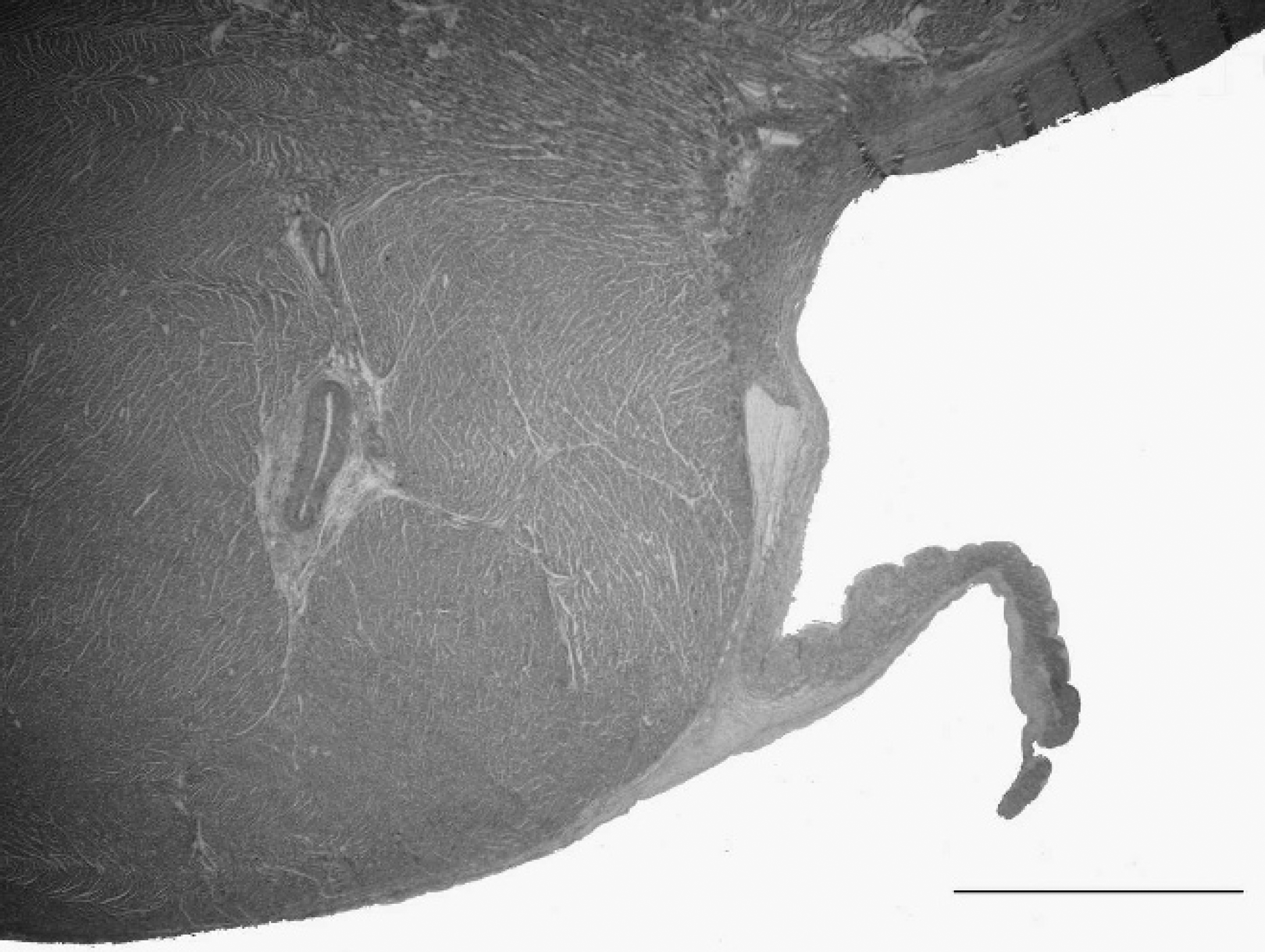
The interventricular septal wall demonstrating subaortic myocardial hypertrophy and mild supra-aortic intimal fibrosis. Trichrome stain. Bar = 2 mm.
Although the linear velocity of air increases exponentially with decreasing luminal diameter resulting in increased respiratory work, it is thought that most dogs are able to compensate for tracheal hypoplasia by modulating their activity level. In fact, contrary to early supposition that tracheal hypoplasia predisposes dogs to secondary respiratory tract infection and bronchopneumonia, 10 a recently published case series demonstrated pneumonia in a minority of the population of dogs with tracheal hypoplasia. 3
Longitudinal section of trachea illustrating concentric ring overlap. Hematoxylin and eosin. Bar = 2 mm.
Transverse section of the trachea illustrating narrowing and plication of the dorsal tracheal membrane with apposition of the dorsal end of the tracheal rings. Hematoxylin and eosin. Bar = 1 mm.
Ultimately, left heart failure resulting from a narrowed subaortic outflow tract and resulting pulmonary edema was thought to be the most likely cause of death in the Rottweiler puppy in the current study. The Rottweiler breed is known to have an increased risk of congenital subaortic stenosis, 6 and the association of a heart defect with tracheal hypoplasia has been previously documented. 3,11 There is a wide variation in the manifestation of subaortic heart defects in humans and dogs, and the defect varies according to age, myocardial perfusion, and progression of shear stresses on the left ventricular outflow tract. 2,4,5,7 Numerous classification systems have been proposed in humans, many of which rely on documentation of dynamic changes on echocardiography. 2,5,7 Classically, subaortic stenosis is defined as an abnormal ring or ridge of tissue that projects from the endocardial surface and encroaches into the lumen of the left ventricular outflow tract, thereby reducing the cross-sectional area through which blood can travel. 9
Although the lesion seen in this puppy is not what is typically described for subaortic stenosis, it is thought that the narrowing caused by hypertrophy of the high interventricular septum caused rheological changes and aortic valvular insufficiency based on the changes in the aortic intima (roughening) suggestive of postvalvular turbulence and pulmonary changes compatible with left heart failure. Whether this lesion represents early changes that would eventually lead to classic subaortic stenosis or another entity altogether is not known.