
Abstract
Mycoplasma bovis was identified by a specific lesion, conventional bacterial culture, immunohistochemistry, and polymerase chain reaction in 2 feedlot bison found dead with severe, chronic, caseonecrotic pneumonia; polyarthritis; and laryngitis. On microscopic examination, pulmonary lesions were characterized by prominent, well-defined areas of caseous necrosis and bronchiectasis. Immunohistochemical analysis of lung exhibited staining in bronchiolar epithelium and in random areas of caseous necrosis. On gross examination, the laryngeal lesion observed in 1 animal was typical of changes seen in cases of calf diphtheria. Nasal swabs taken from 6 clinically ill bison from the same feedlot revealed 1 animal shedding M. bovis by the nasal route. No other pathogens were recovered from the pulmonary or laryngeal lesions; however, Mannheimia haemolytica was cultured from the nasal swabs of 2 clinically ill bison, although not from the animal found to be shedding M. bovis. Several other affected bison had swollen joints and exhibited lameness and a reluctance to move. Changes observed in dead and clinically ill bison from this feedlot are similar to what has been described in the literature as chronic pneumonia and polyarthritis syndrome in feedlot cattle caused by M. bovis. Based on the severity of the lesions, and the number of dead and affected animals, bison in a feedlot setting appear to exhibit sensitivity to infection with M. bovis.
The carcass of a 480-lb, 18-month-old American bison (Bison bison) heifer was delivered to the veterinary diagnostic laboratory at North Dakota State University (NDSU-VDL) in September of 2007. The animal had been sick for 10 days, gradually lost condition, developed diarrhea, and exhibited swelling in both carpal joints. Lameness led to difficult ambulation. The animal eventually became recumbent and refused to stand. Subsequently, it was euthanized. Twenty fatalities of 900 feedlot animals had occurred before the submission of this animal to the laboratory. Another 7 animals were lame and had loss of body condition (Fig. 1).
At necropsy, both lungs contained randomly distributed, irregular, sometimes raised, variably sized foci of caseous necrosis (Fig. 2). In addition, both stifle joints were swollen and fluctuant on palpation. Upon incision, both joints oozed purulent to caseous exudate and exhibited marked inflammation of the joint capsules, synovial tissue, and tendon sheaths (Fig. 3). There was a localized area of necrosis in the cervical musculature on the right side of the neck (injection site). Tissue samples were collected, fixed in 10% neutral buffered formalin, processed, sectioned at 5 μ, and stained with routine hematoxylin and eosin. Large areas of caseous necrosis and inflammation were noted in the pulmonary parenchyma. Affected areas were characterized by a distinct outer zone of variably dense fibrous connective tissue infiltrated with a mixture of inflammatory cells, a thinner middle zone that consisted of a bilayer of activated macrophages and plasma cells (outer portion) and necrotic neutrophils (inner portion), and an expansive interior zone of caseous necrosis in which necrotic alveolar septa and bronchioles were seen. Normal lung adjacent to the outer fibrous connective tissue capsule had several areas of atelectasis, some accumulation of proteinic material within alveoli, and increased numbers of alveolar macrophages. Sections of synovium, joint capsule, and tendon sheath had a marked necropurulent synovitis, arthritis, and tenosynovitis, respectively.
Lung tissue and joint exudate were placed on trypticase soy agar (TSA) II 5% sheep blood a and incubated in atmospheres of O2, 5% CO2 and 15% CO2, MacConkey II a in an atmosphere of O2, brain heart infusion broth a in an atmosphere of O2, and Mycoplasma agar b in an atmosphere of 5% CO2. Plates and broth were incubated in a moist chamber at 37°C. At 48 hr, Mycoplasma plates examined under an inverted microscope (10X) showed typical friedegg colonies. Polymerase chain reaction (PCR) analysis of Mycoplasma isolates identified the organism as Mycoplasma bovis.
DNA from Mycoplasma cultures was extracted by using a rapid boil and spin procedure. 10 The resulting genomic DNA was then amplified by using a standard PCR protocol. The positive extraction control was an American Type Culture Collection M. bovis strain (no. 25025). Mycoplasma bovis primer sequences used are as follows: MBVF (5′-TGATAGCAATATCATAGCGGC-3′) and MBVR (5′-GTAGCATCATTTCCTATGCTAC-3′). c After boiling, incubation, and centrifuging, the supernatant was used as a DNA template in PCR. A master mix of nuclease-free water (30.75 μl), 5X GoTaq Buffer with 7.5 mM of Mg (10 μl), d 25 mM of MgCI2 (1 μl), d 10 mM of dNTP (deoxyribonucleotide triphosphate) mix (1 μl), d 10 μM of Forward Primer (1 μl), c 10 μM of Reverse Primer (1 μl), c 5 U/μl of GoTaq DNA polymerase (0.25 μl), d and DNA template (5 μl) was used. Polymerase chain reaction protocol was as follows: 94°C for 2 min, followed by 40 cycles each at 94°C for 30 sec, 55°C for 30 sec, and 72°C for 30 sec, and a final extension at 72°C for 5 min. The products were analyzed by 1.5% agarose gel electrophoresis with ethidium bromide (0.05 μg/ml) and ulraviolet transillumination. A single band observed at 415 base pair was interpreted as positive for M. bovis.
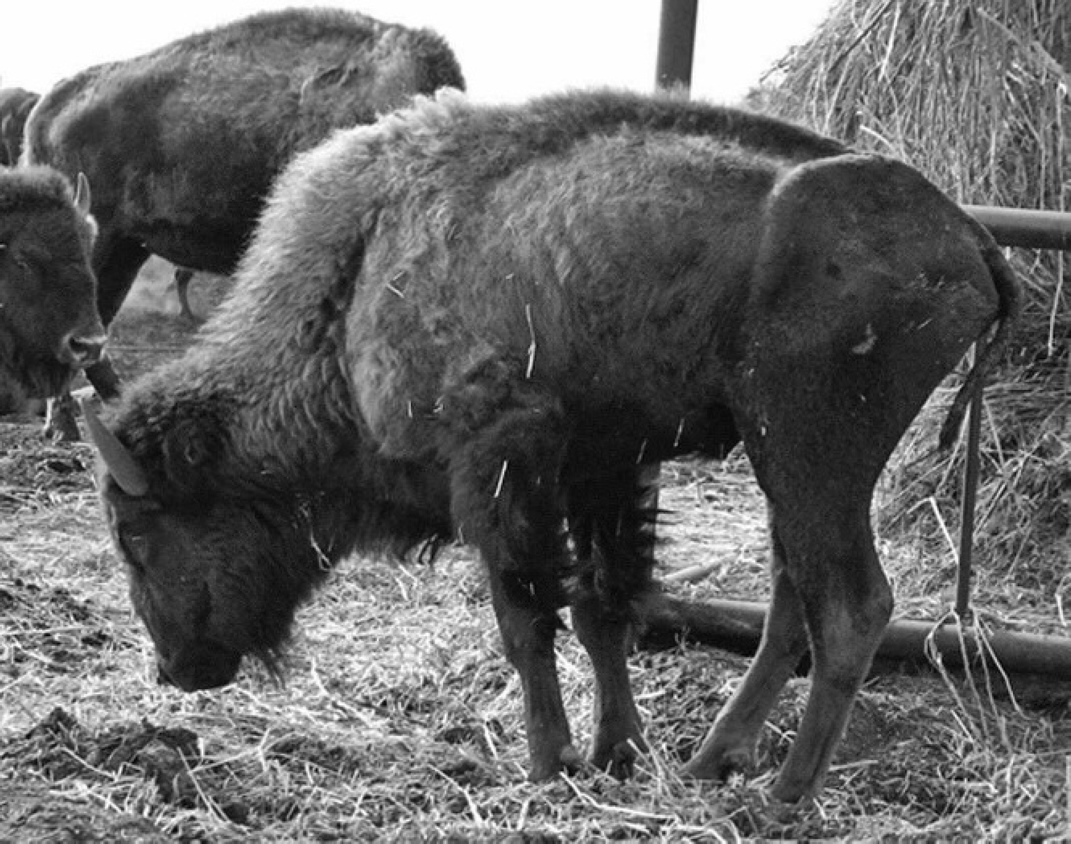
Yearling bison infected with Mycoplasma bovis. Note loss of condition and abnormal stance.
An antibody (1:800) was applied to deparaffinized sections of lung after target retrieval with steam and peroxidase treatments with appropriate Tris-buffered saline solution washes. The marker was applied with En Vision+ e and romulin red. f Specifically, abundant positive staining was observed in bronchiolar epithelium, as well as random, less frequent, multifocal staining in caseonecrotic foci.
Thirty days after the laboratory necropsy of the first animal, a diagnostician from the NDSU-VDL visited the feedlot to collect additional samples. Nasal swabs (16) were taken from clinically healthy, unvaccinated, untreated bison ready for market (10), and from the clinically ill, vaccinated, antibiotic-treated bison (6). One bull in the clinically ill group had significant swelling of the left carpal joint, and a fine needle aspirate was taken from a fluctuant region on this swelling. In addition, a necropsy was performed on a heifer that had died the previous night. This animal had severe, primarily unilateral, pneumonia characterized by massive areas of caseous necrosis throughout all lobes of the right lung. There was also severe, bilateral, fibrinopurulent laryngitis typical of the lesion seen in cases of calf diphtheria. Joint infections were not present in this animal. Fresh and formalin-fixed lung and fresh trachea were taken back to NDSU-VDL for further processing. Histopathology and immunohistochemistry (IHC) results were similar to those seen from the first bison necropsy. Culture of lung yielded Mycoplasma spp., Pseudomonas aeruginosa, and mixed contaminants, and culture of trachea yielded a Mycoplasma sp. Culture of individual nasal swabs from the 6 clinically ill animals yielded a Mycoplasma sp. Mannheimia haemolytica was recovered from two of the affected animals. Cultures of swabs from the clinically normal group yielded no pathogenic bacteria. Polymerase chain reaction identified all Mycoplasma spp. isolates (lung, larynx, and nasal swab) as M. bovis, except the isolate from the carpal joint of the lame bull.
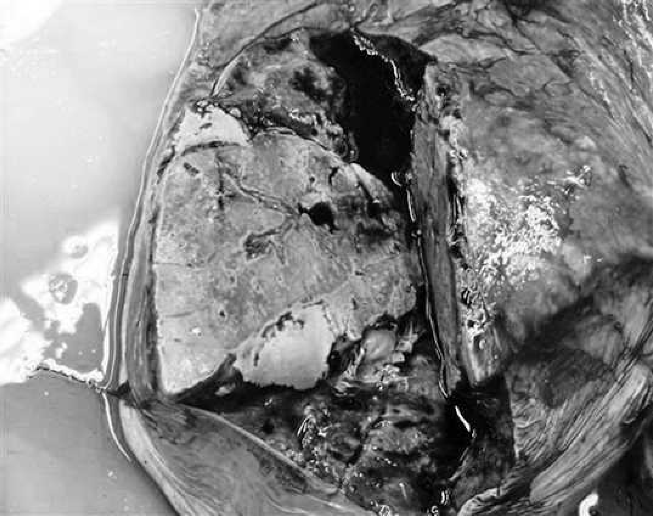
Lung from an 18-month-old bison heifer showing a pale, well-defined, zone of caseous necrosis caused by Mycoplasma bovis.
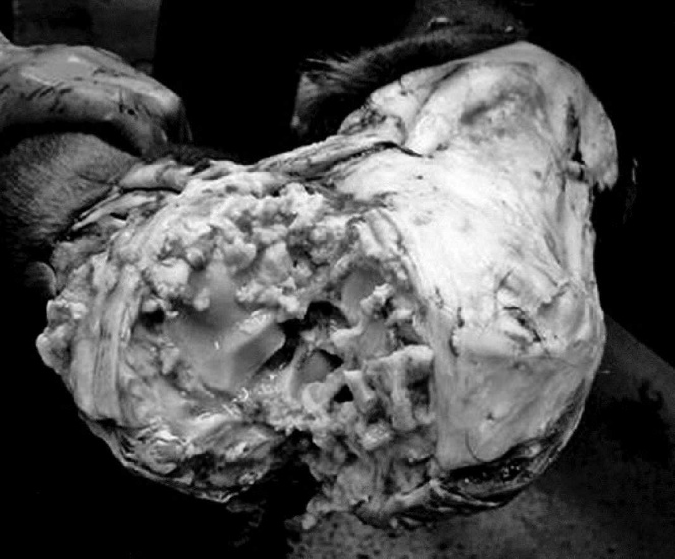
Incised carpal joint from an 18-month-old bison heifer, showing marked purulent to caseonecrotic arthritis and tenosynovitis caused by Mycoplasma bovis.
Mycoplasma bovis–associated disease manifests itself in variety of ways in cattle, including pneumonia and arthritis, 2,7 tenosynovitis, 2 keratoconjunctivitis, 11 otitis media, 12,16 and mastitis. 17 A condition caused by M. bovis characterized by chronic pneumonia and polyarthritis was recognized in feedlot cattle. 5,9 Coinfection with Bovine viral diarrhea virus 6,14 and common bovine respiratory viruses 3 appears to occur with some frequency. Previous studies that used IHC to examine the pattern of bacterial colonization in M. bovis–infected lungs described staining in bronchiolar epithelial cells, inflammatory cells, and abscessed airways (naturally infected animals), 1 random staining in areas of both coagulative and caseous necrosis with peripheral zones of purulent to pyogranulomatous inflammation (naturally infected animals), 8 and staining in areas of coagulative necrosis and bronchiolar epithelium (naturally infected animals) and in inflammatory cells in alveoli and septal walls (experimentally infected animals). 13 Staining patterns in naturally infected animals were consistent with the type of staining seen in bison from the feedlot in the present study. The most intense staining was observed in bronchiolar epithelial cells.
The recovery of M. bovis from 1 nasal swab in the clinically ill group of bison sampled suggests that the bacterium is shed by the nasal route. Because it was shown that bronchoalveolar lavage is a better method then nasal swabbing for isolating a deep respiratory pathogen such as M. bovis, 4,15 it is likely that additional clinically ill animals were capable of shedding the bacterium into the environment and were a source of infection for herd mates. Characteristics of this M. bovis outbreak indicate that bison are susceptible to severe infections by this pathogen and that M. bovis is capable of causing primary disease in this species.
Acknowledgement. The authors would like to thank Lisa Piche for assistance in preparing the manuscript.
Footnotes
a.
Becton, Dickinson and Company, Sparks, MD.
b.
Myco Plate, Vet Med Biological Media Services, University of California, Davis, CA.
c.
Integrated DNA Technologies, Coralville, IA.
d.
Promega Corp., Madison, WI.
e.
Dako North America Inc., Carpinteria, CA.
f.
Biocare Medical, Concord, CA.