
Abstract
A flock of Indian Ringneck parakeets (Psittacula krameri manillensis) was imported to the United States from Australia. Soon after, 1 parakeet suddenly died, and a second parakeet died after a 2-day course of illness, which consisted of anorexia, lethargy, emaciation, and dyspnea. At necropsy, the affected birds had diffuse consolidation and red discoloration of the lungs, as well as thickened, congested air sacs. The microscopic examination revealed multifocal, necrotizing bronchitis, parabronchitis, and interstitial pneumonia. The lumen of the affected airways contained numerous, large syncytial cells with up to 15 nuclei. The nuclei of these syncytial cells often contained large, eosinophilic inclusion bodies, consistent with herpesvirus. The epithelium of the trachea and air sacs was hypertrophied and contained syncytial cells with intranuclear inclusion bodies similar to the bronchi. In addition, a few intranuclear inclusion bodies were also present in the epithelial cells that line the air capillaries. On ultrastructural examination, the nuclei of degenerating epithelial cells contained clusters of viral nucleocapsid proteins and unenveloped, icosahedral, viral particles that were approximately 90 nm in diameter. In addition, some epithelial cells contained clusters of enveloped viral particles approximately 105 nm in diameter, within the cytocavitary network. These lesions are characteristic of those caused by respiratory herpesvirus of parakeets.
Herpesviruses comprise a large family of enveloped DNA viruses. They have a wide variety of hosts, and nearly every mammalian and avian species is a natural host of one or more herpesvirus strains. This report documents a novel lower-respiratory-tract disease in 2 Indian Ring-neck parakeets caused by a herpesvirus.
Tissues from 2 Indian Ringneck parakeets from a flock imported to the United States from Australia were submitted to the Veterinary Pathology Department of Iowa State University (Ames, IA) for diagnostic evaluation. The first bird was presented dead to the referring veterinarian. The second bird was presented with lethargy, emaciation, and dyspnea, and died after a 2-day course of illness. The trachea, lung, air sacs, and liver were collected at necropsy and were fixed in 10% neutral-buffered formalin. According to the referring veterinarian, the gross lesions in the first bird consisted of the markedly and diffusely thickened air sacs, heavy and diffusely red lungs, and disseminated pale foci in the liver. The second parakeet had similar lung and air sac lesions, with no hepatic involvement. Tissues received by the referring veterinarian were routinely processed, embedded in paraffin, and 5-μm thick sections were stained with hematoxylin and eosin.
By light microscopy, the lung and airway sacs of both parakeets had similar lesions. The airway lumina, airway epithelial lining, and air capillaries contained numerous, large syncytial cells. The syncytial cells contained up to 15 identifiable nuclei, with occasional eosinophilic and intranuclear inclusion bodies, both of which are often associated with foci of necrosis (Fig. 1). The airway epithelium was multifocally eroded or lined by hypertrophied cells, and the airway lumina often contained mucus, necrotic cell debris, and heterophils. There were scattered foci of congestion and hemorrhage throughout the pulmonary parenchyma. The epithelium of air sacs and the trachea contained syncytial cells similar to the lung airways. The lung tissue of the first bird contained numerous fungal hyphae, which extended through the mucosa of the large airways into the adjacent parenchyma. The fungal hyphae had parallel walls, septa 5–10 μm in diameter, and dichotomous branching, which is most consistent with Aspergillus sp. In addition, the liver of the first parakeet contained scattered foci of coagulative necrosis but lacked syncytial cells or intranuclear inclusion bodies. The second bird did not contain any identifiable hepatic lesions.
Specimens of lung and air sac from the second bird were further submitted to the National Animal Disease Center (Ames, IA) for electron transmission microscopic evaluation. The tissues were routinely processed, embedded, and stained with lead citrate and uranyl acetate. On ultrastructure examination, the nuclei of degenerating epithelial cells contained clusters of viral nucleocapsid proteins and unenveloped, icosahedral viral particles that were approximately 90 nm in diameter (Fig. 2). In addition, some epithelial cells contained clusters of enveloped viral particles approximately 105 nm in diameter, within the cytocavitary network (Fig. 3). These features are characteristic of herpesviruses, thus the diagnosis was necrotizing bronchitis, bronchiolitis, and air sacculitis because of infection with herpesvirus.

Syncytial cell (arrow) with intranuclear inclusion bodies (open arrows).
Herpesvirus subfamilies include alpha-herpesviruses, beta-herpesviruses, and gamma-herpesviruses. Alpha-herpesviruses are associated with rapid viral replication, host cell lysis, and the ability to establish latent infection. 5 An example of avian alpha-herpesvirus is Gallid herpesvirus 1 (family Herpesviridae, subfamily Alphaherpesvirinae, genus Iltovirus), commonly known as Infectious laryngotracheitis virus (ILTV) of chickens, which causes upper-respiratory-tract infection manifested as necrotizing pharyngitis, laryngitis, tracheitis, and, occasionally, mild pneumonia. 23
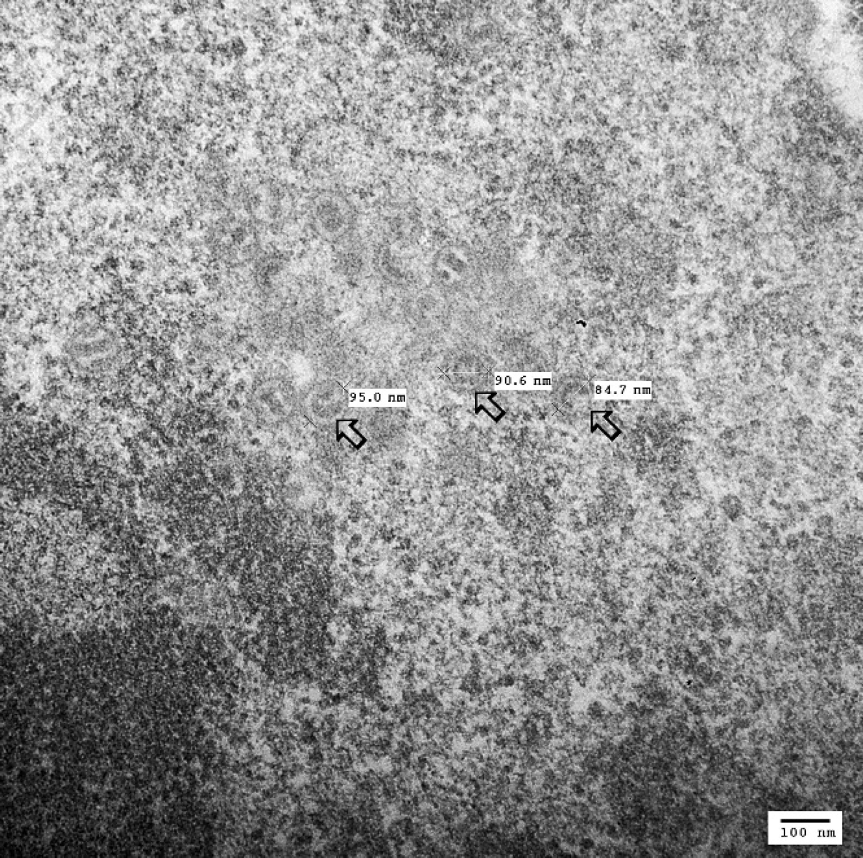
The intranuclear viral particles are not yet enveloped and, therefore, are approximately 95 nm in diameter (open arrows).
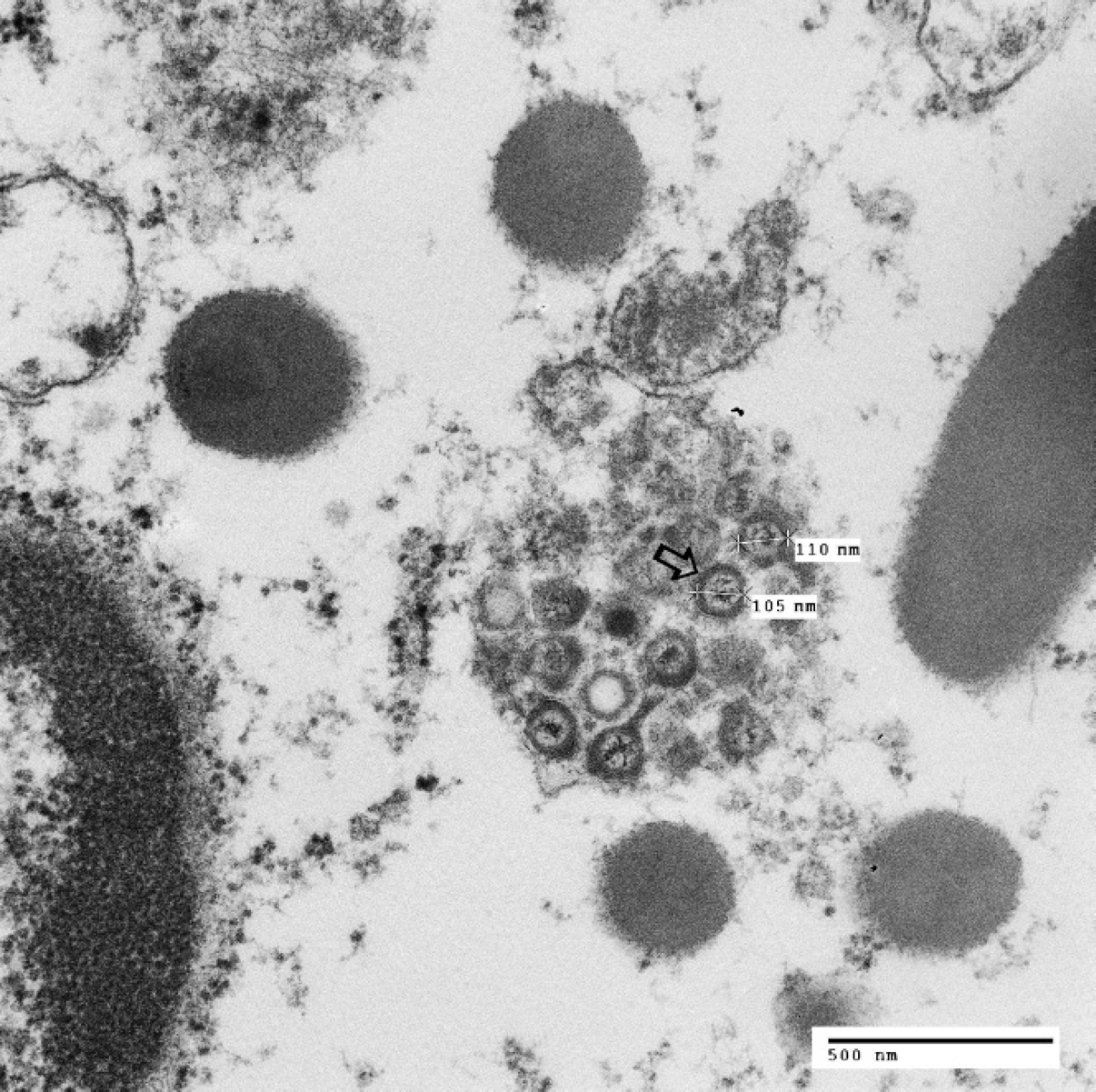
Electron micrograph of epithelial cell. The cytoplasm contains a cluster of enveloped viral particles within cytocavitary network, which approximately measure 105 nm in diameter (open arrow).
In Psittaciformes, there are several recognized diseases associated with avian alpha-herpes viruses. Infection with Psittacid herpesvirus 1 (PsHV-1), formerly known as Pacheco's disease (PD), is characterized by massive hepatic necrosis with formation of syncytial cells. 18 Amazon tracheitis virus (ATV) is a cause of upper-respiratory-tract lesions similar to ILTV in chickens. 18 There are several other herpesviruses associated with more chronic, non-life-threatening skin and/or mucosal lesions. It is speculated that the feather abnormalities referred to as “feather dusters” in European Budgerigars are caused by a herpesvirus. 18 The isolated virus is unrelated to those strains found in psittacine birds, and the significance of its occurrence in these feather lesions remains to be determined. There are reports of possible herpesvirus involvement of wart-like skin lesions on the feet of cockatoos and macaws; however, this remains to be confirmed. 18 Recently, a novel Psittacid herpesvirus strain was isolated from the mucosal papillomas of neotropical parrots 21 and from cloacal and cutaneous papillomas of African grey parrots. 22
However, there have been reports of a different herpesvirus of parakeets that has tropism for the lower respiratory tract, with no hepatic or significant upper-respiratory-tract involvement. One was from the United States, 9 in a Bourke's parakeet, and the other was from Japan. 26 This virus is referred to as “respiratory herpesvirus of parakeets” and represents an unusual manifestation of herpes-virus-induced disease in parakeets. In these 2 parakeets, the herpes-like inclusion bodies were identified within epithelial cells and syncytial cells of the trachea, bronchi, parabronchi, air capillaries, and air sacs. The necrotic foci in the liver of the first bird lacked syncytial cells and intranuclear inclusion bodies. Based on the similarity of the lesions, the authors believe that the herpesvirus in the present case is very similar to, if not the same as, the respiratory herpesvirus of the 2 parakeets previously reported in the United States 9 and Japan. 26 The histological lesions in the lung of the Bourke's parakeet included segmental necrosis, cuboidal metaplasia, and syncytial giant cells in the parabronchial epithelium. On transmission electron microscopy, the location, morphology, and size of the viral particles are typical of a herpesvirus. No histological lesions were found in the liver. The report from Japan describes disease in 14 parakeets that died of herpesvirus-induced pneumonia and air sacculitis. 9 The histological lesions included necrotizing tracheitis, pneumonia, and air sacculitis, with syncytial cells and herpes-like inclusion bodies in the lung. Many affected birds had secondary pulmonary aspergillosis, chlamydiosis, or candidiasis.
Because of its respiratory tropism, it is speculated that this virus may be a variant of ILTV in chickens or ATV in Psittaciformes. However, both of those viruses primarily have tropism for the upper respiratory tract manifested as necrotizing pharyngitis, laryngitis, tracheitis, and occasional pneumonia. In addition, pneumonia is characterized by epithelial cell hyperplasia, edema, and airway mucosal infiltration by moderate numbers of mixed inflammatory populations, without syncytial cell formation.
Another possibility is that this lower respiratory disease is an unusual manifestation of PD. Pacheco's disease occurs worldwide, 1,3,4,6,10,12,13 and once recognized, it was speculated that PD is caused by a single PsHV serotype. Today, there are at least 5 different PsHV serotypes 25 recovered from Psittacid birds, with serotype 1 (PsHV-1) being the most common. 24 Pacheco's disease is primarily characterized by a massive hepatic necrosis, 14 variably accompanied by necrotizing lesions in intestines, spleen, kidney, pancreas, and endocrine glands; 17,18 there have been infrequent reports of unusual manifestations of PD characterized by lung 15,18,20 lesions, which would imply a possible variation in PsHV tissue tropism. In another report, an unusual outbreak of necrotizing esophagitis 2 occurred in Pionus sp. and Amazon parrots. The liver contained only focal areas of necrosis with rare intranuclear inclusion bodies. In the current study, the authors speculate that the esophageal and hepatic lesions might be a manifestation of PD caused by serologically distinct variant of PsHV. Herpesviruses have been isolated from many other bird orders, including Columbiformes (pigeons and doves), 11,19 Falconiformes (falcons), 7 and Anseriformes (ducks). 8 In these birds, the herpesvirus-associated lesions were mainly localized within the gastrointestinal tract. However, in Passeriformes (songbirds), the herpesvirus lesions were confined to the conjunctiva and the respiratory tract. 16 Upon DNA sequencing, this virus was related to ILTV in gallids.
By considering the distribution and characteristics of the lesions, as well as electron microscopy findings, it is hypothesized that this virus is very similar to the virus previously reported in Japan 9 and the United States. 26 Although there are several publications that reported the genomic properties of different herpesviruses, there are no reports that provide the DNA sequencing information on the virus referred to as “respiratory herpesvirus of parakeets.” To confirm the authors' hypothesis and to determine the exact genetic phylogeny of this lung-associated herpesvirus, nucleic acid sequencing is necessary.