
Abstract
The aim of this study was to investigate the presence of dermatophytes and yeasts in healthy and diseased dogs. A total of 633 samples were collected from 26 healthy animals (104 samples), 131 with dermatitis (343 samples), 74 with otitais (148 samples), and 19 with ocular diseases (38 samples). Cultures from healthy animals were positive for Malassezia pachydermatis in 13.5% (7/52) of samples from skin, 42.3% (11/26) from ear, and 3.8% (1/26) from eye. Fungal growth was observed in 20.4% (70/343) samples from animals with dermatitis. Microsporum canis was the most isolated fungus (n = 39), followed by M. pachydermatis (n = 30) and Malassezia sp. (n = 3). Of the 148 samples from dogs with otitis, 90 (60.8%) were positive for M. pachydermatis, and of the clinical specimens from the conjunctiva of animals with ophthalmic disease, 2.6% (1/38) presented positive cultures for M. pachydermatis. Only 14.3% (2/14) of the positive cultures for M. pachydermatis and 40.9% (9/22) of those for M. canis were positive in the direct exam. Direct exams were positive in 84.3% (70/83) of the culture positive samples from affected ears of dogs with otitis. Malassezia pachydermatis may act as an aggravating factor in the occurrence of cutaneous diseases, or the isolation of M. canis may be associated with the onset of dermatophytosis. Fungal culture, rather than microscopic examination, should be used as the definitive diagnostic test for dermatomycoses and otitis.
Keywords
Introduction
Fungi have either unicellular or multicellular structures and are classified according to their morphology into filamentous fungi, yeasts, and dimorphic fungi. The filamentous form consists of a group of tubular structures while the yeasts are fungi whose unicellular structure presents only 1 nucleus per cell. The dimorphic group has either the filamentous or the yeast form (or spherule in the case of Coccidioides spp.), depending on the temperature as well as other environmental factors. 26 The most common pathogenic fungi isolated from dogs and cats belong to the filamentous group, especially dermatophytes, followed by yeasts from the genus Malassezia, particularly M. pachydermatis. 16
Dermatophytes are infections of keratinized structures, such as nails, hair, and the stratum corneum of the skin, and are the most common fungal diseases diagnosed by veterinarians. 5,22 They are caused by dermatophytes belonging to the genera Microsporum, Trichophyton, and Epidermophyton, which utilize keratin as a nutrient substrate. 10 In dogs and cats, M. canis is the most frequently isolated species and plays a major role as a constant reservoir of zoonosis. 3,10,15
Yeasts of the genera Candida and Malassezia are usually isolated from the skin and mucosa of healthy cats and dogs but may become pathogens whenever there are alterations to the host's defenses or skin surface microenvironment. 4,14,18 Otitis externa and dermatitis in dogs and cats are usually associated with these yeasts as a primary agent or in combination with bacteria, with M. pachydermatis being the most commonly found. 8,13,16,18,20,21 In the state of Ceará in northeastern Brazil, a study 24 reported the presence of this yeast in the conjunctival sac in both healthy dogs and those with corneal ulcers. In addition, other species, such as M. furfur, M. obtusa, and M. sympodialis, have been described as agents of otitis externa in cats and dogs. 11 Infections caused by Candida spp. in animals are infrequent, 19 but in Brazil C. albicans has been detected in cattle with otitis externa 12 and in cases of dermatomycosis in dogs. 25 The aim of this study was to investigate the presence of fungi in the axillas, groins, ears, and eyes of healthy dogs, as well as in dogs with dermatitis, otitis, and ocular diseases.
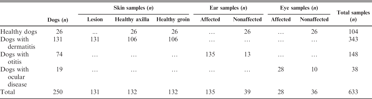
Number of animals and samples collected from each group of dogs (healthy, with dermatitis, with otitis, with ocular diseases).
Materials and methods
Animals
This study included 26 healthy dogs, 131 with dermatitis. 74 with otitis, and 19 with ophthalmic diseases. The samples were collected between July 2005 and April 2006. with the collaboration of 3 veterinary clinics located in the city of Fortaleza, Ceará, Brazil. The age, breed, sex, living conditions, and clinical data of the animals were recorded. After clinical examination the dogs were arranged in 4 groups, which are listed in Table 1.
Specimen collection
Samples from healthy skin of the axilla and groin and from suspected dermatitis lesions were obtained by plucking the fur with forceps, and scraping the epidermal scales with a scalpel. The samples from each animal were placed in separate sterile plastic containers and labeled. 3 Clinical specimens from each healthy and/or diseased ear canal were obtained using a sterile dry cotton swab. Each swab was then replaced in the plastic storage tube and labeled. 13 Samples from healthy or diseased eyes were collected from the conjunctival sac with a calibrated platinum loop (1 μl) and then placed directly on modified Dixon agar. a , 24 All samples were then transferred to the Medical Mycology Specialized Center at the Faculty of Medicine (Federal University of Ceará).
Laboratory methods
The clinical specimens from healthy or diseased ears of dogs with otitis and from skin lesions of animals with dermatitis were initially examined for the presence of fungal elements. From the samples of the ears, slides were stained with Gram stain for immersion microscopic examination. For the plucked fur and scraped scales, slides were flooded with 30% potassium hydroxide and Parker Quink ink b (3:1) for microscopic examination at a 40 × magnification. All the samples were cultured on modified Dixon agar. The skin samples were additionally incubated on Sabouraud dextrose agar with or without 0.05% chloramphenicol and 0.05% cycloheximide. The Sabouraud's dextrose agar was incubated at room temperature (28°C) and the Dixon's agar at 32°C, for up to 10 days, and examined on a daily basis.
The preliminary identification of dermatophytes was based both on the macroscopic appearance of colonies and microscopic features. In addition, special studies such as nutrition requirements, in vitro hair perforation tests, urease tests, growth on rice medium, and growth on Borelli's lactritmel agar were performed. 3 Malassezia pachydermatis was identified microscopically by its morphology as well as by its ability to grow when subcultured on Sabouraud dextrose agar (medium without lipid supplementation). 13 The identification of Candida spp. was based on phenotypic features, such as description of the macro- and micromorphology, and through fermentation of carbohydrates and auxanographic typing. 4
Statistical analysis
The Fisher exact test was performed to determine whether there was a statistically significant difference among the sampled groups. Differences of P < 0.05 were considered significant.
Results
A sample was considered positive if a microorganism was isolated on the culture. A total of 104 clinical specimens from the axilla, groin, ear, and eye (n = 26 for each anatomic region) from 26 healthy dogs were collected (Table 2). The only fungus isolated from these animals was M. pachydermatis. Of the 52 skin samples (axilla and groin), the culture was positive in 7 (13.5%) of the samples. Five dogs were culture positive for M. pachydermatis: in 1 dog this yeast was isolated only from the axilla, in 2 dogs only from the groin, and in 2 dogs from both sites. Of these 5 dogs with positive cultures, 3 were male, and only 1 was less than 1 year old; 3 of them were kept indoors, but only 2 had access to sand. Of the 26 samples of healthy ears, 11 (42.3%) were positive for fungal growth. Of the 11 healthy dogs with positive cultures for M. pachydermatis, 5 (45.5%) were from Poodles and 6 (54.5%) were from other breeds; 7 (63.6%) were from males and 4 (36.4%) from females; and only 2 (18.2%) were from dogs less than 1 year old while 9 (81.8%) were from dogs older than that. Only 1 sample from conjunctival sac was positive for M. pachydermatis. It was a sample from a male Poodle aged 2 years and 11 months. The frequency of positive culture from the ear was significantly higher than axilla (P = 0.0215), groin (P = 0.0391), and eye (P = 0.0063).
Frequency and comparison of isolation of Malassezia pachydermatis from different anatomic regions of healthy dogs.
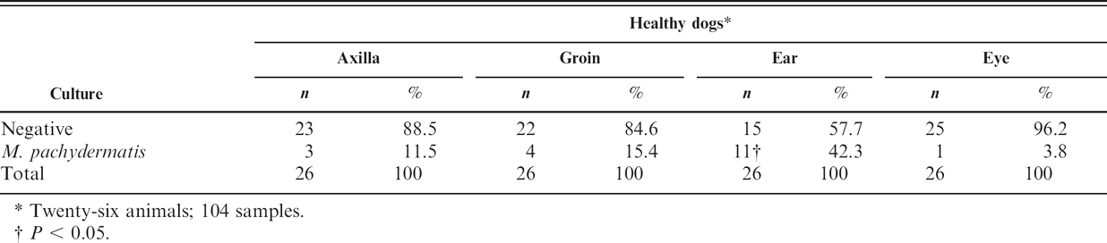
Twenty-six animals; 104 samples.
P < 0.05.
A total of 343 samples were collected from 131 dogs with dermatitis: 131,106, and 106 from skin lesions, axillas, and groins, respectively (Table 3). Fungal growth was observed in 20.4% (70/343) of all samples. The 70 positive samples were from 42 dogs with dermatitis, and the most representative breeds were Poodle (38.1%), followed by English Cocker Spaniel (7.1%) and Yorkshire terrier (7.1%). Only 9 dogs (21.4%) were less than 1 year old. Two samples from skin lesions of dogs were positive in the direct exam for dermatophyte, but was culture negative. Of the 14 positive cultures for only M. pachydermatis from skin lesions of dogs, only 2 (14.3%) were positive in the direct exam. On the other hand, only 9 (40.9%) of 22 dog skin lesions positive by culture for Microsporum canis were also positive by direct examination. In addition, the 2 samples that were culture positive for both M. canis and M. pachydermatis could only be identified as dermatophytes in the direct exam.
From the 148 otitis clinical specimens, M. pachydermatis was isolated in 90 (60.8%) samples (Table 4). The most common breed in the group of dogs with otitis was Poodle (49.0%), followed by English Cocker Spaniel (11.8%). Concerning the shape of the ears, 42 animals had pendulous ears while only 7 had erect ears. Data were not recorded for 2 dogs. Of the 61 dogs with bilateral otitis, 7 presented unilateral and 33 bilateral positive cultures for M. pachydermatis. In addition to this yeast, in 1 dog's ear (Maltese, female, 1 year old) other Malassezia sp. was also isolated; and in 1 dog's ear (Poodle, male, 1 year old) Candida tropicalis was also isolated. Of the 13 dogs with unilateral otitis, 10 were culture positive for M. pachydermatis in the diseased ear and 7 in the healthy ear. Of the 83 positive samples from affected ears and the 7 from contralateral healthy ears, 70 (84.3%) and 3 (42.9%), respectively, were positive in the direct exam for the yeast. In addition, 7 (13.5%) and 1 (16.7%) negative cultures from affected (n = 52) and nonaffected (n = 6) ears showed positive results from direct exam. Of the dogs with M. pachydermatis, the most common breed was the Poodle (n = 25; 33.8%), followed by the English Cocker Spaniel (n = 6; 8.1%).
A total of 28 specimens from diseased eyes and 10 from contralateral healthy eyes were collected (Table 5). Of a total of 38 samples, only 1 yielded positive growth of M. pachydermatis, from a diseased eye. It was from a female Boxer (aged 3 years and 1 month) that had a corneal ulcer.
Frequency of isolation of fungi from different anatomic sites of dogs with dermatitis.
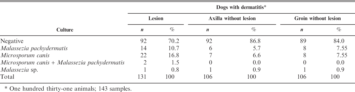
One hundred thirty-one animals; 143 samples.
Frequency of isolation of fungi from the ears of dogs with otitis.
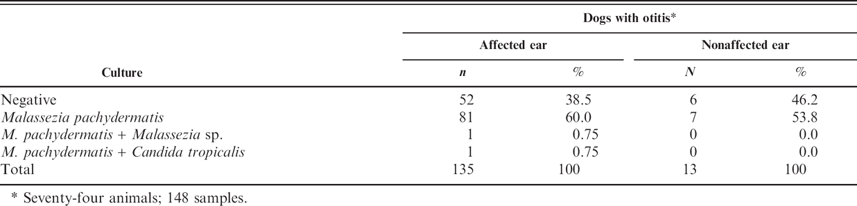
Seventy-four animals; 148 samples.
Discussion
Direct microscopic examination was less sensitive than fungal culture. For example, 15.7% and 57.1% of the clinical specimens from otitis externa cases and from the unaffected ears, respectively, were negative by direct microscopic examination but positive by culture. Less cerumen on the swab from unaffected ears, and consequently a slide with less material for direct examination, may explain the higher frequency of false-negative results. The direct microscopic examination of the samples from dermatitis was less sensitive for yeasts (87.5% false negatives) than for dermatophytes (50.0% false negatives). This was probably because of the fact that the technique chosen (slides with 30% potassium hydroxide and Parker Quink ink with 40 × magnification) was not the best to visualize yeast structures but was better for dermatophytes. Therefore, although direct examination gives an immediate diagnosis, more accurate results are obtained following fungal culture.
Although different studies have isolated dermatophytes from asymptomatic dogs and cats, 7,9 in this study the only fungus isolated from healthy animals was M. pachydermatis. This yeast is often recovered from healthy animals, from different anatomic regions, but more frequently in dogs than cats. 14 In the present study, the frequency of positive culture from the left ear was significantly higher than from the axilla, groin, and eye of healthy dogs (P < 0.05). Culture from the conjunctiva of healthy animals was positive in only 1 animal (3.8%). These data agree with the findings from a previous study 24 in which M. pachydermatis was cultured in 3% of ocular samples collected from healthy dogs.
Dermatophytosis is a common skin disease in small animals, and various studies have been described in the international literature, with M. canis being the most common causative agent. 6,10,15 These data also correspond to the situation in Brazil, where this species is the most common seen in dogs and cats. 3 However, the proportion of positive cultures in relation to the number of examined clinical specimens is variable. 6 In this study, the percentage of positive animals for M. canis, the only isolated dermatophyte, was 18.3% (24/131) of those with dermatitis. According to other studies, there is a higher prevalence of M. canis infection in dogs and cats aged less than 1 year. 9,10,23 However, this was not observed in this study, and there was no indication that age played a significant role, corroborating the findings from previous studies. 2,15
Although carnivores can be colonized by lipid-dependent species of Malassezia, 1 only 1 dog, a female Dachshund, aged 2 years and 4 months, had a positive culture for a lipid-dependent Malassezia. In 1 previous study, 20 Malassezia spp. was isolated from at least 1 skin site of all atopic dogs while in the present study only 15.3% (20/131) of the animals were culture positive for Malassezia yeasts. This difference may be explained by the fact that the study included animals with any signs of dermatitis, and not only dogs with atopy, which could modify the skin microenvironment, thus enhancing the growth of this yeast. Moreover, the previous study 20 examined samples from 16 anatomic sites for each animal, while the samples collected in this study were from only 3 different sites.
Frequency of isolation of fungi from the eyes of dogs with ocular diseases.
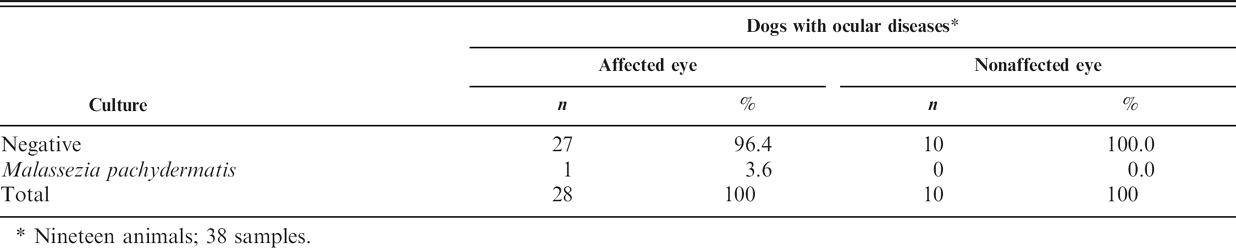
Nineteen animals; 38 samples.
In the present study, Microsporum canis isolation from dogs was possible only from the lesion or the lesion plus axilla and groin. However, of the 20 animals from which M. pachydermatis was isolated, this yeast was isolated from the lesion or the lesion plus axilla and groin in 16 dogs (80.0%). In 4 animals (20.0%), M. pachydermatis was not isolated from the lesion, and 2 dogs were culture positive for both M. canis and M. pachydermatis. From these results, it would appear that this yeast acts as an aggravating factor in the occurrence of cutaneous diseases, as a consequence of the alteration of the skin microenvironment, rather than playing a role in the onset of infection. On the other hand, the isolation of M. canis may be automatically associated with the appearance of lesions and the onset of dermatophytosis.
Otitis externa is 1 of the most commonly diagnosed diseases in dogs, and M. pachydermatis is the most commonly isolated organism and complicating factor. 13,17 However, the isolation of this yeast in the external ear canal does not necessarily mean it is the cause of disease, since it is frequently cultured from samples of healthy or dogs with otitis. 11,13 In this study, a higher frequency of M. pachydermatis in ears affected by otitis was found, rather than in samples from dermatitis or ophthalmic conditions, possibly because of the high levels of fatty acids present in that microenvironment, which may create a favorable habitat for the growth of this yeast. Of the positive dogs, 60.7% were male and 33.0% were female (data were not recorded in 6.3% of the animals), thus showing that gender may be a predisposing factor. With regard to the shape of the ear, the findings in this study corroborate other reports 8,11,13 and suggest that dogs with pendulous ears (82.4%) are predisposed to otitis externa associated with M. pachydermatis.
In summary, the results showed M. canis as the only dermatophyte and the most frequently detected fungus from animals with dermatitis in this investigation. It was also found that M. pachydermatis is present in healthy animals, animals with dermatitis and ophthalmic diseases, and with a high frequency in dogs with otitis. However, it remains to be determined if this yeast participates as the primary etiology of dermatitis, otitis, and ophthalmic diseases. The results of the present study also indicated that fungal culture, rather than microscopic examination, should be used as the definitive diagnostic test for dermatomycoses and otitis.
Footnotes
a.
Oxoid Ltd., Basingstoke, Hampshire, United Kingdom.
b.
Solv-X, Parker, United Kingdom.