
Abstract
An indirect enzyme-linked immunosorbent assay (ELISA) based on baculovirus recombinant P30 protein of Ehrlichia canis and the 1BH4 anticanine IgG monoclonal antibody was developed and evaluated by examining a panel of 98 positive and 157 negative sera using the indirect fluorescent antibody (IFA) test as the reference technique. The P30-based ELISA appeared to be sensitive and specific (77.55% and 95.54%, respectively) when qualitative results (positive/negative) were compared with those of the IFA test; the coefficient of correlation (R) between the 2 tests was 0.833. Furthermore, it was possible to establish a mathematical formula for use in comparing the results of both techniques. These results indicate that recombinant P30 antigen-based ELISA is a suitable alternative of the IFA test for simple, consistent, and rapid serodiagnosis of canine ehrlichiosis. Moreover, the use of this recombinant protein as antigen offers a great advantage for antigen preparation in comparison with other techniques in which the whole E. canis organism is used as antigen.
Keywords
Introduction
Canine monocytic ehrlichiosis (CME), caused by Ehrlichia canis, is a tick-borne disease transmitted by the brown dog tick Rhipicephalus sanguineus. In Spain, E. canis has recently been isolated. 1 Classically, 3 phases are described in the pathogenesis of CME: acute phase, subclinical phase, and chronic phase. Clinical signs of canine ehrlichiosis are often non-specific and include fever, anorexia, lymphadenomegally, lethargy, depression, splenomegaly, and hemorrhagic tendencies. 7,22 Different clinical abnormalities, such as anemia, thrombocytopenia, and hyperproteinemia due to hypergammaglobulinemia, can be found in dogs infected by E. canis.
Diagnosis of canine ehrlichiosis can be performed using various techniques: demonstration of inclusions compatible with E. canis within monocytes and lymphocytes, culture of the agent in specific cell lines, different serologic techniques, and polymerase chain reaction (PCR). The indirect fluorescent antibody (IFA) test is the most widely used serologic assay for the diagnosis of infection with E. canis. The IFA test has been considered the “gold standard” technique for detection and titration of E. canis antibodies and has been used to evaluate other diagnostic assays. 2,3,6,20 However, the IFA can only be performed in specialized laboratories by qualified personnel to prevent results from being read in a subjective way. In addition, the IFA test is time consuming, cumbersome for testing multiple samples simultaneously, and not a technique easy to perform in a standard laboratory. For these reasons, other serologic assays for the diagnosis of E. canis infection in dogs need to be developed.
Some commercial serologic tests are available for detection of E. canis antibodies based on whole E. canis organisms as the source of antigen. These include dot enzyme-linked immunosorbent assay (ELISA) test Immunocomb® a , 21 IgG MIF antibody test Kit®, b and Dip-S-Ticks®. c Another test, immunochromatography Snap3®Dx assay, d based on the specific P30 protein cloned and expressed as a fusion protein in the Escherichia coli system, has been recently modified and now uses synthetic E. canis peptides (P30, P30–1) derived from E. canis immunodominant epitopes. 15 Several groups have reported the expression of different E. canis recombinant proteins (major antigenic protein 2, 2,10 P43, 12 P30, 13,17 P120, 23 gp36, and gp194) in E. coli and their use in developing diagnostic tests. However, to the authors' knowledge, none of these tests are commercially available. The use of a recombinant protein instead of whole agent in the ELISA is expected to greatly improve the assay because of the high quality of the test antigen. Indeed, the specificity of this kind of test eliminates the subjectivity of dot blot, Western blot, or IFA tests.
The purpose of the present study was to develop and evaluate an antibody detection ELISA based on a highly specific baculovirus-expressed E. canis recombinant antigen. The cloning and expression of P30 protein expressed in the baculovirus expression system is reported. The value of the recombinant protein for E. canis serodiagnosis purposes was tested by indirect ELISA using field dog sera, and the results were compared with the IFA test, which is considered the reference technique for detecting E. canis antibodies.
Materials and methods
Organisms, cells, and viruses
E. canis isolated from a naturally infected dog was grown on monolayers of the continuous canine cell line DH82 e as described previously. 1 Spodoptera frugiperda cell line Sf9 f was used to propagate recombinant baculovirus. The cells were grown in suspension or monolayer culture as previously described. 8 Recombinant baculovirus AcHLT-p30, which expresses the P30 protein of E. canis (see below), was used for antigen preparation.
DNA isolation and cloning of p30 gene in pAcHLT baculovirus expression vector
E. canis genomic DNA was extracted from DH82 cells infected with E. canis using DNAzol®reagent.
g
The complete E. canis p30 gene was amplified by PCR using the forward primer EH30+ (5′ GGTTAT
Transfection and selection of recombinant baculoviruses
Sf9 cells were transfected with a mixture of transfer vector (2 μg) and linearized BacPak 6 DNA 1 (500 ng) in the presence of Jet PEI™. m After 5 to 6 days, culture supernatant was plated for baculovirus isolation in the presence of X-Gal n (5-bromo-4-chloro-3-indolyl-β-D-galactopyranoside), white plaques were recovered, and the recombinant baculoviruses were plaque purified. 8
Expression and purification of P30 protein in the baculovirus system
Sf9 cells (0.8 × 10⁶ cells/ml) were infected at a multiplicity of infection of 2 plaque-forming units per cell. Cells were collected at 96 h postinfection with a clear cytopathic effect. Sf9 cells were lysed by hypotonic shock with 25 mM NaHCO3 pH 8.3 (1 ml/20 × 106 cells) for 30 min at 4°C. Then cells were pelleted by centrifugation, and the insoluble P30 protein was extracted by solubilization of the pellet with the elution buffer (Na2HPO4 20 mM, guanidinium hydrochloride 6M, and NaCl 0.5M). Then sonication was performed in 5 ml of the elution buffer added to 20 × 106 cells (5 times for 10 s in ice), and finally the soluble fraction was recovered by centrifugation. This simple treatment allowed a quick purification of the recombinant protein to more than 85% purity determined by densitometric assay. The partially purified P30 was highly purified by chromatography with nickel-charged0 high-density resin. Briefly, the soluble fraction in the guanidine hydrochloride 6M buffer was applied to a Ni++-charged resin and, after washing with imidazole 50 mM, the P30 protein was eluted with elution buffer (imidazole 500 mM, NaCl 0.5 M, Na2HPO4 20 mM [pH 7.4]). The refolding of the purified protein was achieved by sequential dialysis in phosphate-buffered saline (PBS) containing 0.3 M arginine. Purified protein was stored at −70°C until use.
The identity of P30 protein was confirmed by sodium dodecyl sulfate polyacrylamide gel electrophoresis (SDS-PAGE) and immunoblotting with a commercial anti-His monoclonal antibody (MAb) p and with serum from E. canis-infected dogs by standard procedures. Protein concentrations were estimated with the Bio-Rad protein assay reagent q following the manufacturer's instructions.
Serum samples
A total of 255 canine serum samples submitted for E. canis antibody testing to the Ehrlichiosis Diagnostic Service, College of Veterinary Medicine, Complutense University of Madrid, were used. Of the 255 sera, 98 were from dogs with naturally occurring ehrlichiosis and positive IFA test results (≥ 1:80). Twenty-one of the IFA-positive samples had a titer of 80, 8 had titers of 160, 11 had titers of 320, 14 had titers of 640, 1 had titers of 1,280, 13 had titers of 2,560, and the remaining 20 samples had titers of 5,120 or higher. One hundred and fifty-seven of the 255 serum samples were from clinically healthy dogs that were seronegative to E. canis. In addition, 4 canine serum samples that were positive to Anaplasma phagocytophilum and 19 serum samples positive to Leishmania infantum were also analyzed using the IFA test. All these samples (except 1 from a dog with concurrent ehrlichiosis and leishmaniasis) were seronegative to E. canis using the IFA test. All serum samples were tested blindly in the ELISA test.
Immunofluorescent antibody test
The antigen used was the DH82 cell line infected with E. canis (Madrid strain) as previously described. 1 An immunofluorescent IgG antibody titer of ≥ 1:80 was considered positive. 6
Indirect ELISA using recombinant P30 protein
The reactivity of the dog sera with P30 recombinant protein was determined by indirect ELISA. Polystyrene microtiter plates r were coated with 0.3 μg of antigen overnight at 4°C in 0.05 M sodium carbonate buffer (pH 9.6). Washes between consecutive steps were performed with 0.05% Tween 20 in PBS. Plates were incubated with the dog sera diluted 1/100 in PBS containing 0.35 M NaCl, 0.05% Tween 20 for 10 min at room temperature. A horseradish peroxidase-labeled anti-dog IgG MAb designated 1BH4 previously developed in the authors' laboratory was used as conjugate. After incubation for 10 min at room temperature, bound antibodies were detected by adding TMB-MAX s as substrate. The reaction was stopped with H2SO4 0.5 M, and the absorbance was measured at 450 nm in a Multiskan Ascent ELISA reader.
Data analysis
Analysis of the data was performed using Statgraphics plus 5.1 and Excel (Microsoft) computer programs. Repeatability was calculated by comparing replicates in the same plate using Pearson's variation coefficient (CV: 100 × [SD of replicates/mean of replicates]), where less than 20% indicates adequate repeatability. Intra- and interplate variability was analyzed by testing the same positive sample 16 times in the same plate and 1 row per plate in 10% of plates of the final batch, respectively. The results were expressed as CV. Reproducibility was assessed by Pearson's correlation coefficient (r2), comparing the data obtained from the same samples, analyzed twice by the same laboratory in different plates and at different times.
Sensitivity, specificity, predictive values, prevalence, and accuracy for the ELISA test were calculated in relation to the IFA test, considered the gold standard technique for the diagnosis of canine ehrlichiosis. 20 Variables measured included the number of true positives (TP), number of true negatives (TN), number of false positives (FP), and number of false negatives (FN). Sensitivity was calculated as 100 × TP/(TP + FN), specificity was calculated as 100 × TN/(TN + FP), positive predictive value was calculated as 100 × TP/ (TP + FP), negative predictive value was calculated as 100 × TN/(FN + TN), and prevalence was calculated as 100 × (TP + FN)/total sample. Test accuracy, the proportion of all tests that gave a correct result, was defined as 100 × (TP + TN)/total sample.
The level of agreement between IFA and ELISA test results was evaluated and expressed as a percentage of overall agreement and as a kappa (κ) value, defined as the proportion of agreement beyond chance exhibited by the 2 tests. By convention, κ values of 0.0 to 0.2 = slight, 0.2 to 0.4 = fair, 0.4 to 0.6 = moderate, 0.6 to 0.8 = substantial, and 0.8 to 1.0 = almost perfect agreement. Correlation between IFA titers and the absorbance at 450 nm obtained by indirect ELISA test were assessed by Pearson's correlation coefficient (r2), analyzing only positive samples. Different mathematical equations (linear, logarithmic, polynomic, etc.) were applied to relate optical densities and antibody titer by IFA. By using the model best adapted to the data, the correlation factor of the curve (R2) and the mathematical equation was calculated.
Results
Cloning, expression and purification of P30 recombinant protein
To construct the baculovirus transfer vector, the complete sequence of the p30 gene was generated by PCR with specific forward and reverse primers and cloned into the baculovirus transfer vector. The plasmid obtained was called pAcHLT-p30.
Sf9 cells infected with the recombinant baculovirus AcHLTA-p30 were collected at 96 hours postinfection, and proteins were analyzed after SDS-PAGE by Coomasie blue staining and immunoblotting analyses. The protein was expressed as a fusion protein with 41 extra amino acids in the amino terminus, including a 6 His-tag. As shown in Figure 1A, the size of the expressed protein agreed with the expected size for the P30 plus the fusion. The identification of a protein of approximately 34 kDa as the P30 protein was confirmed by immunoblotting analysis using a commercial anti-His MAb (Fig. 1B) and specific dog sera from animals naturally infected with E. canis, diluted 1:500 in PBS-Tween20 0.05% (Fig. 1C). The protein was expressed as an insoluble form, and the maximum expression was observed at 96 hours postinfection. After semipurification of the protein from the infected cell pellet, the production of the P30 protein expressed in the baculovirus expression system was analyzed by SDS-PAGE and Western blot. P30 constituted approximately 50% of the total protein, and the yield was about 14 μg/1 × 108 Sf9 cells estimated by densitometric assay with known quantities of bovine serum albumin performed with 1D Image Analysis Software 2.0.1. 5
The purity of the protein after purification by Ni+-conjugated column was >90%, and the final yield of the P30 using this procedure was 1.3 μg/1 × 108 Sf9 cells (Fig. 1). When P30 purified by Ni+-conjugated column was stored at −70°C, precipitation of the protein was observed after 2 to 3 weeks. Therefore, only P30 protein stored in guanidine hydrochloride was subsequently used as ELISA antigen.
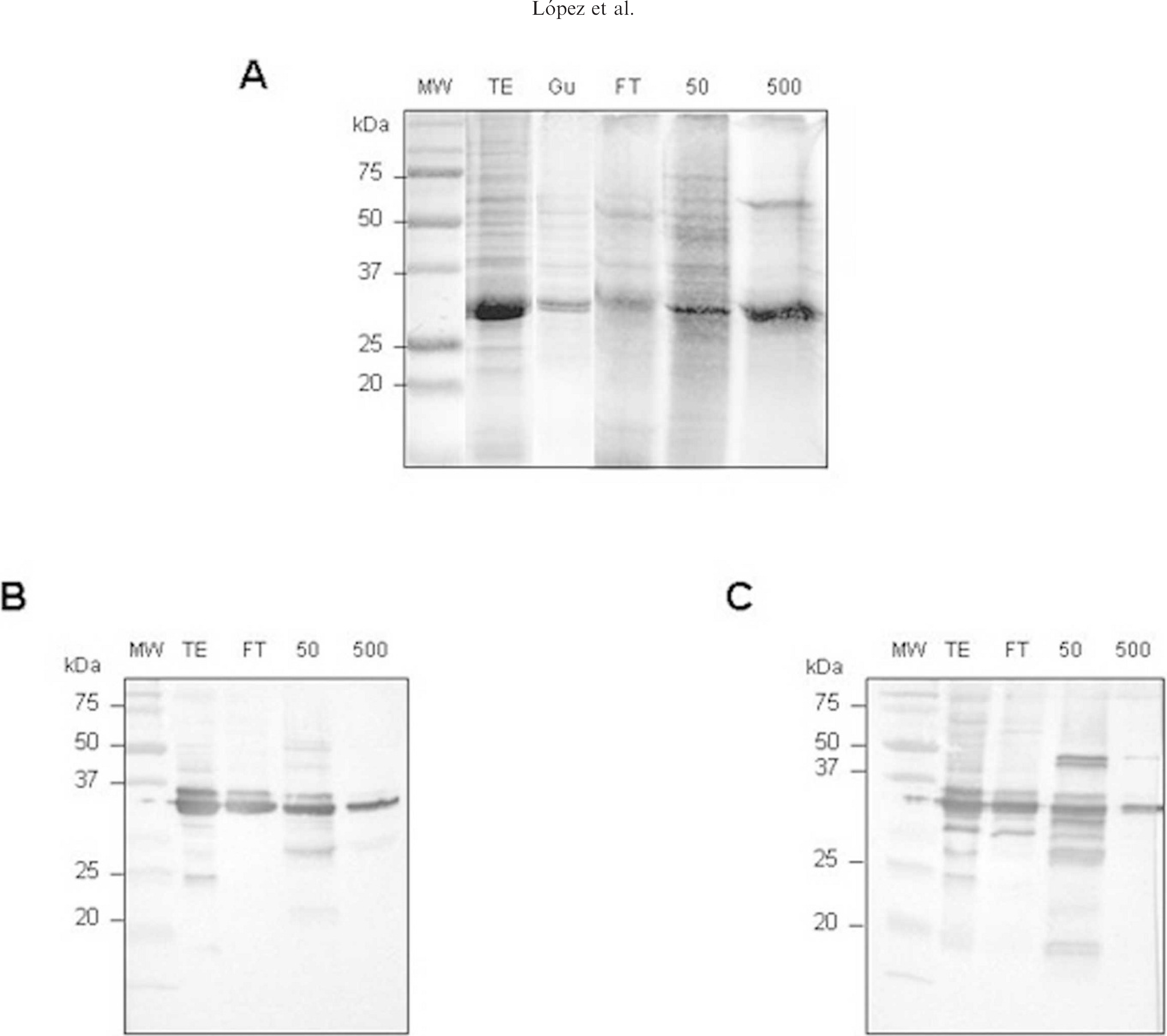
Expression of P30 protein in insect cells. Sf9 cells were infected with the recombinant baculovirus at a multiplicity of infection of 2. Cell extracts were harvested at 96 hours postinfection, and P30 was purified as described in the Materials and Methods section. Different fractions of P30 were separated by 11% sodium dodecyl sulfate polyacrylamide gel electrophoresis and stained with Coomasie blue,
Development of the indirect ELISA
The ELISA was initially developed by using a panel of 32 negative and 16 positive serum samples previously tested by IFA as the reference technique. Optimal concentrations of the antigen, serum dilution, conjugate, and substrate solution were determined in a series of checkerboard titrations of each reagent against all other reagents. Additional experiments determined the optimal temporal, chemical, and physical variables of the protocol, including incubation temperatures and durations; composition, pH, and molarity of diluent; and washing and blocking buffers. Final ELISA conditions were as described in the Materials and Methods section. Under those conditions, the ELISA yielded 100% sensitivity and specificity on the initial panel of 32 negative and 16 positive sera.
Indirect ELISA validation and comparison with IFA
The reliability of the newly developed P30 indirect ELISA for detecting E. canis antibody in several sets of well-defined sera was determined, using the IFA as the reference test. A total of 98 positive sera from dogs naturally infected with E. canis and 157 negative serum samples were tested.
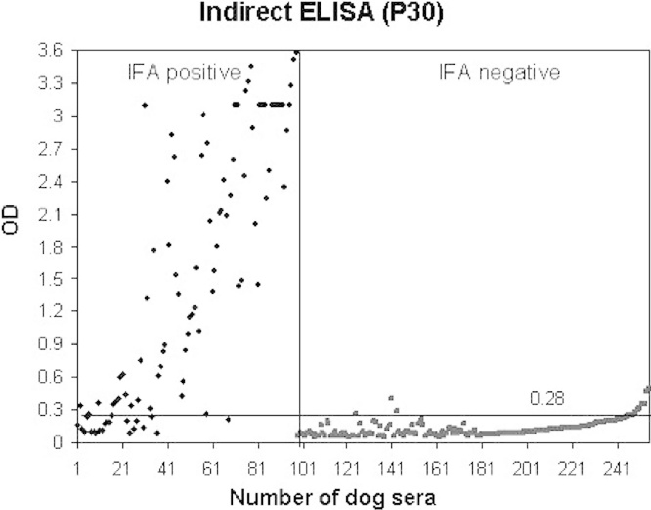
Scatter plot analyses of 255 sera measured by P30bac indirect enzyme-linked immunosorbent assay. The horizontal line indicates statistical cutoff in this assay (0.3). Sera numbers of 1 to 98 and of 99 to 255 on an X-axis represent indirect fluorescent antibody-positive and -negative samples, respectively.
The average optical density value for the positive samples was calculated separately for each group of sera with different IFA titers. For example, the average was 1.47 ± 0.7 for samples with IFA titer 1:640 and that of the negative ones was (0.129 ± 0.078), which provides a cutoff (mean negative value + twice the SD) of 0.285 (Fig. 2).
Evidence of repeatability was accomplished by evaluating results from replicates of several samples in each plate. When the same samples were analyzed twice in different plates and at different times, the results showed an excellent correlation with an r2 of 0.992. Coefficients of variation of ELISA tests were less than 15% in all cases, except in 4 samples (2 seropositive and 2 seronegative).
When intraplate variability was measured by testing a single positive serum sample 16 times in different plate positions, a CV of 1.88% was obtained. For interplate variability, the same serum tested 8 times in 10% of all plates yielded a CV of 2.75%.
Of the 255 sera analyzed, 226 yielded identical qualitative results (positive or negative) in both tests (indirect ELISA and IFA), corresponding to an accuracy value of 88.62% (Table 1), with a κ index of 0.75. The sensitivity of the ELISA was 77.55, and the specificity was 95.54. It should be noted that 14 of 22 positive samples with discordant qualitative results had IFA titers of less than 160. In 21 samples with an IFA titer 1/80 (positive samples), only 6 were positive by ELISA. If the technique was evaluated using samples with IFA titers greater than 80, a significant increase on the ELISA sensitivity from 77 to 89 was observed, which improves to 95 when only serum samples with IFA titers greater than 160 are considered.
Summary of the statistical evaluation of indirect enzyme-linked immunosorbent assay (ELISA) in relation to indirect fluorescent antibody (IFA) test results.∗
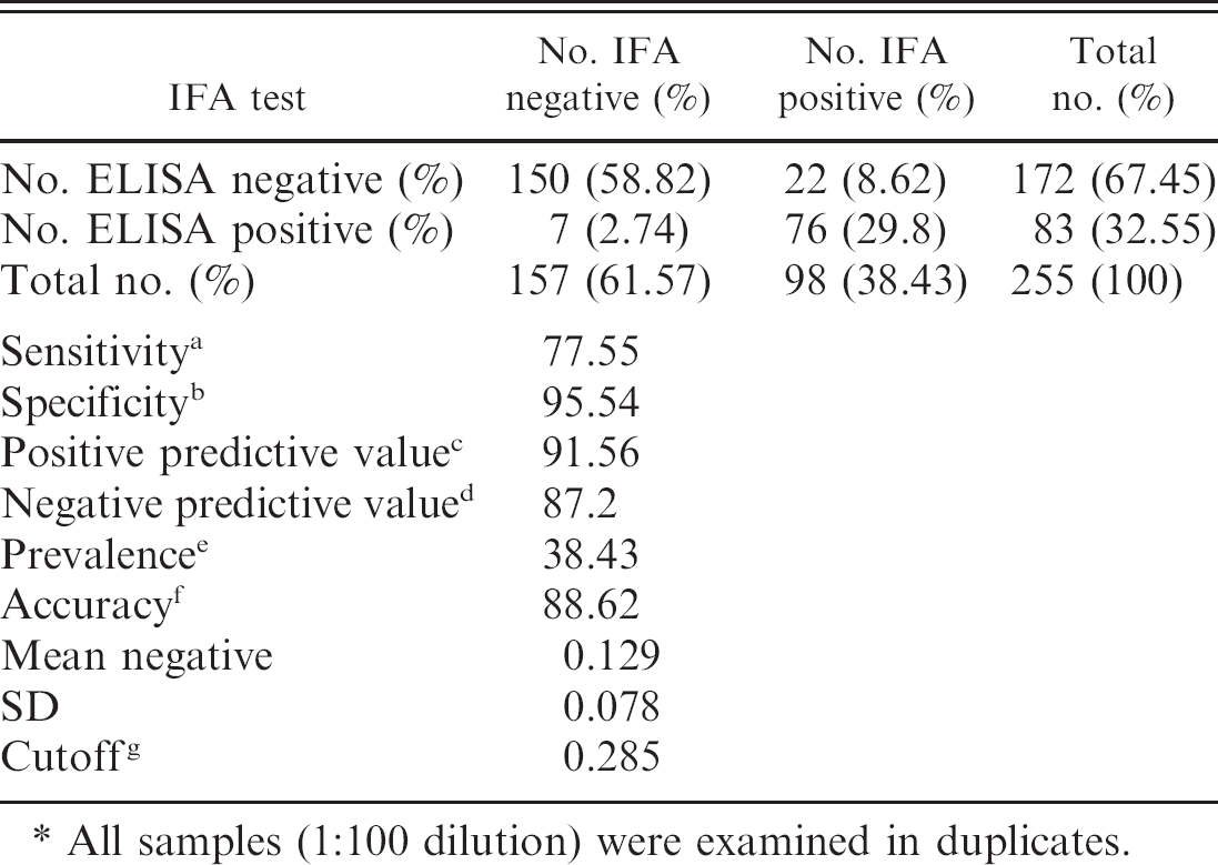
All samples (1:100 dilution) were examined in duplicates.
To determine the relation between the optical density of the indirect ELISA and IFA titer, regression analysis and calculation of Pearson's correlation coefficient were carried out. A significant correlation between both techniques was observed (r = 0.833). The results were analyzed by multiple equation models, and a higher regression factor (R2) was found using the exponential model. By using the aforementioned model, a mathematical formula (IFA titer = 69.59 e1.59×OD) to calculate the IFA titer from optical density data was established, with R2 = 0.985 (Fig. 3).
Discussion
The 30-kDa major outer membrane protein of E. canis is a highly conserved major immunodominant antigen recognized by both naturally and experimentally infected dog sera. 9,13,18 This protein is encoded by a polymorphic multigene family consisting of more than 20 paralogs with some level of expression differences. In this study, we used the p30 gene, because it is one of the most widely represented in dogs, ticks, and cell cultures. 16,19
The baculovirus system has been extensively used to express large quantities of proteins that are antigenically similar to their native counterparts and can be used in standardized assays to provide consistent results. In fact, the system has been widely used to produce recombinant proteins for use in diagnostic assays. 5,11 Initially, different genes of E. canis (p30, p43, and MAP2) were cloned and expressed (data not shown), but the best results were obtained with P30 protein.
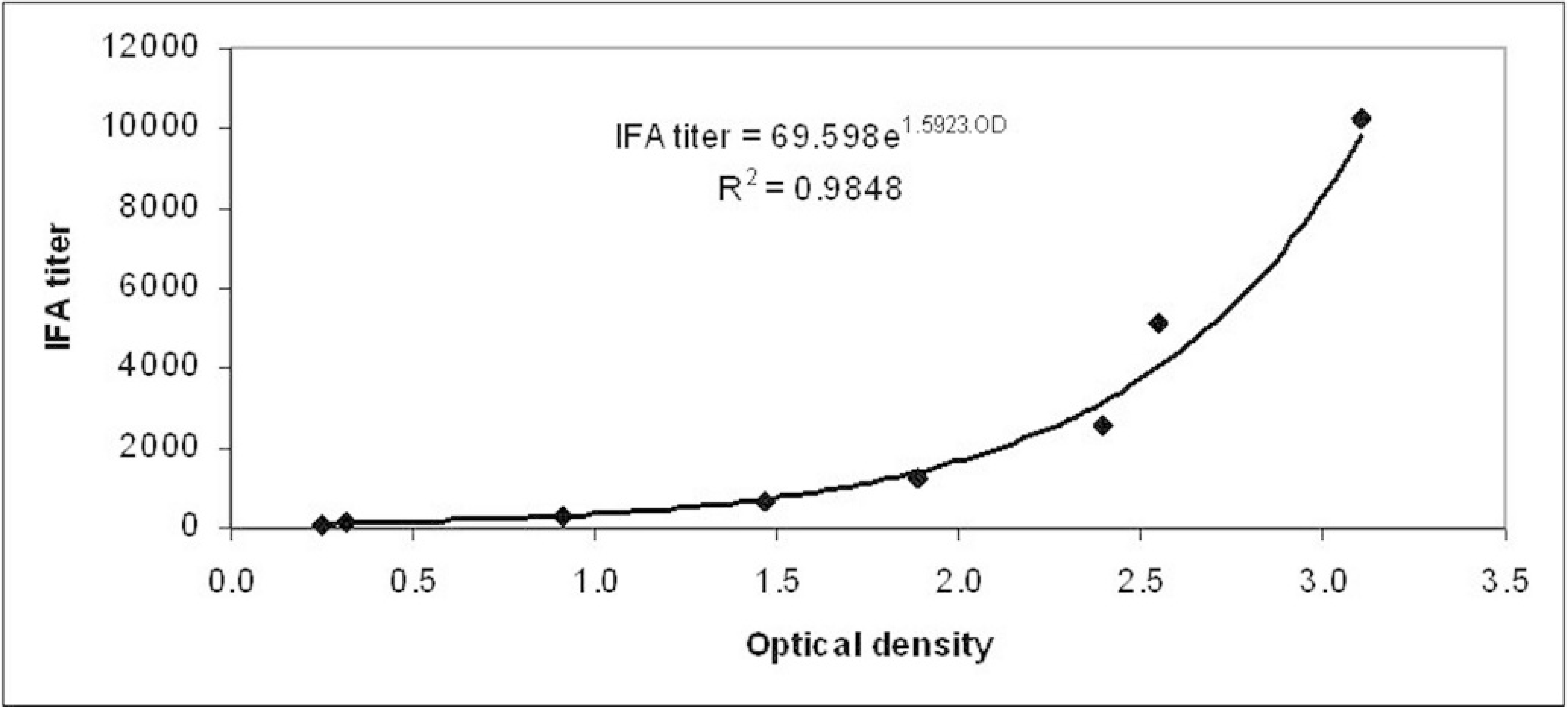
Determination of indirect fluorescent antibody titer by indirect p30bac enzyme-linked immunosorbent assay. The regression curve was calculated using Excel (Microsoft).
The P30 recombinant protein expressed in the baculovirus system preserved its immunogenic and antigenic characteristics after expression because it satisfactorily immunized mice, eliciting a strong and specific immune response. Those mice were used for producing anti-P30 monoclonal antibodies (data not shown).
The level of recombinant P30 protein expression was high, and, although the protein was only present in the insoluble fraction, it was possible to solubilize it in guanidine hydrochloride for further purification by Ni+-conjugated column. Both protein fractions (P30 in guanidine hydrochloride and P30 purified by column) were assessed as antigen, and it was determined that it is not necessary to perform the purification to chromatographic level to develop a reliable indirect ELISA. This fact is very important in a production process due to the simplification of the purification procedures and product cost reductions.
The accuracy and repeatability of a diagnostic test are essential for effective evaluation of results, and both parameters were very high in the present assay. When the P30 indirect ELISA was compared with the IFA test as a reference technique using a panel of well-characterized sera, the Pearson's variation coefficient (0.833) and the agreement quotient, κ (0.75), indicated high correlation and agreement between the 2 tests. Further evidence for a close correlation between the 2 techniques is the possibility to establish a mathematical formula that relates IFA titer to ELISA optical density. This is a very important point given that the IFA technique is considered the gold standard for E. canis diagnosis. The specificity of the indirect ELISA was very high (95.54). The sensitivity was 77.55 but improved considerably, to 95%, when results of sera with IFA titers higher than 1/160 were considered. Titers of 1/80 using IFA are generally considered positive. 14,21 However, when absorbance data from P30 indirect ELISA of sera with an IFA titer of 1/80 were analyzed in detail, the authors observed that the ELISA method was clearly able to differentiate between 2 groups, 1 with absorbances under 0.25 and 1 with absorbances higher than 0.35 (ELISA cutoff 0.285). Probably this group should be considered as a doubtful group. To clarify this point, it would be very interesting to perform an experimental infection in dogs with E. canis to analyze well-controlled canine sera at different times and with different titers. These observations suggest that with dogs included in this group, it would be preferable to combine other diagnostic methods, or to repeat the analysis after several weeks. Other studies 20 underline the importance of establishing criterion to define “definite” or “probable” cases of the disease and suggest that by using IFA antibody titers, a “definite” case of canine ehrlichiosis can be defined as one with clinical signs and hematologic parameters suggestive of the disease and an antibody titer ≥ 1:256. On the other hand, a “probable” case of ehrlichiosis would be a case in which the clinical signs and symptoms are suggestive of the disease with an IFA titer of 1:64 to 1:128. Different laboratories use different cutoff values to differentiate positive and negative results. For that reason, some authors have suggested that titers <1:80 should be deemed suspect and that repeated serologic testing within 2 to 3 weeks should be considered. 14 Finally, a commercial antibody screening test d has calibrated a titer of approximately 1:100 or greater to be positive.
Several positive samples to A. phagocytophilum and L. infantum were also tested to determine the possible cross-reaction of the P30 indirect ELISA with these microorganisms. The 4 seropositive samples to A. phagocytophilum were seronegative to E. canis using ELISA. Two of the 10 seropositive samples to L. infantum were P30 ELISA positive, but 1 of them was also positive to E. canis using IFA. With the exception of only 1 serum sample, no cross-reaction was detected with antisera against other pathogens (A. phagocytophilum and L. infantum) by ELISA, but the authors have not found any clear explanation for this discordant result.
The ELISA-based system has many distinct advantages over the methods traditionally used. The amount of E. canis-specific antigen can be standardized for each assay, antibody to P30 can be measured, and the results are based on defined criteria rather than subjective determinations of a positive result in an immunofluorescence study or in a dot-blot assay. Another advantage of using a recombinant protein as antigen in the ELISA is that is possible to avoid the purification of E. canis, which is time consuming and expensive and after serial passage in cell culture may produce batch-to-batch variation in the antigen.
The authors are now working on a new competition ELISA assay based on P30 recombinant protein and anti-P30 MAbs developed in their laboratory. On the basis of these results, the recombinant P30 protein expressed in the baculovirus system and purified according to the method described herein appears to be a suitable candidate for the development of a diagnostic kit for detection of E. canis-specific antibodies in field samples. This technique presents other benefits, such as the possibility of rapidly evaluating a large number of samples and methodology standardization.
Acknowledgements
This study was carried out as a collaborative project between Inmunología y Genética Aplicada S.A. (INGENASA) and the Service of Diagnosis of Ehrlichiosis (College of Veterinary Medicine, Complutense University of Madrid). This study was partially supported by a grant (Ref. 09/0099/2001) from the Consejería de Educación de la Comunidad de Madrid. The authors thank Dr. Antonio Sanz for critical review of the manuscript.
Footnotes
a.
dot-ELISA test Immunocomb®, Biogal, Israel.
b.
IgG MIF antibody test Kit®, Fuller Laboratories, LabClinics, BCN, Spain.
c.
Dip-S-Ticks®, PanBio InDx, Inc., TDI, Madrid, Spain.
d.
Snap3®Dx assay, IDEXX Laboratories, Inc., Barcelona, Spain.
e.
Cell line DH82, CRL10389, ATCC, Barcelona, Spain.
f.
Cell line Sf9, CRL1711, ATCC, Barcelona, Spain.
g.
DNAzol® reagent, Invitrogen, Barcelona, Spain.
h.
Taq DNA polymerase, BioTools, Madrid, Spain.
i.
pGEMT-easy vector, Promega, Madrid, Spain.
j.
pAcHLT-A, Pharmingen, San Diego, CA.
k.
ABI PRISM® 3130 DNA automatic sequencer, Perkin-Elmer Applied Biosystems, Foster City, CA.
l.
BacPAK 6 viral DNA, Clontech, Mountain View, CA.
m.
Jet PEI™ transfection reagent, Polyplus transfection, Illkirch, France.
n.
X-Gal (5-bromo-4-chloro-3-indolyl-β-D-galactopyranoside), Fermentas, Quimigen, Madrid, Spain.
o.
His-Bind Resin, Hispanangar, Burgos, Spain.
p.
Anti-His MAb, SIGMA-Aldrich Química S. A, Madrid, Spain.
q.
Bio Rad Protein Assay, Madrid, Spain.
r.
Polystyrene microtitre plates, Labsystem, Barcelona, Spain.
s.
TMB (MAX), Neogen, IL.
t.
1D Image Analysis Software 2.0.1, Eastman Kodak Co., Rochester, NY.