
Abstract
Leiomyosarcoma was diagnosed in the uterus surgically removed from a 3-year-old pet Suffolk ewe with a history of bleeding from the vulva, spontaneous lactation, and nursing behavior. The uterus contained multiple well-circumscribed, soft, intraluminal polypoid masses of variable sizes (0.5–4 cm). The masses were red, with white, smooth, and glistening cut surfaces. Histologically they comprised variably dense sheets of moderately pleomorphic, plump spindle cells embedded in richly vascularized stroma. The mitotic index was usually low (0-1/high-power field), but in some polyps there were up to 10 mitoses/high-power field. Neoplastic cells stained positive for alpha smooth muscle actin (α-SMA) by immunohistochemistry. Ultrastructural features of neoplastic cells included the presence of basal lamina, scant microfilaments, contracted nuclei with blunt ends, and flat intercellular junctions. Uterine leiomyosarcoma was diagnosed based on cellular morphology and atypia and positive immunohistochemistry for α-SMA.
Uterine leiomyosarcomas are malignant smooth muscle neoplasms of the uterine myometrium, with a general incidence of 10% among all smooth muscle neoplasms of the female genital tract. 5 These tumors can be solitary or multiple, with occasional multicentric involvement of the genital tract. 5 Uterine leiomyosarcomas have been sporadically described in cattle and goats. 1 , 3 , 6 , 10 , 13 , 14 In sheep, a few cases of leiomyomas and leiomyosarcomas have been reported in the uterus, 1 , 3 , 6 , 9 but detailed case descriptions of such tumors are rare. This article describes a unique case of polypoid uterine leiomyosarcomas in a sheep, with no known counterpart in the English literature.
A surgically removed uterus and ovaries from a 3-year-old pet Suffolk ewe with a history of recurrent hemorrhage from the vulva, spontaneous lactation, and nursing behavior were submitted for gross and histological evaluation to the Animal Disease Diagnostic Laboratory (ADDL) at Purdue University (West Lafayette, IN). The uterus contained about 5 ml of red, transparent fluid and multiple variably sized (0.5–4 cm), pink to red, soft to fleshy, pedunculated, intraluminal masses with smooth, white, and shiny cut surfaces (Fig. 1). Multiple sections of the masses were fixed in 10% neutral-buffered formalin, paraffin embedded, sectioned at 5-μm thickness, and stained with hematoxylin and eosin and Masson trichrome. For immunohistochemical (IHC) examination, dewaxed sections of uterine masses were incubated with mouse monoclonal antibody against alpha smooth muscle actin (α-SMA). 11 For transmission electron microscopy, formalin-fixed tissues were postfixed in 1% buffered osmium tetraoxide and embedded in Epon following dehydration in graded alcohols and propylene oxide. Uranyl acetate- and lead citrate-stained thin sections were examined using a Philips EM 201 transmission electron microscope.
Histologically the pedunculated masses were continuous with the myometrium, were composed of closely packed sheets of spindle cells embedded in richly vascularized stroma, and were covered by a discontinuous endometrial epithelium. Variably abundant clear to collagenous matrix separated the neoplastic cells, which had scant to moderate, fibrillar, frequently vacuolated eosinophilic cytoplasm with indistinct borders (Fig. 2). The nuclei of these cells were irregularly round to oval with blunt ends, stippled chromatin and indistinct nucleoli. There was moderate nuclear and cellular pleomorphism, with pleomorphic cells being more abundant in the larger masses. The mitotic index was usually low (0-1/high-power field), but in some polyps there were up to 10 mitoses/high-power field. A few lymphocytes, plasma cells, macrophages, and hemosiderophages were scattered among the spindle cells. Several macrophages that contained dark-brown pigment (melanin) were present within the epithelium and superficial portions of the masses. Immunohistochemistry for α-SMA revealed uniform, moderate cytoplasmic staining of the tumor cells (Fig. 3). Multiple occurrences of the tumor involving the entire uterus; a variable, often low to occasionally high mitotic index; moderate cellular and nuclear atypia; and positive IHC for α-SMA supported a diagnosis of low-grade uterine leiomyosarcoma.
Transmission electron microscopy was performed in order to further characterize the neoplastic cells. Ultrastructurally the cells exhibited elongated or contracted nuclei with blunt ends and irregular contours. Basal lamina, membrane densities, and flat intercellular junctions were frequently present. In the cytoplasm there were abundant dilated cisternae of rough endoplasmic reticulum (RER), scant microfilaments, and moderate numbers of mitochondria (Fig. 4). The neoplastic cells also exhibited numerous cell processes, which were separated by homogeneous electron-lucent to moderately electron-dense extracellular matrix occasionally interspersed with collagen.
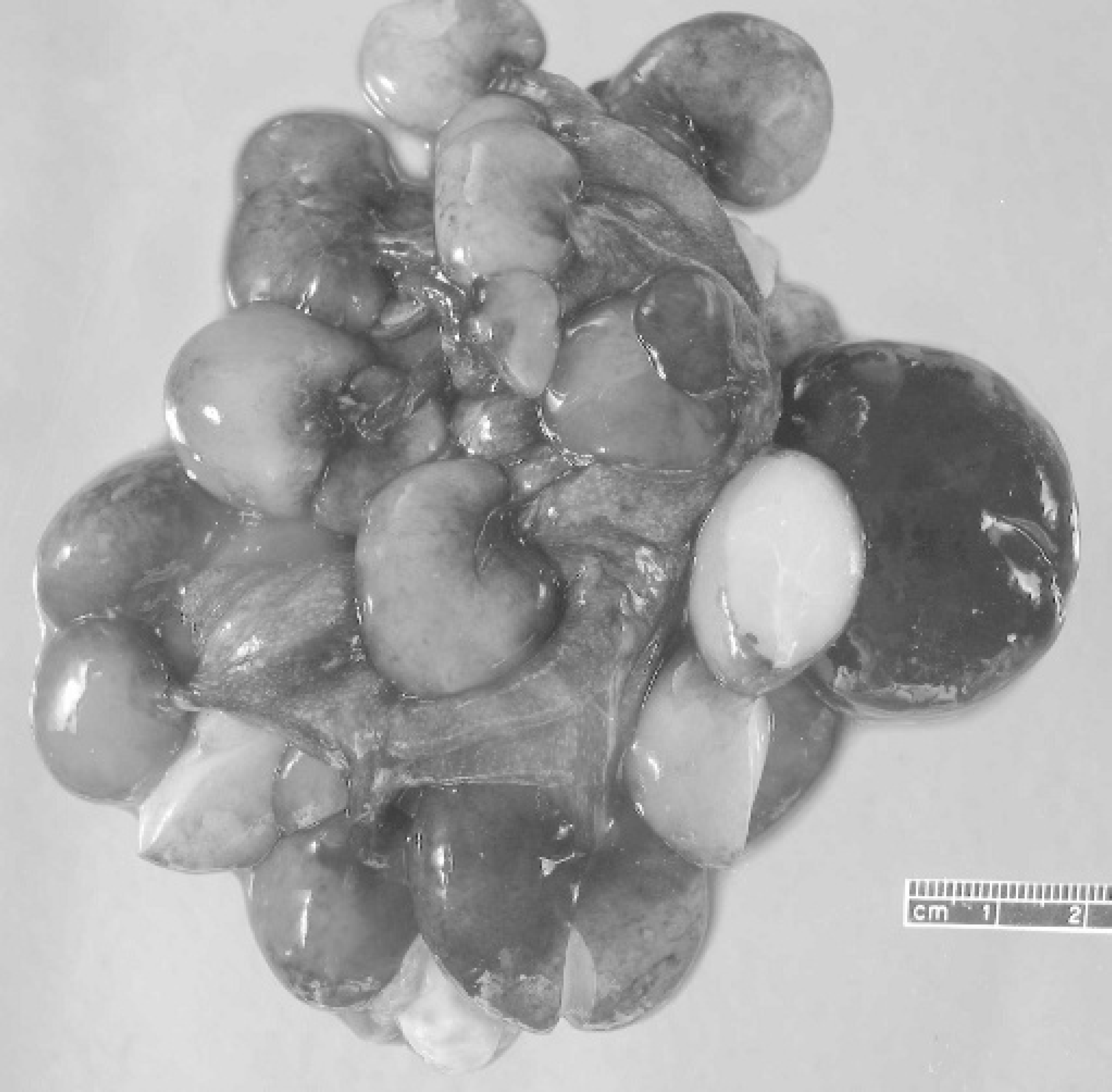
Uterus, sheep. The lumens of both uterine horns were filled with multiple pink to red, pedunculated masses. The cut surfaces of the masses were smooth, white, and shiny.
Smooth muscle tumors account for up to 50% of the reportedly rare neoplasms of the female genital tract in ruminants, with leiomyosarcomas being far less common than leiomyomas. 1 , 3 , 6 Most of the genital tract smooth muscle neoplasms in female ruminants from abattoir surveys have been limited to the uterus. 1 , 3 , 6 Similarly, the tumor in the present ewe also exclusively affected the uterus, extensively involving both horns. Individual examples of these neoplasms have also been described in other locations in the female genital tract, including the cervix, vagina, and vulva. 2 , 8 , 9 , 12–14
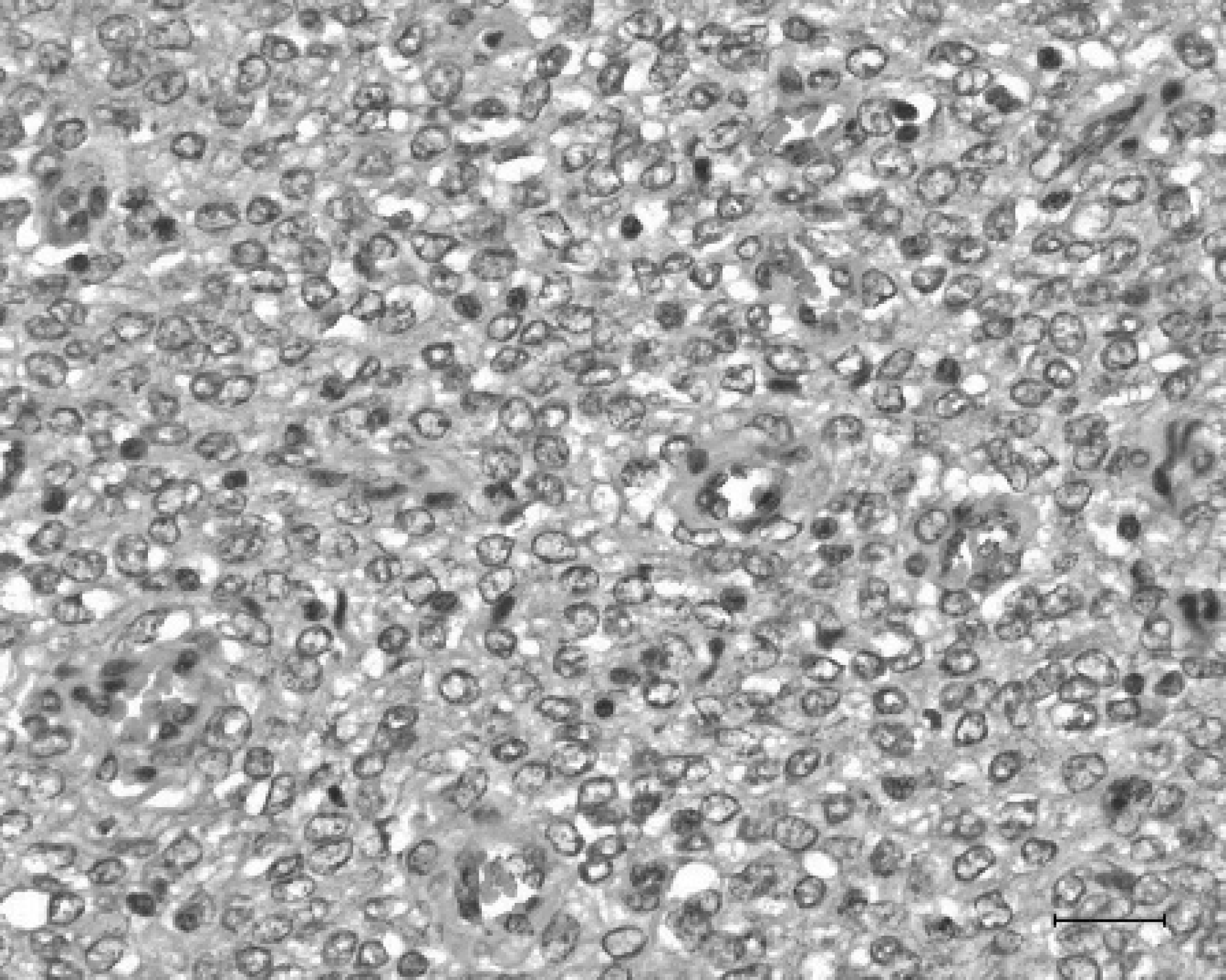
Uterus, sheep. Histologically, these masses were composed of sheets of spindle cells with moderate amounts of eosinophilic cytoplasm and round to oval nuclei. Hematoxylin and eosin (HE). Bar = 25 μm.
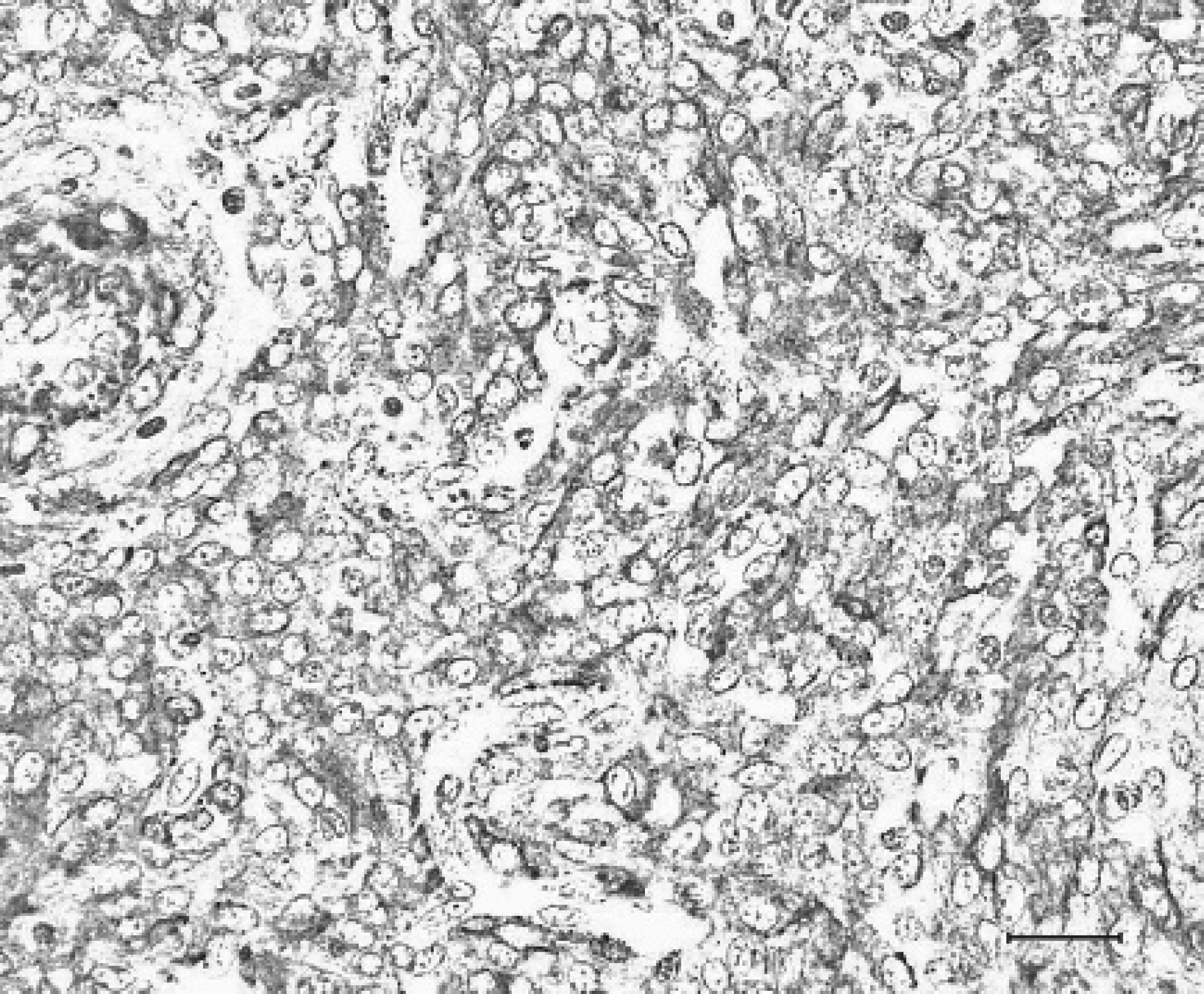
Uterus, sheep. Approximately 70% of the neoplastic cells showed positive cytoplasmic staining with an antibody against alpha smooth muscle actin. IHC. Bar = 25 μm.
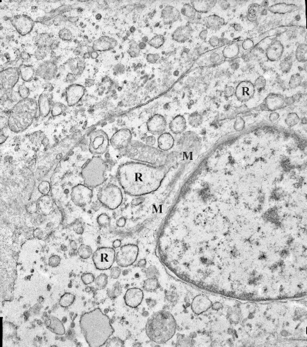
Uterus, sheep. Ultrastructurally, the neoplastic cells contained nuclei with blunt ends, abundant dilated RER (R), a few mitochondria, scant microfilaments (M), and basal lamina. Magnification = 15,000X.
Uterine leiomyosarcomas in cattle and goats have been reported to be locally invasive or capable of metastasis to distant organs. 1 , 10 , 14 Even genital smooth muscle tumors with little cellular atypia and a low mitotic rate seem to be capable of malignant behavior in the goat. 14 The tumor in the present case was not invasive but was classified as a low-grade malignant variant on the basis of the variable mitotic index and moderate cellular pleomorphism. As a possible differential diagnosis for the current neoplasm, leiomyoma of the uterus can be multiple and frequently presents with a history of vaginal bleeding 5 ; however, in this neoplasm tumor cells essentially resemble normal smooth muscle cells with few mitoses and limited cellular pleomorphism. 5 In addition to leiomyoma, another differential diagnosis for multiple myometrial masses, considered in humans, is diffuse leiomyomatosis. 4 In this condition, although the masses are histologically benign, they are ill-defined compared to a leiomyoma and are disseminated throughout the myometrium, resulting in a symmetrical thickening. 4 The masses from the currently described ewe were multiple and continuous with the myometrium but formed intraluminal polypoid projections rather than masses embedded within the myometrium.
A recently published case report 14 indicates that older goats are predisposed to develop uterine smooth muscle tumors. In sheep, age-related predisposition to development of these tumors is not well established, but sporadic cases of genital smooth muscle neoplasms in young ewes have been reported. 6 , 9 Saanen does have been overrepresented in the reports of genital leiomyosarcomas compared to other caprine breeds, indicating a possible hereditary component in this breed. 14 No information about the siblings of the ewe described in this report that would indicate a genetic predisposition to the development of this type of tumor was available. In one of the affected Saanen does in the goat study there were multiple polypoid uterine masses similar to the ones described in the present case. In contrast, the only more-detailed report of a smooth muscle tumor in the female genital tract of a sheep 9 describes a single neoplastic mass in the uterus accompanied by multiple microscopic foci in the cervix, interpreted by the authors to indicate metastasis (although it could also indicate a multicentric origin of the tumor).
Hormonal influences have been implicated in the development of genital smooth muscle tumors in some animals, including ruminants. 5 , 8 , 14 An association between ovarian follicular cysts and mammary hyperplasia with vaginal leiomyofibromas in bitches is well established. 5 Similarly, multiple ovarian follicles, ovarian follicular cysts, and endometrial hyperplasia were described in conjunction with vaginal leiomyofibromas and uterine leiomyosarcomas in goats. 8 , 13 The ovaries of the currently described ewe were grossly and histologically normal; however, a possible hormonal influence cannot be entirely eliminated because of the history of spontaneous lactation and nursing behavior and the multicentric origin of the tumors. Clinical signs in this ewe resolved with ovariohysterectomy.
Benign smooth muscle tumors containing abundant collagenous stroma have also been called “fibroleiomyomas.” 5 These tumors exhibit histologic evidence of fibroblastic and smooth muscle differentiation and have been reported in the uterus of a sheep, the cervix of a cow, and the vagina of a goat. 1 , 8 , 12 Fibroblasts producing collagen can be differentiated from smooth muscle fibers in microscopic tissue sections using the Masson trichrome technique. Similarly stained sections of the uterine masses in the present case did not indicate the presence of collagen and, thus, fibroblastic differentiation of neoplastic cells.
Electron microscopy can be useful in determining the nature of spindle cell neoplasms. Fibroblasts have abundant RER and scant microfilaments in the cytoplasm. 7 In contrast, the cytoplasm of smooth muscle cells contains many actin filaments with focal densities, pinocytotic vesicles, and some mitochondria. 7 Other features of smooth muscle cells include flat intercellular junctions, basal lamina, and round-ended, contracted nuclei. 7 The neoplastic cells in this sheep demonstrated ultrastructural features of both fibroblasts (abundant RER) and smooth muscle cells (basal lamina, elongated or contracted nuclei, and flat intercellular junctions). Smooth muscle cells with ultrastructural features similar to those of the currently described tumor were named as myofibroblasts in the report of a neoplasm from the vulva of a cow. 2 Other authors 5 have proposed that similar tumors with a fibroblastic component should be classified as leiomyomas or leiomyosarcomas if smooth muscle differentiation is apparent. The neoplasm in the present case was diagnosed as being of smooth muscle origin based on tumor cell morphology, positive staining with α-SMA, and lack of evidence of fibroblastic differentiation by Masson trichrome stain.
In summary, this report describes the gross, histologic, immunohistochemical, and ultrastructural features of polypoid uterine leiomyosarcomas in a 3-year-old Suffolk ewe. This study confirms previous observations that in female ruminants presented with a history of bleeding from the vulva, uterine leiomyosarcoma should be considered in the differential diagnosis. 14 The ewe that was the subject of this report is alive, without clinical evidence of recurrence 6 months after surgery, indicating that the identification of these tumors may be associated with a good prognosis.
Acknowledgements. The authors would like to thank Dr. Larry W. Smith for submission of the case and Diane Hostether for providing additional clinical history. Histotechnicians at the ADDL histology laboratory and Phyllis Lockhard at the ADDL electron microscopy laboratory are acknowledged for their technical help.