
Abstract
In vitro toxin production is an important tool not only for diagnostic purposes but also for the study of pathogenesis of Clostridium perfringens infections. The present study was carried out to compare the level of toxin production by several strains of C. perfringens type A, isolated from the intestine of animals, when cultured in 3 different conventional culture media. Six strains of C. perfringens type A isolated from the small intestine of healthy sheep were cultured in commercial cooked meat medium (CMM), brain heart infusion (BHI), and tryptone glucose yeast (TGY). Intravenous lethality in mice and phospholipase C (PLC) activity were measured in filtered culture supernatants. Lethality of culture supernatants was highest for all isolates when grown in BHI, followed by CMM. No supernatants from any isolates grown in TGY produced lethality in mice. Phospholipase C activity was highest when the isolates were grown in BHI and CMM and significantly lower when grown in TGY.
Clostridium perfringens is a Gram positive, spore-forming, anaerobic rod that is classified into 5 types (A, B, C, D, and E) according to the production of 4 major toxins (α, β, ɛ, ι). 4 This microorganism is widely distributed in the environment and is responsible for several diseases of animals and humans. 1,8 Although several reports describe the isolation and toxinotyping of C. perfringens from the intestine of healthy animals, 1,5,10 information about the lethal potential of C. perfringens strains isolated from the intestine of nondiseased animals is scant. In vitro toxin production evaluation is an important tool not only for diagnostic purposes but also for the study of pathogenesis of C. perfringens infections. The present study was carried out to define which culture media could optimize toxin production by C. perfringens type A obtained from clinically healthy sheep.
Six strains of C. perfringens type A (Table 1) isolated from the small intestine of healthy sheep at slaughter were used. The intestinal contents were cultured on 5% sheep blood agar a at 37°C in anaerobiosis for 24 hours. Colonies compatible with C. perfringens were Gram stained and then processed by a multiplex polymerase chain reaction (PCR) technique to amplify the genes that encode the 4 major toxins of C. perfringens plus the genes that encode beta 2 toxin and enterotoxin as previously described. 6 The 6 isolates were then freeze-dried and stored for 6 years until they were reconstituted in cooked meat medium (CMM) b before being cultured in 10 ml of commercial CMM, brain heart infusion (BHI), b or tryptone glucose yeast (TGY) b anaerobically at 37°C to late log phase. Monitoring and determination of the growth phase was achieved by measuring the optical density at 620 nm (OD600 nm) of the cultures at hourly intervals. Bacteria were then removed from the cultures by centrifugation at 3,000 × g for 30 minutes at 4°C, and supernatants were filter sterilized through 0.45-μm filters. The toxic activity of the filtered culture supernatants was evaluated by several assays as follows.
Comparison of 3 culture media for the characterization of Clostridium perfringens type A isolated from healthy sheep. *
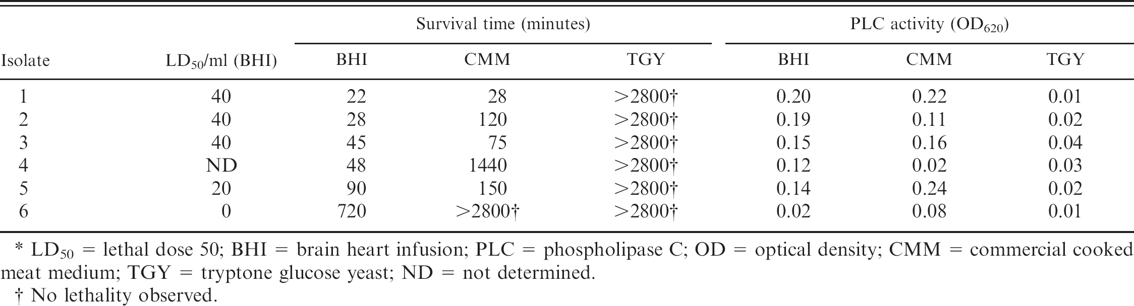
LD50 = lethal dose 50; BHI = brain heart infusion; PLC = phospholipase C; OD = optical density; CMM = commercial cooked meat medium; TGY = tryptone glucose yeast; ND = not determined.
No lethality observed.
Surviving time was recorded in groups of 3 mice inoculated intravenously with 0.5 ml of a 1/2 dilution of culture supernatant in 1% peptone water. The survival time until assay endpoint (see definition of assay endpoint below) was recorded for each mouse, and the average survival time of each group was calculated. Lethal dose 50 (LD50)/ml was determined as previously described.
2
In brief, pairs of mice received IV injections containing 2-fold dilutions (between 1/2 and 1/80) of filtered culture supernatants in 1% peptone water. LD50 was calculated as the reciprocal doses that produced a transition from total survival to death in mice within 48 hours. To prove that lethality was due to alpha toxin, 0.6-ml volume of undiluted type A culture supernatants were mixed with 0.1 ml of a solution containing 2 mg/ml of an alpha toxin-neutralizing monoclonal antibody (MAb),
c
brought to 1.2 ml with 1% peptone water and incubated for 30 minutes at room temperature. A 0.5-ml aliquot of each mixture was injected intravenously into 2 mice, while an additional pair of mice received a similar IV injection of the same supernatant that had been identically prepared, except for the omission of alpha toxin MAb. The mice were observed until assay endpoint (see definition of assay endpoint below). To determine phospholipase C (PLC) activity, an egg yolk solution (10% vol/vol) was prepared by diluting freshly obtained egg yolk in phosphate buffered solution (PBS) pH 7.4 and by filtering the solution through 0.45-μm filters. Samples of filtered supernatants were diluted 2-fold in 100 μl of PBS; and after the addition of 10% (vol/vol) egg yolk solution, they were incubated at 37°C for 3 hours. After incubation, samples were diluted 1/100 in PBS and optical density at 620 nm was measured in a Spectronic D20 spectrophotometer. A positive control consisting of semipurified C. perfringens alpha toxin
d
at 3 different concentrations (2, 20, and 200 LD50, respectively) and a negative control consisting of sterile, nontoxic media culture were included in the assay.
For all experiments involving mice, assay endpoint was defined as severe clinical signs necessitating euthanasia, spontaneous death, or survival without clinical alterations for 48 hours when the animals were euthanized. Euthanasia was performed with CO2.
Table 1 shows the results of characterization of the 6 C. perfringens isolates grown in the 3 culture media. The levels of PLC in culture supernatants were inversely correlated to the survival times in mice (r = 0.8, P < 0.05). Survival times were shortest in BHI culture supernatants, followed by CMM, while no lethality was observed in TGY. Since BHI produced the shortest survival times, LD50/ml was measured by growing these isolates in BHI culture medium only. Levels up to 40 LD50/ml were detected in BHI culture supernatants (Table 1). The lethality of all culture supernatants was neutralized when the supernatants were previously incubated with anti-alpha toxin MAb, indicating that alpha toxin was responsible for the lethality observed in mice. Phospholipase C levels were similar in BHI and CMM culture supernatants, both of which were higher than in TGY. Culture supernatants were collected at late log phase because previous reports showed that for most C. perfringens toxins the highest level of production occurs at this stage. 2,6
The analysis of toxicity of C. perfringens supernatants indicates that the culture medium is an important factor in determining the toxigenicity of this microorganism. Tryp-tone glucose yeast and CMM are recommended in different laboratory manuals for toxinotyping of C. perfringens. 3,7,9 However, under the conditions used in this study neither TGY nor CMM were the ideal culture media for lethal toxin production by C. perfringens type A. Although it seems that BHI and CMM are the best media for PLC activity expression, BHI produced higher lethal activity than CMM. The results show that although CMM is a suitable medium, under our conditions BHI was the best of the tested media for the assessment of toxicity of most C. perfringens type A strains isolated from the intestine of healthy sheep.
The pathogenesis of C. perfringens infections is mostly related to powerful toxins, which are produced in variable levels by different strains. However, detailed information about the toxigenic potential of C. perfringens isolated from healthy animals is scant. It is possible that earlier attempts to evaluate C. perfringens toxicity established the absence or low toxic activity of many isolates based on the use of culture media that were not ideal for toxin production.
Acknowledgements. This research was supported by Fondo Nacional para la Ciencia y la Tecnología (PICT-01-3591), Argentina, and Public Health Service Grant AI56111 from the National Institute of Allergy and Infectious Diseases, National Institutes of Health, Bethesda, MD. We thank Dr. Bruce McClane from Pittsburg University Medical School for his suggestions.
Footnotes
a.
Hardy Diagnostics, Santa Maria, CA.
b.
Anaerobe Systems, Morgan Hill, CA.
c.
Dr. P. Hauer, Center for Veterinary Biologics, Ames, IA.
d.
CSL Ltd., Melbourne, Victoria, Australia.