
Abstract
A rapid and simple latex agglutination test (LAT) for the detection of avian influenza virus (AIV) subtype H5N1 in chicken allantoic fluids, tracheal swabs, and tissues was developed. Monoclonal antibodies against the hemagglutinin glycoprotein of H5N1 were covalently coupled onto the surface of carboxylated latex bead using a water-soluble carbodiimide to obtain sensitized latex particles (SLP). These SLPs strongly agglutinated in the presence of allantoic fluid containing H5N1, but not fluids containing other AIV sub-types such as H1N1, H3N2, H4N6, and H9N2. Using this LAT, the virus was detectable in tracheal swabs 24 hours to 30 days after inoculating chickens with H5N1, with detection rates ranging from 45.5 to 79.2%. Much higher rates of detection were obtained from tissues collected postmortem from H5N1 experimentally infected chickens; lung tissue yielded the highest detection rate (96.7%), followed by kidney, spleen, brain, and liver tissues (90%). Lower detection rates were achieved with heart (41.7%) and cloacal tissues (26.8%). When the LAT was compared with other detection methods, the agreement with the viral isolation, H5 antigen immunochromatographic test, and H5 real-time RT-PCR test was 93.97, 95.18, and 87.95%, respectively. The test was highly specific for H5N1 in chickens and water fowls and had sensitivity comparable to other diagnostic tests evaluated.
Introduction
Avian influenza A viruses (AIV) are the causative agents of the currently most important poultry disease. Of the 16 AIV subtypes, H5N1 is of particular concern because of its ability to infect humans and the potential for person-to-person transmission. 14 Although H5N1 is currently believed to be transmitted only through direct contact with infected birds, this virus may mutate to allow efficient transmission among mammals. The virus could also reassort its gene segments with human influenza viruses during co-infection, resulting in a new virus that can be both highly lethal and easily transmissible from person to person. Therefore, variants of H5N1 have the potential to cause the next influenza pandemic because of the lack of natural immunity to H5N1 in humans. 10 Wild migratory birds— historically the reservoir of all influenza A viruses— are now dying in large numbers from the highly pathogenic H5N1 and have spread the virus from Asia to Europe and Africa along their migratory routes. 11 In contrast, domestic ducks can excrete large quantities of highly pathogenic virus without showing signs of illness. 6 The insidious role of domestic birds in maintaining transmission further complicates prevention and control of the infection in poultry and humans. Thus far, the transmission of H5N1 from birds to humans has resulted in 127 fatalities in 261 infections in Indonesia, Vietnam, Thailand, Cambodia, China, and several other countries. 1
The recent emergence and re-emergence of influenza viruses with pandemic potential is of great concern to both the veterinary and public health communities. Early diagnosis of influenza virus infection is therefore essential, especially for the highly pathogenic avian influenza (HPAI) virus H5N1. A rapid, sensitive and inexpensive diagnostic test is also required for H5N1 surveillance.
Presently, the identification of the H5N1 can be accomplished using various diagnostic assays, such as virus isolation (VI) in embryonated chicken eggs, direct antigen detection using enzymatic, fluorescent, optical, or chromatographic immunoassays, 4 and nucleic acid detection assays (reverse transcription-PCR [RT-PCR], 8 real-time RT-PCR, 9 and nucleic acid sequence-based amplification 3 ). The objectives of this study were: 1) to develop a rapid latex agglutination test (LAT) for detecting H5N1 in chicken tracheal swabs and tissues; and 2) to compare sensitivity and specificity of the newly developed LAT in relation to VI, immunochromatographic assay, and real-time RT-PCR.
Materials and methods
Viruses. The AIV strains used in this study included A/ Chicken/HuBei/327/2004 (H5N1), A/duck/HuBei/425/2004 (H9N2), A/duck/HuBei/505/2004 (H4N6), A/swine/Shanghai/306/2003 (H3N2), and A/Swine/HuBei/327/2002(H1N1). These viral isolates were obtained from the Animal Infectious Diseases Unit of the National Key Laboratory of Agricultural Microbiology at Huazhong Agricultural University a . All strains were cultured in the allantoic cavity of specific-pathogen-free (SPF) chicken eggs. Other respiratory disease viruses were also included in the study, including Newcastle disease virus (NDV), infectious bursal disease virus (IBDV), infectious bronchitis virus (IBV), egg-drop syndrome virus (EDSV), and avian paramyxovirus type 2 (PMV-2). All laboratory work involving H5N1 was performed in a biosafety-3 laboratory facility.
Monoclonal antibody preparation. Six-week-old female BALB/c mice were immunized with A/Chicken/HuBei/327/ 2004(H5N1). The spleen of immunized mice was harvested and the lymphocytes isolated were used in the generation of hybridomas as previously described. 5 Hybridoma culture supernatants were screened for reactivity against H5N1 by hemagglutination inhibition (HI) assay, and hybridoma cells from wells containing HI titers of 1:32 were expanded and cloned by limiting dilution. To ensure that the monoclonal antibodies (MAbs) secreted by the hybridomas were specific for AIV H5N1, the hybridoma culture supernatants were analyzed by HI assay for reactivity against H9N2, H4N6, H3N2, and H1N1 viruses. Seven hybridoma cell lines secreting MAbs specific for the HA protein of H5N1 were established. The IgG antibodies in ascites were purified using protein A affinity chromatography.
Preparation of sensitized latex particles (SLPs). Affinity-purified antibody was coupled to carboxylated latex particles of 0.75 μm in diameter by modification of procedures recommended by the manufacturer b . Briefly, 500 μl of 2.5% (w/v) latex particles were washed 3 times in 0.1 M carbonate buffer (pH 9.6) and 3 times in 0.02 M phosphate buffer (pH 4.7) in 1.5-ml microcentrifuge tubes. All washes were done at 14,000 × g for 6 min at 25°C unless otherwise stated. Following the final wash, the particles were re-suspended in 625 μl of the 0.05 M 2-[N-morpholino] ethane sulfonic (MES) acid, pH 4.7 and transferred into 5-ml centrifuge tubes. Afterwards, 625 μl of freshly prepared 2% 1-ethyl-3-(3-dimethylaminopropyl) carbodiimide · HCl (EDC) was added drop by drop while the solution was slowly vortexed. The tubes were further incubated at 25°C for 4 hr while being rotated slowly end-to-end. The sensitized latex particles (SLPs) were then transferred into microcentrifuge tubes, washed 3 times in 0.02 M phosphate buffer (pH 4.7), and re-suspended in 3 ml of 0.01 M borate buffer (pH 8.0). Concentrations of IgG ranging from 200 to 2,400 μg/500 μl of particles were added to the SLPs and the tubes were rotated slowly end-to-end for 8 hr at 25°C. To block nonspecific binding, 50 μl of 0.1 M ethanolamine was added to the SLPs and rotated for additional 30 min. The SLPs were then transferred into 1.5-ml microcentrifuge tubes and centrifuged at 14,000 × g for 10 min. The supernatant was saved for protein determination and the SLPs were re-suspended in 1 ml 0.01 M borate buffer (pH 8.0, containing 1% bovine serum albumin) and rotated at 25°C for 30 min. Finally, the SLPs were washed one more time in BSA in 0.01 M borate buffer (pH 8.0) and re-suspended in 0.5 ml of latex storage buffer (0.1% BSA, 5% glycerol, and 0.1% NaN3 in 0.01 M PBS, pH 7.4).
Latex agglutination assays. Latex agglutination tests were performed by mixing 10 μl of SLPs with 10 μl of sample on a black-coated glass slide. The slide was then rotated manually for 30 sec. Test results were scored as follows: 4+, rapid agglutination of 100% SLPs, with formation of a ring; 3+, agglutination of >75% of SLPs, with some ring formation; 2+, agglutination of ≥50% of SLPs, with no ring formation; 1+, agglutination of ≤25% of SLP; and -, no visible agglutination.
Sample preparation for testing by LAT. Tracheal swabs from birds were collected in 1.0 ml of 0.3 M borate buffer (pH 8.4, 0.85% NaCl, 0.5% N-acetyl-cysteine, 0.1% NP-40, 5,000 IU/ml penicillin G, 5 mg/ml streptomycin), and the suspensions were frozen and thawed 3 times and centrifuged at 14,000 × g for 10 min before testing by LAT. All tissue specimens were homogenized to give a 50% suspension (w/v) in sample buffer (PBS, pH 7.4, containing penicillin G 5,000 IU/ml and streptomycin 5 mg/ml). The suspensions were centrifuged at 14,000 × g for 10 min before testing by LAT.
Virus isolation (VI) in embryonated chicken eggs. Isolation of influenza virus was performed by inoculating 9-day-embryonated chicken eggs with 0.2 ml tissue suspensions or swab suspensions via the allantoic cavity. The eggs were incubated for 4 days and candled daily for viability; embryos that died within 24 hr of inoculation were discarded as nonspecific. Allantoic fluid from dead and surviving embryos was tested for Hemagglutinin (HA) activity. Samples that yielded no hemagglutination were re-inoculated for a second passage.
Hemagglutinin (HA) and hemagglutination inhibition (HI) assays. HA and HI assays were performed in V-bottomed microtiter plates that contained 1% chicken erythrocytes solution following standard protocols recommended by International Animal Health Organization (OIE). 13
Animal infection experiments. An experiment was designed to determine whether the LAT could be used in direct analysis of tracheal swabs and bird tissues. Seventy-two 8-wk-old White Stone chickens were experimentally infected with H5N1; these birds were divided into 3 groups of 24 chickens based on pre-existing H5N1 HI titers resulting from vaccination. Group I chickens had HI titers of ≥1:64, group II HI≤1:32, and group III HI = 0. Each group of 24 chickens was further divided into 6 subgroups of 4. Sub-groups 1, 2, 3, 4, and 5 received inoculation doses of 1,000, 100, 10, 1, and 0.1 50% egg infectious doses (EID50), respectively. Subgroup 6 was used as control and inoculated with virus-free PBS (0.01 M, pH 7.4). All birds were inoculated intramuscularly through the brisket muscle, housed in ventilated stainless steel isolation cabinets in a room with high efficiency particulate air under negative-pressure, and illuminated under continuous light for the duration of the experiment. Feed and water were provided ad libitum. All experiments were conducted in a biosafety level 3-plus laboratory. Tracheal swabs were collected daily after inoculation and treated as described above. Internal organs (heart, liver, spleen, lung, kidney, brain, pancreas, muscle, abd cloaca) of chickens that died were collected. The remaining chickens were euthanized 30 days postinoculation and internal organs were collected. All specimens were frozen at −70°C until tested.
Comparison of LAT with other diagnostic tests. Two commercial kits were compared with LAT: a colloidal gold-based immunochromatographic test (H5 AIV Ag Test Kit c ) and a real-time RT-PCR test (H5 AIV real-time RT-PCR Test Kit d ). Ninety field specimens (23 livers, 41 lungs, 22 kidneys, 10 brains, and 4 tracheal swabs) were collected from birds during an H5N1 outbreak in Hubei Province, China in 2004. All specimens were tested by LAT, colloidal gold-based immunochromatographic test, real-time RT-PCR test, and VI. This work was conducted with the assistance of the Veterinary Diagnostic Center of Hunan Province, P. R. China and Shenzhen CIQ, P. R. China.
Field evaluation of LAT using outbreak specimens. A total of 828 tissue specimens (89 hearts, 102 livers, 102 spleens, 102 lungs, 102 kidneys, 23 pancreas, 27 tracheas, 77 cloacas, 102 brains, and 102 bone marrows) were obtained from chickens in the 2004 H5N1 outbreak in Hubei Province, China. These specimens were previously tested by VI for H5N1, and were used in this study for field evaluation of the LAT using procedures described previously.
Field evaluation of LAT using routine clinical specimens. Six hundred and forty chicken specimens (160 tracheal swabs and 480 lung specimens) and 750 duck specimens (210 tracheal swabs and 540 lung specimens) were collected from slaughterhouses that processed animals from large commercial poultries. In addition, 260 lung specimens were collected from ducks on small family farms. All the specimens were tested for H5N1 using the LAT.
Results
Optimization of the LAT assay. Titration of various dilutions of IgG for binding to latex particles indicated that about 1,000-1,200 μg of IgG/0.5 ml of 2.5% (w/v) latex particles was optimal concentration of IgG needed for obtaining a maximum sensitivity. The sensitivity of the reagent (SLPs) increased as the percentage of IgG bound to the latex particles increased but not as the total amount of IgG bound increased. When 1,000 μg of IgG was added, 489±14 μg of IgG was bound (49±1.4%), but when 1,600 and 2,400 μg of IgG were used, the sensitivity decreased significantly as the percentage of IgG bound reduced respectively to 25±0.5% and 23±1%, even though the total amount of bound IgG increased. When 400 μg of IgG was added to the latex particles, 136±9 μg of IgG (34±2%) was bound, which also resulted in a reduced sensitivity.
Analytical sensitivity and specificity of LAT. When H5N1 isolate Chicken/HuBei/327/2004 with a HA titer of 1:256 was serially diluted 2-fold (from 1:2 to 1:512), the highest dilution to produce agglutination (2+) was the 1:128 dilution. The specificity of LAT was evaluated using AIV subtypes H9, H4, H3, and H1 and other avian respiratory pathogens such as NDV, IBDV, IBV, EDSV-76, and PMV-2. No agglutination was observed with any viruses with the exception of AIV strain H5N1. Thus, LAT was highly specific for AIV H5N1.
Detection of H5N1 in experimentally infected chickens by LAT. All chickens inoculated with 1,000 and 100 EID50 of A/Chicken/HuBei/327/2004 died within 36 hours of inoculation and those in the 5 subgroups of group III (HI = 0) died within 96 hours (Table 1). Members of group I (HI≥1:64) inoculated with 1 EID50 and 0.1 EID50 survived the infection and were euthanized 30 days postinoculation. Internal organ specimens were collected from all animals and tested using the LAT. Results of the LAT tests are described in the following 2 sections.
Detection of H5N1 in tracheal swabs of inoculated chickens. The highest percentage (79.2%) of positive specimens for H5N1 was found in swabs taken from chickens in the 1 EID50 subgroup of group I, whereas the lowest percentage (45.5%) was found in those taken from the 100 EID50 sub-group of group II. Animals inoculated with 1 EID50 (76.0-79.2%) and 0.1 EID50 (70.9-72.0%) had much higher LAT positive rates than those that received higher doses (Table 2).
Detection of H5N1 in internal organs of inoculated chickens by LAT. The highest percentage (96.7%) of positive specimens for H5N1 was obtained with the lung (Table 3). This was followed by the kidney, spleen, brain, and liver (90.0-91.7%) and the pancreas (80%). The lowest detection rate was obtained with the cloacae (26.8%). H5N1 was not detectable in muscle tissue using the LAT.
Comparison of the performance of LAT and other diagnostic techniques. Ninety specimens were obtained from chickens and ducks in an H5N1 outbreak in 2004 and analyzed for H5N1 by the LAT and other diagnostic techniques. Using VI as the gold standard, LAT had a sensitivity of 83.3% (25/ 30), specificity of 100% (60/60), positive predictive value of 100% (25/25), and negative predictive value of 92.3% (60/65). Similarly, the H5 AIV Ag Test Kit had sensitivity, specificity, positive and negative predictive values of 86.7% (26/30), 100% (60/60), 100% (26/26), and 93.8% (60/64), respectively. In contrast, the sensitivity, specificity, positive and negative predictive values for the H5 AIV real time RT-PCR tests were 53.3% (16/30), 100% (60/60), 100% (16/16), and 81.2% (60/74), respectively. The correlation between VI and LAT, immunochromatographic test, or real-time RT-PCR was 94.4% (85/90), 95.6% (86/90), and 84.4% (76/90), respectively (Table 4).
Survival rates of chickens after inoculation with H5N1.
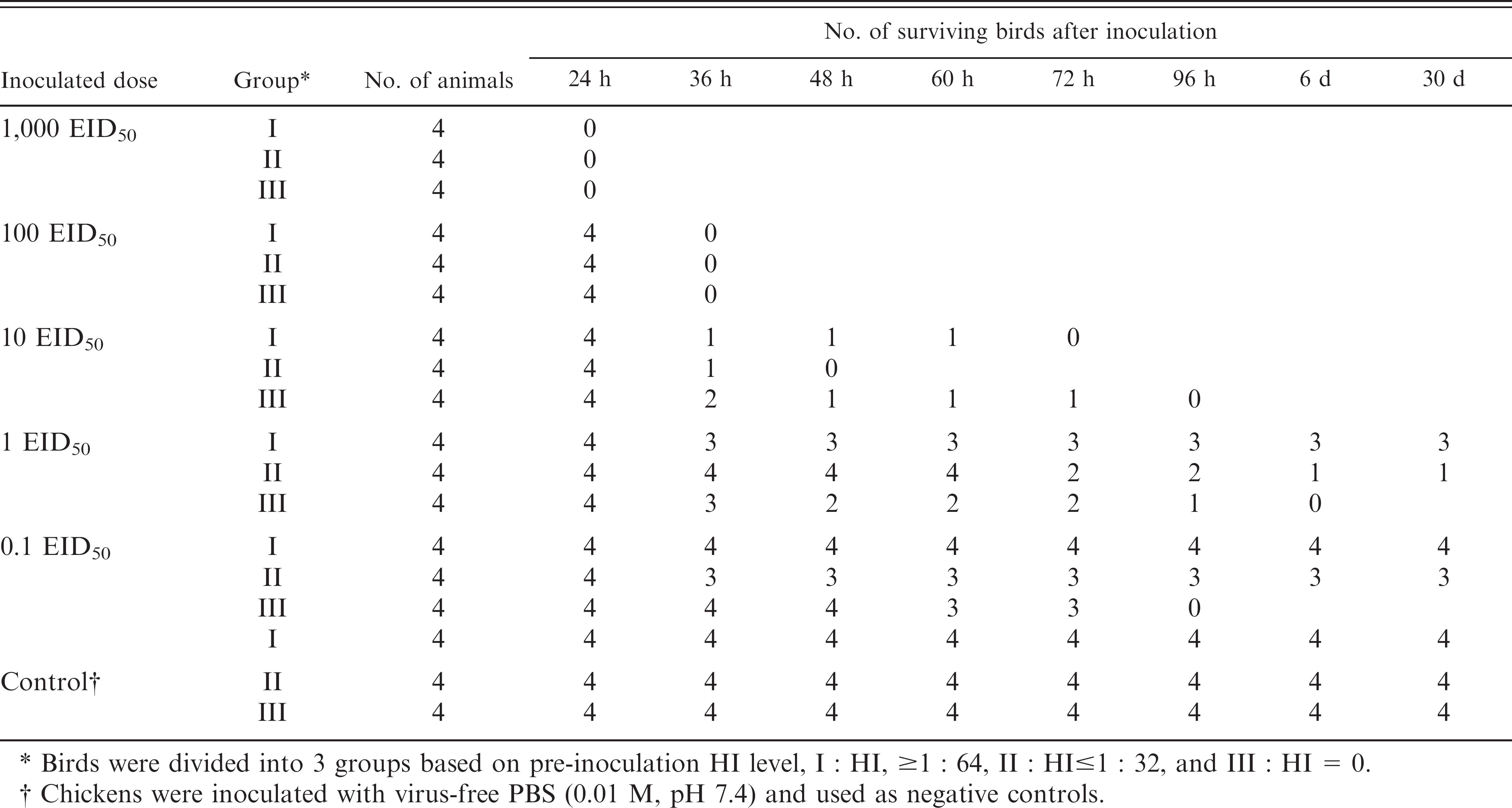
Birds were divided into 3 groups based on pre-inoculation HI level, I:HI, ≥ 1:64, II:HI≤1:32, and III:HI = 0.
Chickens were inoculated with virus-free PBS (0.01 M, pH 7.4) and used as negative controls.
Detection of H5N1 in tracheal swabs of experimentally infected chickens using LAT. *
All tracheal swabs were treated with 0.3 M borate buffer (0.85% NaCl, 0.5% N-acetyl-cysteine, 0.1% NP-40, 5000 IU/ml of penicillin G, 5 mg/ml of streptomycin, pH 8.4) to prevent nonspecific agglutination.
All chickens inoculated with 1,000 EID50 of H5N1 died within 24 hr postinoculation, thus no specimens were collected from them.
Chickens were inoculated with virus-free PBS (0.01 M, pH 7.4) and used as negative controls.
Of the 90 specimens analyzed by LAT and the immunochromatographic test, 87 had identical results by both tests (24 positives and 63 negatives), with a correlation of 96.7% (87/90). In contrast, the correlation between LAT and real-time RT-PCR test was 87.8% (15 positives and 64 negatives) (Table 4).
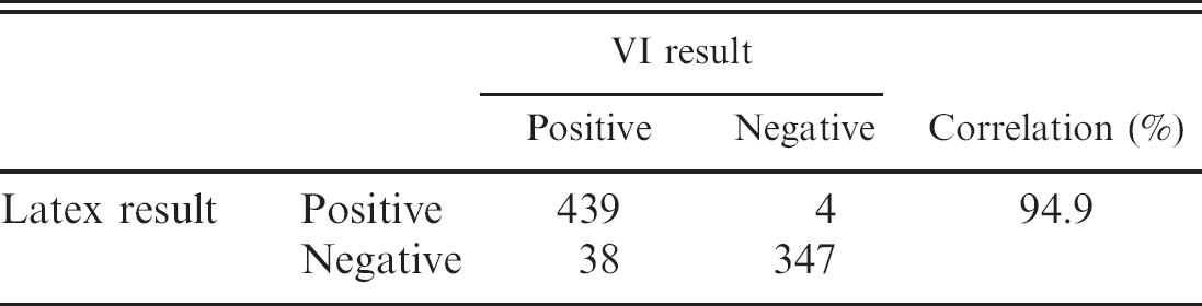
Field evaluations of LAT using outbreak specimens. The sensitivity and specificity of the LAT were compared to those of the current gold standard for AIV detection, VI, using 828 chicken specimens obtained during the 2004 H5N1 outbreak. The performance characteristics of the LAT compared to the VI assay are shown in Table 5. A total of 477 were positive by VI, of which 439 were also positive by the LAT. In addition, 4 specimens were negative by VI but positive by the LAT. Thus, LAT had a sensitivity of 92.0%, specificity of 98.9%, positive predictive value of 99.1%, and negative predictive value of 90.1% for H5N1. The correlation between VI and LAT was 94.9%, and there was no significant difference in H5N1 diagnosis between the two techniques (P > 0.05).
Detection of H5N1 in internal organs of experimentally infected chickens. *
+ = Positive result.
Evaluation of LAT using routine clinical samples. All 640 chicken and 750 duck specimens that were collected from slaughterhouses were negative in the LAT. Of the 260 lung specimens obtained on some family farms where the animals were not immunized, 32 were found to be positive when tested with the LAT. In all cases, the positive diagnosis by the LAT was confirmed by VI.
Discussion
Previous studies have shown that the covalent protein coupling technology using carboxylated microparticles is a very good mechanism for linking proteins to microparticles. 2,7,12 Carboxylate-modified latex particles are superior to unmodified latex particles for use in agglutination tests because they are more hydrophilic, more stable in aqueous solutions, and contain reactive sites for covalent conjugation of proteins.
In this study, IgG concentration affected the sensitivity of SLP. The best sensitivity was obtained when the largest percentage of the starting amount of IgG was bound to the latex beads, which was achieved using 1,000 μg of IgG per 0.5 ml of 2.5% latex particles. Although the total amount of IgG bounded to latex particles increased when more IgG was added, the rate of agglutination was significantly reduced when 1,800 μg of IgG or more per 0.5 ml of latex particles was used in conjugation.
Comparisons in the performance of H5N1 diagnosis among the LAT, VI, the H5 AIV Ag Test and the H5 AIV real time RT-PCR Test using 90 specimens collected from chickens in an outbreak. *
+ = Positive; — = Negative.
To increase the specificity of the LAT, MAbs were used instead of polyclonal antibodies. Seven hybridoma cell lines were established that secreted MAbs specific for different regions of the HA protein of H5N1 virus. The reactivity of MAbs in culture supernatants was assessed by HI and the HI titer of culture supernatants was generally ≥32, indicating that high affinity MAbs were produced.
In the animal infection experiments, the chickens were divided into 3 groups based on the pre-inoculation HI titer and inoculated with varying virus doses (ranging from 1,000 EID50 to 0.1 EID50) in order to mimic field conditions in the laboratory but control other variables that might occur in the field, such as: medication, stress, and co infection with multiple bacterial and viral agents. Data obtained suggested that the LAT could detect H5N1 in tracheal swabs (Table 2). However tracheal swabs taken from the chickens inoculated with H5N1 at 100 EID50 that died within 36 hours without obvious clinical signs, had a low positive rate (52.8%). It is suggested that this low positive rate might be due to the likelihood that the high dose of the inoculum might have attacked the brain and lung directly and killed the chickens before systemic replication, proliferation, and subsequent discharge of viruses through the trachea. In contrast, chickens inoculated with 1 EID50and 0.1 EID50 lived longer and had higher positive rates (average 77.9% and 71.4%). The difference in the detection rates from various internal organs might be due to differential tropism. As expected, the detection rate was much higher in lung tissue than that in the heart, cloaca, and muscle. The low detection of H5N1 in the cloaca suggests that the current practice of using fecal droppings of waterfowls for AIV detection may seriously under-estimate the prevalence of H5N1 in these reservoir animals.
Performance characteristics of the LAT compared to VI in the analysis of H5N1 outbreak specimens from chickens.
The evaluation results of the LAT when applied to 828 specimens of chickens collected during an outbreak (Table 5) and 90 specimens from naturally infected animals show excellent performance characteristics (sensitivity, specificity, and correlation) in comparison to VI, immunochromatographic test, and real-time RT-PCR test. Like immunochromatographic test, LAT results are read visually. However, unlike the former, the latter is less affected by the presence of blood in the specimens. Compared to real-time RT-PCR, LAT is much less expensive, less technically demanding, and can be performed in the field.
When LAT was evaluated in the field using specimens from large commercial operations that mandatorily immunize their birds and specimens from small family poultries that did not immunize, all specimens from large commercial operations were negative whereas those from family farms had a relatively high positive rate. This apparent discrepancy could be due to the following 3 reasons. First, these farms were far away from cities and widely scattered, which made governmental supervision very difficult. Second, veterinary services in China are relatively less developed in rural areas where the small family farms abound. Third, current poultry production systems in China vary tremendously in scale and technology, from large-scale intensively managed mega farms to backyard flocks with a small number of chickens, ducks, geese, and pigs housed in close contact. In the latter situation, no obvious severe losses would be felt when birds are infected with AIV and there are no significant incentives to the farmers for vaccination. Therefore, the occurrence of H5N1 in birds in small operations in the absence of apparent outbreaks demonstrates that the prevention and control of AIV in China is a very formidable task. It also suggests that AIV infections can only be controlled by wide-scale immunization and testing.
From 2003 to date, H5N1 has caused great losses to the poultry industry in China. As demonstrated in the study, the newly developed LAT test can accurately detect AIV subtype H5 without cross reactivities to the other viruses that were evaluated in the study. This test is applicable to internal organs and tracheal swabs. It is rapid, simple, and easy to perform and does not require expensive equipment or skilled personnel. Thus, this test is appropriate for use in field investigations and in rural animal clinics for the rapid detection of H5N1.
Acknowledgements
This work was supported by the Chinese National “973” Program grant 2005CB523003. We thank the Veterinary Diagnostic Center in Hunan Province P. R. China for analyzing field specimens by the immunochromatographic test and the Shenzhen CIQ P. R. China for real time RT-PCR analysis.
Footnotes
a.
Unit of Animal Infectious Diseases, National Key Laboratory of Agricultural Microbiology, Huazhong Agricultural University, Wuhan 430070, Hubei Province, P.R. China.
b.
Polysciences, Inc. Corporate Headquarters, Warrington, PA.
c.
Animal Genetics, Inc. Jangan-gu, Suwon, Kyonggi-do, Korea.
d.
Shenzhen Taitai Genomics, Inc. Shenzhen 518057, P.R. China.