
Abstract
Introduction:
Isolated distal ulnar fracture is an uncommon injury. Only a few case series that look into the management of ulnar fracture nonunion have been reported in the literature.
Case presentation:
A middle-aged manual worker presented to us for isolated fracture of his left distal ulna. He received open reduction and fixation of the distal ulnar fracture using distal ulna locking plate. It was complicated with nonunion and he complained of persistent wrist pain. This distal ulnar fracture nonunion was finally treated by Sauve-Kapandji procedure with good functional outcome.
Discussion:
Displaced distal ulnar fracture should be reduced anatomically with rigid fixation to prevent disruption of the distal radioulnar joint. Fracture nonunion was traditionally managed with revision osteosynthesis and bone grafting. It is, however, technically difficult at the distal ulnar region because of the poor bone stock and lack of soft tissue coverage.
Conclusion:
We have demonstrated that the Sauvé-Kapandji procedure is a good treatment alternative for distal ulnar fracture nonunion.
Introduction
Isolated distal ulnar fracture is an uncommon upper limb injury. 1 It is usually the consequence of a direct blow against the soft tissue-deficient ulnar border. Although the best treatment option for displaced distal ulnar fracture remains a subject of debate, most surgeons aim for anatomical reduction and stable fixation to avoid disruption of the distal radioulnar joint. 1 Fixation of the distal ulnar is technically demanding and complications could arise. Only a few case series that look into the management of ulnar fracture nonunion have been reported in the literature.
We report a case of isolated distal ulnar fracture that was treated with open reduction and fixation with locking plate which subsequently developed fracture nonunion. We present our management strategy, treatment outcome, as well as review of the literature.
Case presentation
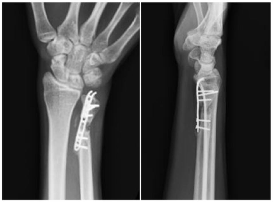
A 47-year-old, right-hand dominant electrical worker was admitted to the emergency department of our hospital, after injury of the left wrist by a fallen metal bar at work. Clinical examination revealed swelling and tenderness at the left distal ulna. Distal neurovascular status was intact. Anteroposterior and lateral wrist radiographs demonstrated an isolated distal ulnar fracture with displacement (Figure 1). Open reduction was performed using a direct ulnar approach. Intraoperatively, there was comminution over the fracture site with small bone fragments. Anatomical reduction could not be achieved. Allowing for bone contact over the fracture ends, suboptimal alignment was accepted. The fracture was fixed with a distal ulnar locking plate using three locking screws distally and two cortical screws proximally (Figure 2).

Anteroposterior and lateral X-ray showing fracture distal ulnar.
Anteroposterior and lateral X-ray showing fracture fixation with locking plate immediately after initial fracture fixation.
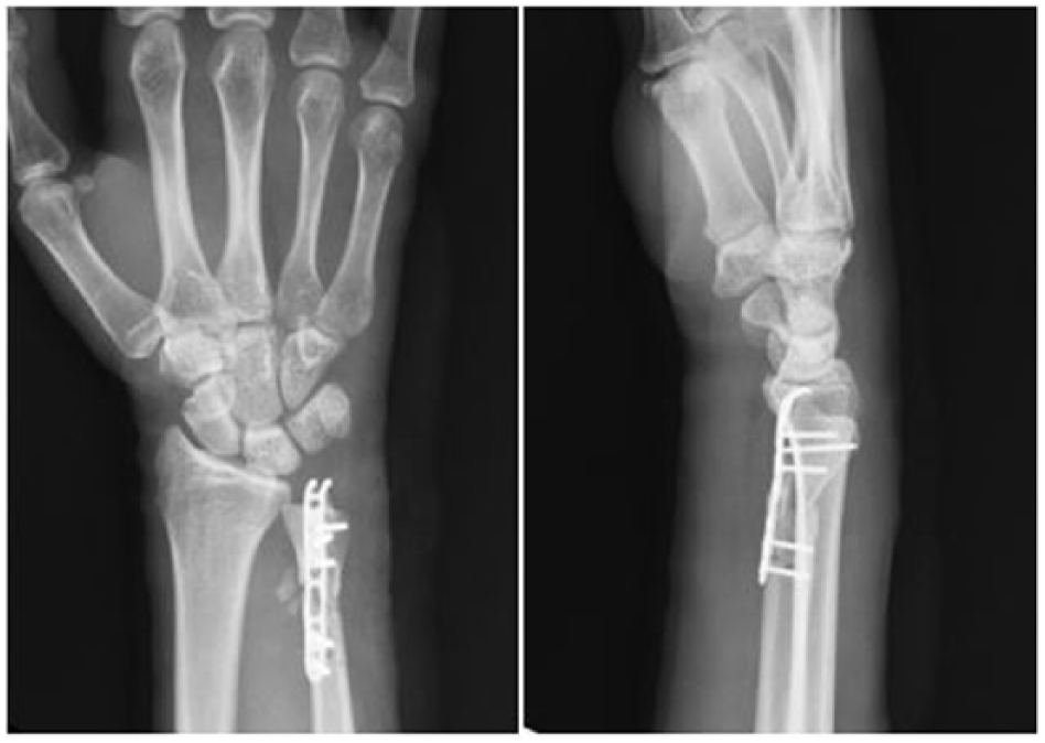
The patient complained of persistent pain at the fracture site and limited forearm rotation at 6-month follow-up. There was no sign of infection and the inflammatory markers were normal. Radiographs of the left wrist showed fracture nonunion with bone loss and implant loosening (Figure 3).
Anteroposterior and lateral X-ray showing fracture nonunion with loosening of implant at 6 months after the initial operation.
Sauvé-Kapandji procedure was performed through incision on the previous scar. The locking plate was found to be loosened and there was a 5-mm fracture gap filled with fibrous tissue. The plate and screws were removed. The distal ulnar head was reduced and fused with the sigmoid notch of the radius using two 3.5-mm cannulated screws. The fracture site was debrided and the proximal bone end was cut short to create a 15-mm pseudoarthrosis gap. Half of the extensor carpi ulnaris tendon was harvested at wrist level and was attached to the proximal bone end to stabilize the ulnar shaft. Pronatus quadratus was sutured to the tendon sheath of extensor carpi ulnaris to fill the pseudoarthrosis gap (Figure 4). A below-elbow splint was applied to protect the wrist for 4 weeks. The hand and forearm were then gradually rehabilitated with motion and strengthening exercise under the supervision of a hand therapist.
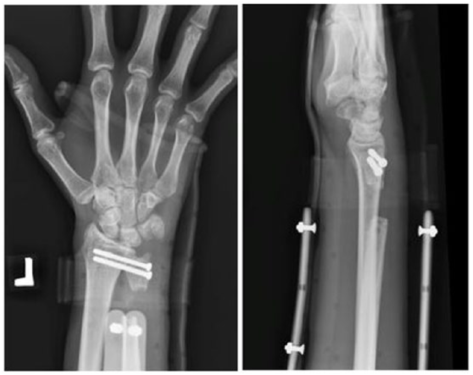
Anteroposterior and lateral X-ray taken immediately after revision operation by Sauvé-Kapandji procedure.
At 2-year follow-up, the patient was pain free and returned to pre-injury level of activity. His left wrist flexion and extension range reached 50° and 40°, respectively. Supination and pronation of the forearm was 80° and 90°, respectively. The grip strength of his left hand was 38 kg, compared to 46 kg on the right side. Radiographic examination reviewed solid fusion of distal radioulnar joint (Figure 5).
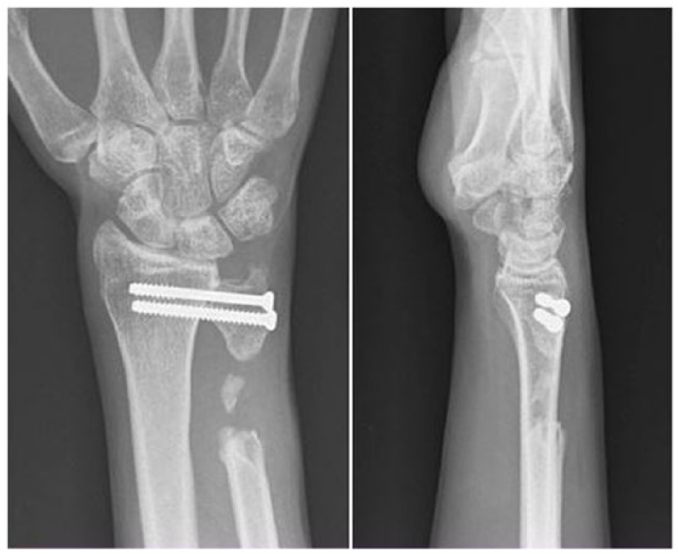
Anteroposterior and lateral X-ray taken at 2 years after revision operation by Sauvé-Kapandji procedure.
Discussion
Most, if not all, distal ulnar fractures are associated with fracture distal radius. Isolated distal ulnar fracture is rare and a universally accepted treatment protocol has not been developed. A systematic review reported excellent results of casting in managing minimally displaced distal ulnar fracture, which was defined as a fracture with less than 50% translation and 15° of angulation. 2 Displaced fracture should, however, be reduced anatomically to prevent disruption of the distal radioulnar joint, which is important for maintaining forearm rotation and wrist stability. Fixation of distal ulnar fracture can be achieved by Kirschner wire, blade plate, and lag screws supplement with tension band wiring. 3 The newly designed LCP distal ulna plate consisting of pointed hooks to grip the ulnar styloid and angled locking screws to hold the ulnar head is advocated to be the implant of choice for distal ulnar fracture; fixation failure, however, could occur as in our patient.
The union rate for distal ulnar fracture is determined by the fracture characteristic and the fixation method. Comminuted fracture with suboptimal reduction and extensive periosteal stripping is more likely to result in nonunion. There is currently no literature that looks specifically into the management of distal ulnar fracture nonunion; only few case series report the outcomes of ulnar shaft fracture nonunion managed by revision osteosynthesis and bone grafting. Kloen et al. 4 reviewed 33 isolated ulnar shaft fracture nonunion managed with compression plate fixation and autologous bone graft, all nonunion healed within a median of 7 months. Ring et al. 5 reported 11 patients with atrophic nonunion of the ulnar diaphysis treated by autologous cancellous bone grafting and plate fixation, all patients achieved fracture union. Pagnotta et al. 6 described successful treatment of two ulnar nonunion using dorsal distal radius vascularized bone graft. Although revision fixation and bone grafting were shown to produce excellent results in ulnar shaft fracture nonunion, the success could not be simply translated to the management of distal ulnar fracture nonunion. Revision fixation of the distal ulnar at the metaphyseal region is particularly challenging because of its poor bone stock and lack of soft tissue coverage. Moreover, there is no ideal implant for fixation of distal ulnar at the metaphysis. The hooked locking plate may not have sufficient length for proximal fixation, while other standard plates are bulky and may not provide adequate fixation distally. Another limiting aspect of this particular anatomic location is the intimate relationship with the distal radioulnar joint; malreduction may give rise to incongruity of the joint, resulting in poor functional outcomes. Restoring normal anatomy is, however, difficult in revision osteosynthesis because of bone loss, scarring, and soft tissue contracture. We decided to perform Sauvé-Kapandji procedure in our patient because it is technically feasible and the outcome is more predictable.
Sauvé-Kapandji procedure describes the arthrodesis of distal radioulnar joint and creation of a pseudarthrosis proximal to the arthrodesis site to preserve forearm rotation. Louis de Gonzague Sauvé and Mehmed Kapandji described the technique in 1936 in treating a patient with isolated distal ulnar dislocation. Sauvé-Kapandji procedure is now used in the treatment of ulnar impaction syndrome and a variety of disorders causing distal radioulnar joint arthrosis and instability, particularly in rheumatoid arthritis. We are now extending the indication to manage failure fixation of distal ulnar fracture. Compared with revision fixation, Sauvé-Kapandji procedure is a relatively simple procedure that achieves good pain relief, restores pronation–supination movement of the distal forearm, and allows early rehabilitation. However, the procedure requires creation of a pseudoarthrosis, which might give rise to rare complications such as stump impingement, extensor tendon rupture, and ossification of pseudarthrosis. 7 To achieve good postoperative range of motion, we must limit the time of immobilization to at most 4 weeks, followed by intensive mobilization exercise under supervision of a hand therapist.
In conclusion, displaced isolated distal ulnar fracture is a rare upper limb injury. It should be suspected in patients with direct trauma against the soft-tissue-deficient ulnar border. Minimally displaced distal ulnar fracture, which is defined as fracture with less than 50% translation and 15° of angulation, can safely be managed with casting. Displaced fracture should, however, be admitted and treated with anatomical reduction and rigid fixation to prevent disruption of distal radioulnar joint. Fracture fixation could be challenging and it is not a fracture for a beginner. Implant selection should be well thought out beforehand, with locking plates and small screws available. Fracture nonunion should be suspected if there is persistent pain over the fracture site after 6 months of injury. Once nonunion of the distal ulnar metaphysis is established, it is a difficult problem, due to poor bone stock, bone loss, and requirement of a congruent distal radioulnar joint. Revision osteosynthesis and bone grafting is one option. It is however, technically demanding and bone healing is uncertain. We have demonstrated that the Sauvé-Kapandji procedure is a good treatment alternative for distal ulnar fracture nonunion.
Footnotes
Acknowledgements
The manuscript is the original work of the authors. The manuscript has not been and will not be published elsewhere or submitted elsewhere for publication.
Declaration of conflicting interests
The author(s) declared no potential conflicts of interest with respect to the research, authorship, and/or publication of this article.
Funding
The author(s) received no financial support for the research, authorship, and/or publication of this article.
Availability of data and materials
All data, tables, and figures used in the manuscript were prepared originally by the authors; if otherwise, the sources are cited and reprint permission is attached.
Informed consent
Written informed consent was obtained from the patients for their anonymized information to be published in this article.