
Abstract
Background
Meniscus injuries are familiar sources of knee pain, with meniscus repair sometimes yielding unsatisfactory results. Microfracture is a standard procedure for treating articular cartilage damage in the knee that promotes the formation of fibrocartilage over damaged cartilage. Microfracture enhanced the healing rate of meniscus repair in animal models.
Objective
Investigate the effectiveness of microfracture augmentation on the wall of femoral condyle in meniscus healing among patients with meniscus repair.
Methods
The current study, conducted in a single center, involved patients with red-white zone meniscal tears due to sports injury who underwent arthroscopic meniscus repair between January 2018 and December 2023. Meniscal repair was performed without microfracture in the control group but with microfracture augmentation in the intervention group.
Results
Functional outcomes, assessed using the Lysholm score, were significantly better in the microfracture group (p = 0.000). Both groups showed significant reductions in tear size according to its intensity on magnetic resonance imaging (MRI) (p = 0.002 in the control group and p = 0.001 in the microfracture group), with a notably better meniscus healing rate (35.7%) in the microfracture group compared to the control group.
Conclusion
This initial study highlights substantial effectiveness between meniscus repair augmented with microfracture and enhanced healing compared to repair without microfracture. Functional scores were notably higher, and MRI signal intensity decreased to grade 1 in a significant portion of patients in the microfracture group.
• Microfracture augmentation on the wall of femoral condyle stimulates bone marrow cells that will form growth factor • The patients in the microfracture group has a higher the functional outcome (Lysholm Score) compared to the control group • The patients in the microfracture group has a higher healing rate according to the MRI compared to the control group • Microfracture augmentation on the wall of femoral condyle increases meniscal healing among patients with meniscus tears after sports injury
Introduction
Meniscus injury is a prevalent cause of knee pain. The meniscus is a semilunar fibrocartilage structure found on the medial and lateral sides of the knee. The meniscus absorbs impact and protects the articular cartilage during weight-bearing activities, joint stability, proprioception, and nutrition. This has a significant effect on knee biomechanics.1,2 Several adjuvants have been proposed to improve meniscus healing. However, a study by Nepple et al. showed that the failure rate of meniscus repair increased from 22% to 24% at 5 years. 3
The healing rate of meniscus repair, accompanied by anterior cruciate ligament (ACL) repair, is more significant, 93% compared to 50%. Hemarthrosis that occurs after creating a bone tunnel during ACL reconstruction creates a fibrin clot and an environment rich in factors that promote healing of the newly repaired meniscus. The fibrin clot provides the structural foundation for meniscus healing. Transcription factors, such as fibronectin and growth factors, are also found. 4
Microfracture is a standard procedure for treating articular cartilage damage in the knee. This procedure is used to stimulate fibrocartilage production in the injured cartilage area. Microfractures are performed by creating one or more small channels (1 to 3 mm in size) that penetrate the subchondral bone and release bone-forming components into the joint. Microfractures serve to promote the formation of fibrocartilage over damaged cartilage. A study in 2016 by Howarth et al. used an animal model (Capra hircus) to investigate the effectiveness of the microfracture technique on the rate of healing of meniscus tears. They found significant healing in meniscal repair using the microfracture technique (65% vs 12%). 5 Given the context, this study aimed to investigate the efficacy of microfracture in the wall of femoral condyle augmentation in promoting meniscus healing among individuals undergoing meniscus repair surgery.
Materials and Methods
This single-center study included patients with red-white zone meniscal tears who underwent arthroscopic meniscus repair between January 2018 and December 2023. The patients who only underwent meniscus repair surgery were included in the current study. All patients underwent clinical evaluation, functional score assessment, and MRI examination 6 months postoperatively. A single senior orthopedic sports surgeon (MS) diagnosed and surgically treated all patients. The study was conducted following the Declaration of Helsinki and approved by the In-stitutional Review Board and Ethics Committee (424/UN4.6.4.5.3 L/ PP36/ 2A23). The study protocol was also registered on ClinicalTrials.gov.
Inclusion and exclusion criteria
Patients were included if they were diagnosed with a meniscus tear of the red-white zone and treated by arthroscopic meniscus repair, which had pre-operative and six-month post-operative data. The patients must also have been involved in sports injuries (recreational athletes). Patients were excluded if they had additional procedures.
Surgical procedure
The patients underwent arthroscopic meniscus repair under spinal anesthesia. The meniscus was repaired arthroscopically using the all-inside technique (ULTRA FAST-FIX Meniscal Repair System). Among the microfracture group, the awl (5 mm diameter) was used to create ten microfracture holes with a 3 – 4 mm depth on the femur condyle’s wall. Microfracture is performed on the outer side of the condyle, adjusted to the position of the torn meniscus. If it is lateral to the meniscus, microfracture is performed on the lateral wall of the condyle, and vice versa, aiming for the growth factor. These mesenchymal stem cells come out directly towards the torn meniscus. (Figure 1) The microfracture was made by awl in the subchondral bone at 3–4-mm intervals to preserve the subchondral bone plate’s structure and function, promoting the natural healing process. (Figure 2).
6
Microfracture augmentation on the wall of femoral condyle. Bone marrow cells are released after microfracture is created on the wall of condyle with a 5 mm awl diameter.

Evaluation
The magnetic resonance imaging of the knee examination was taken two times, pre-operatively and postoperatively. A knee MRI examination was done pre-operatively and postoperatively to find the stage of the meniscal tear, according to Lotysch et al. 7 The Lysholm score, assessed using eight questions and with a maximum score of 100, evaluated the patient’s knee function. 8
Statistical analysis
The pre-operative and post-operative Lysholm score was compared statistically. A paired t test was used for the functional outcome score, and the Wilcoxon Test was used for the meniscal tear stage to find the variables’ significance. All statistical analyses were performed using SPSS Statistics for Windows Version 25 (IBM), with p < 0.05 defined as statistically significant.
Results
Patient’s characteristics
Patient’s characteristics.

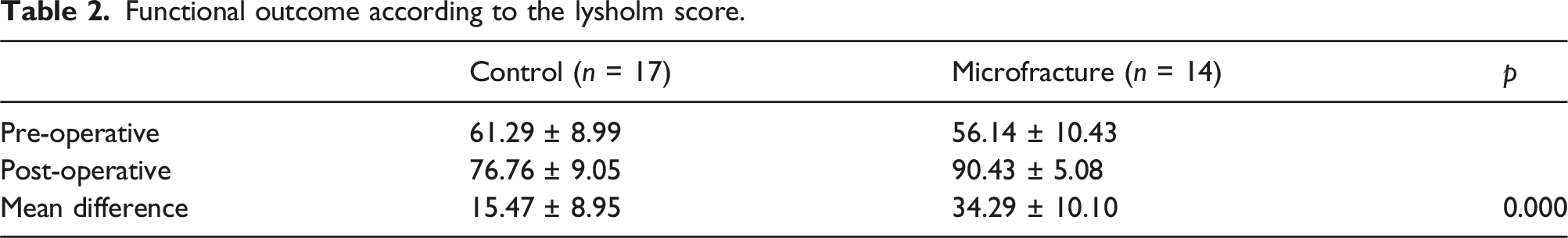
Functional outcome
Functional outcome according to the lysholm score.
Radiological outcome
MRI measured radiological outcomes according to the staging developed by Lotysch et al.
7
Significant tear size reduction was observed in both groups (p = 0.002 in the control group and p = 0.001 in the microfracture group). (Figure 3) There was better meniscus healing in the microfracture group, where five cases (35.7%) were healed to the first degree of tear, yet in the control group. No case was reduced to the first degree of tear. Pre-operative and Post-operative radiological staging of meniscus tear, according to Lotysch
7
; 
Discussion
This study found significant functional and radiological outcomes in the microfracture group compared to the control group. Functional outcomes were assessed using Lysholm scoring with a higher mean difference in the microfracture group compared to the control group (34.3 ± 10.1 vs 15.5 ± 9.0, p = 0.000). Radiological outcomes were assessed using MRI with a significant decrease in both groups (p = 0.002 in the control group and p = 0.001 in the microfracture group). In the group with microfractures, healing of the meniscus was better, reaching grade 1 in 5 cases (35.7%) compared to the group without microfractures (0%). Four patients from grade 3 meniscus tears and one from grade 1 meniscus tear were healed to grade 1 after the surgery (Figure 4). Pre-operative magnetic resonance imaging of a 31-year-old male with a grade 3 meniscus tear (left), meniscus repair was done with microfracture augmentation (center)
The findings were similar to those in research by Howarth et al., who found better meniscus healing outcomes in the microfracture group compared to the control group (65% vs 12%, p < 0.001). 5 Meniscus healing tends to be better because bone marrow cells are produced in the joint. These bone marrow cells will form growth mediators such as platelet-derived growth factor, insulin-like growth factor, and transforming growth factor. This results in increased healing of the meniscus.9,10 These factors are present in the bone marrow and may be responsible for the different healing rates noted between meniscus tears repaired with and without concurrent ACL reconstruction.4,9
Biological augmentation in meniscus repair has shown comparable results with concurrent ACL reconstruction. When comparing the meniscus repair group augmented with a marrow venting procedure at the intercondylar notch and meniscus repair with concurrent ACL reconstruction, Dean et al. found no significant differences in the failure rates between these two groups (12.9% vs 7.8%, p = 4.29). 11 A case series by Ahn et al. also uses biological augmentation through stimulation of bleeding at the intercondylar notch. A 5-mm diameter and 20-mm deep canal was made into the medulla of the bone to induce the bleeding at the intercondylar notch. The study found improvement in VAS score (p < 0.001), Lysholm score (p < 0.001), and Tegner score (p < 0.001). 12
Initial studies have shown promising results for regenerating meniscal tissue with growth factors, but this technique is not readily available for treating meniscal injuries. Studies in other animal models have shown improved meniscus healing with fibrin clots and autologous marrow or blood products.13,14 Autologous growth factor and pluripotent mesenchymal cells are present in the bone marrow and may increase the rate of meniscus healing.9,13
This study contributes to understanding the role of microfractures in the meniscus healing process in patients with meniscus tears in the red-white zone. Although these findings provide valuable insights, the current study has several limitations. The study had a small sample size and a short duration of follow-up, which may have affected the results. In addition, this study used a retrospective design, which has a lower degree of evidence than that of prospective studies and randomized clinical trials. Further research with larger sample sizes and longer follow-ups is needed to corroborate the findings of this study. More strict inclusion and exclusion criteria must be included to reduce the factors that affect the results. It is hoped that the current research can be used as a “stepping-stone” and a reference for further research, especially regarding microfracture as an augmentation for meniscus repair.
Conclusions
This study showed a significant association between the meniscus repair group with microfracture augmentation and meniscus repair compared to those without microfracture augmentation. Meniscus healing was better in the microfracture augmentation group, with a higher increase in functional scores and a decrease in MRI signal intensity to grade 1 in 35.7% of patients.
Footnotes
Author contributions
Conceptualization, M.S., L.C.S., N.H., and D.A.N; Methodology, L.C.S.; software, L.C.S.; validation, M.S., N.H., and D.A.N; formal analysis, L.C.S; investigation, M.S., L.C.S., N.H., and D.A.N; re-sources, M.S.; data curation, L.C.S.; writing—original draft preparation, L.C.S; writing—review and editing, M.S., N.H., and D.A.N; visualization, M.S., L.C.S., N.H., and D.A.N; supervision, M.S., N.H. and D.A.N; project administration, M.S., N.H. and D.A.N; funding acquisition, M.S. All au-thors have read and agreed to the published version of the manuscript.
Declaration of conflicting interests
The author(s) declared no potential conflicts of interest with respect to the research, authorship, and/or publication of this article.
Funding
The author(s) received no financial support for the research, authorship, and/or publication of this article.
Institutional review board statement
The study was conducted in accordance with the Declaration of Helsinki, and approved by the Institutional Review Board (or Ethics Committee) (424/UN4.6.4.5.3 L/ PP36/ 2A23 and 26th June 2023).
Informed consent statement
The hospital’s ethical committee gave consent due to the study’s retrospective nature and the withdrawal of identification information from the data matrix.