
Abstract
Mycobacterium tuberculosis infection has emerged as a global public health issue, predominantly manifesting as pulmonary tuberculosis. Bone and joint tuberculosis, with spinal tuberculosis accounting for approximately 50%, represents a significant form of extrapulmonary tuberculosis. Over the past years, there has been a rise in the incidence of spinal tuberculosis, and research concerning this area has gained significant attention. At present, animal models provide a means to investigate the pathogenesis, drug resistance, and novel treatment approaches for spinal tuberculosis. New Zealand rabbits, possessing a comparable anatomical structure to humans and capable of reproducing typical pathological features of human tuberculosis, are extensively employed in spinal tuberculosis research using animal models. This article comprehensively evaluates the strengths, considerations in strain selection, various modelling approaches, and practical applications of the rabbit model in studying spinal tuberculosis based on pertinent literature to guide fundamental research in this field by providing valuable insights into appropriate animal model selection.
Introduction
Tuberculosis has been a longstanding and significant threat to human life and health since the 19th century. In 2020, around 10 million people globally were infected with Mycobacterium tuberculosis (MTB), resulting in approximately 1.3 million tuberculosis-related deaths, impacting roughly 25% of the global population.1,2 Musculoskeletal system involvement occurs in 1% to 2% of tuberculosis patients, with spinal tuberculosis (STB) being a prevalent form of extrapulmonary tuberculosis, accounting for over 50% of skeletal tuberculosis cases.3,4 Neurological symptoms accompany approximately 23% to 76% of STB patients, and if left untreated, can lead to severe complications, including paralysis, significantly reducing patients’ quality of life.5,6 However, research on spinal tuberculosis faces challenges including unclear pathogenesis, low diagnostic and treatment efficacy for tuberculosis, as well as difficulties in treating multidrug-resistant cases. Consequently, there has been a growing body of research on spinal tuberculosis in recent years, highlighting the increasing recognition of the significance of animal models in fundamental medical research. These models facilitate a more convenient and effective understanding of the patterns of disease occurrence and progression, serving as valuable tools for investigating and designing intervention strategies. 7 Rabbit models currently play a predominant role in spinal tuberculosis research. This article provides an extensive review of the literature on the establishment of rabbit models for spinal tuberculosis, encompassing model selection, strain varieties, methods for modelling infection, current applications, and future prospects. Its main objective is to offer dependable references for selecting animal models to investigate the pathogenesis and mechanisms of drug resistance in spinal tuberculosis in future research endeavors.
Selection of model animals
Rodents
Guinea pig
Guinea pig models have been widely used in infectious disease research. Guinea pigs are highly susceptible to Mycobacterium tuberculosis and can be effectively infected with low doses of M. tuberculosis aerosols. This characteristic makes it possible to establish model conditions for studying strains of bacteria with variable or low virulence.8,9 Recent studies have shown that guinea pig models exposed to high-dose bacterial strains have shorter lifespans compared to those using lower doses of bacteria. 10 Additionally, guinea pigs infected with tuberculosis demonstrate caseous necrosis and granulomas that closely resemble those observed in humans with primary tuberculosis. As a result, they are considered a more reliable model for studying tuberculosis. 11 However, the use of this model in spinal tuberculosis research is limited due to its high cost, expensive rearing requirements, small vertebral structure, heightened sensitivity, and lack of relevant anti-tuberculosis immunity.
Mice
Mice are preferred as model animals because of their affordability, rapid breeding cycles, adaptability to laboratory settings, extended lifespans, low cost, and ease of genetic manipulation. 12 Mice show a certain degree of susceptibility to MTB infection, with colonies forming within their bodies as soon as 4 weeks after transmission. 13 However, the bronchial anatomy of the mice is relatively simple and lacks an accompanying vascular system. This limitation hinders the accurate replication of pathological modifications observed in human tuberculosis cases. 14 Consequently, this limitation weakens their utility in experimental research, prompting researchers to explore the utilization of human haematopoietic stem cells in the development of humanised mice models for tuberculosis research. 15 Humanised mice models provide a valuable approach to investigating the complexities associated with tuberculosis and HIV. These models maintain the advantages of traditional mice models while incorporating a functioning human immune system. 16 Nevertheless, significant progress is required before the widespread implementation of large-scale research can be achieved for large-scale purposes.
Rats
Similar to mice, rats exhibit susceptibility to MTB and possess comparatively large vertebrae among rodents, rendering them manageable. 17 Following MTB infection, rats exhibit the development of granulomatous lesions; however, macrophage infiltration is significantly reduced compared to that of mice. Consequently, this animal model has been extensively employed to assess the effectiveness of tuberculosis vaccines and novel medications.18–20 Additionally, Zhou et al. developed a rat model of tuberculosis ulcers using an attenuated strain of Mycobacterium bovis. Their successful modelling efforts offer a novel option for studying animal models of tuberculosis ulcers. 21 Nevertheless, rat models feature specific limitations, particularly in terms of replicating the progression of spinal tuberculosis in humans. Ultimately, the utility of both rats and mice is limited for spinal tuberculosis research because of substantial disparities in anatomy, pathology, immunology, and other aspects compared with humans.
Non-human primates
Non-human primates (NHP) exhibit a high degree of anatomical, physiological, immunological, and other resemblances to humans, making them potential optimal models to study spinal tuberculosis. 22 Most NHP are naturally susceptible to Mycobacterium tuberculosis infection. After infection, they develop tuberculosis granulomas and extensive caseous necrosis, similar to those seen in humans, making them ideal for tuberculosis research.23–25 Hunter et al. successfully induced tuberculosis in rhesus and cynomolgus monkeys by aerosolizing Mycobacterium tuberculosis (MTB), thus determining the basic cellular composition and spatial distribution of immune cells within monkey lung granulomas. These findings provide valuable insights for future studies on tuberculosis vaccines and therapies. 26 Additionally, rhesus monkeys serve as an ideal model for evaluating the efficacy of tuberculosis vaccines, as their disease progression closely resembles that observed in humans. Rhesus monkeys have been extensively employed to assess the effectiveness of tuberculosis vaccines or drugs. 27 The effectiveness of the MTBVAC vaccine was demonstrated using this model by White et al. 28 However, due to their high cost, expensive maintenance, limited availability, significant individual variations, and complex social behaviors involving ethical concerns, as well as their limited application in spinal tuberculosis research, they still have significant potential for future research.
Rabbit
Rabbit models were initially utilized in molecular immunology research due to their similarities to humans in terms of tuberculosis lesion presentation. Currently, they are widely employed for screening and evaluating tuberculosis vaccines, as well as studying the pathogenic factors and mechanisms of MTB. 29 The primary advantages of using rabbits as models for spinal tuberculosis include moderate cost, gentle temperament, ease of management, comprehensive genetic information, and their capacity to act as hosts for MTB, which provides objective conditions for their extensive application.30–32 Furthermore, research conducted by Popov et al. has demonstrated significant similarities between rabbit tuberculosis strains and human tuberculosis strains, resulting in their pathological changes after MTB infection being more comparable to those in humans. 33 Studies have also shown that rabbits display milder symptoms and longer survival times following MTB infection. 8 Another notable advantage of rabbits as models for spinal tuberculosis is their ability to replicate typical radiological manifestations of STB. 31 Scholars have observed characteristic X-ray findings in rabbit models of spinal tuberculosis, including indistinct and narrowed intervertebral spaces, uneven vertebral density, and new bone formation at the vertebrae edges. CT scans reveal adjacent endplate bone hyperplasia accompanied by bone resorption and osteophyte formation, while MRI demonstrates inflammatory changes at the upper end of the vertebrae, accompanied by intervertebral disc damage. 30 Furthermore, other researchers have also identified similar radiological changes in their rabbit models compared to human cases of STB. 34 These aforementioned studies further confirm that rabbit models can simulate the pathological and physiological changes associated with STB. Moreover, rabbits possess similar spinal anatomical characteristics to humans, with larger vertebrae and intervertebral spaces, resulting in improved surgical visibility and facilitating modelling, wound debridement, lesion clearance, and the administration of sustained-release drugs. 35 Additionally, rabbits can be infected with various strains, such as MTBH37Rv, MTBHN878, MTBErdman, MTBCDC1551, and bovine branch bacillus. 29 These advantages make rabbits the optimal choice as an animal model for spinal tuberculosis.
Scholars have demonstrated the feasibility of using rabbits as a model for spinal tuberculosis. In 1882, Koch et al. conducted the first-ever attempt to create a tuberculosis model in rabbits by implanting MTB, thus confirming the suitability of rabbits as tuberculosis models. 36 Subsequently, other researchers achieved successful modelling of spinal tuberculosis in New Zealand white rabbits by injecting MTB suspension, yielding a modelling success rate of 86.7%. 37 In another study, a different group of researchers used gelatin sponges containing MTB suspension to establish a rabbit model of spinal tuberculosis, which also produced satisfactory results. 38 These findings indicate that rabbits exhibit good compatibility and high success rates in modelling when confronted with various approaches and methods in spinal tuberculosis models.
Despite being regarded as an important and reliable animal model for spinal tuberculosis, the rabbit spinal tuberculosis model has certain limitations. First, being a large animal, it incurs higher maintenance costs and demands more significant living spaces, resulting in biological contamination through the excretion of Mycobacterium tuberculosis in urine, thereby limiting the applicability of the rabbit tuberculosis model. 39 Second, challenges arise in establishing a standardized modelling method, encompassing the MTB inoculation dosage and surgical techniques, necessitating additional research for clarification and refinement. Lastly, the absence of pertinent immune reagents for the rabbit spinal tuberculosis model impedes diverse research in this field.
Advantages and disadvantages of common model animals.

Animal models suitable for various types of tuberculosis research.

Selection of strains for the rabbit model of spinal tuberculosis
MTB standard strains
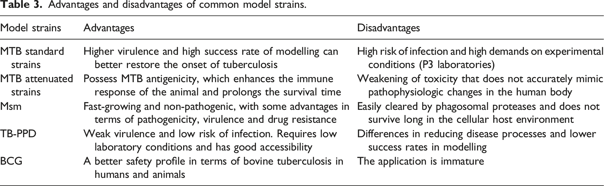
Standard strains of Mycobacterium tuberculosis (MTB) are commonly used for modelling purposes. These bacteria are small (3 μm long, 0.5 μm wide) Gram-positive, acid-fast, facultative aerobic organisms that can partially adapt to facultative anaerobic conditions. 44 The H37Rv strain, a standard isolate of MTB, was clinically derived from strain H37 and is highly invasive and contagious, making it extensively employed in spinal tuberculosis models. H37Rv exhibits relatively slow growth rates but can thrive in diverse culture media, including Lowenstein-Jensen medium (L-J medium), Middlebrook 7H9 broth medium, or blood agar medium.45,46 Research has consistently shown the significant utility of H37Rv in spinal tuberculosis rabbit models. In their study, Geng et al. included 42 New Zealand white rabbits and successfully established 17 spinal tuberculosis models using the H37Rv strain, resulting in a remarkable success rate of 89.5%. 30 Likewise, Liu et al. achieved successful establishment of 23 models on New Zealand white rabbits utilizing the H37Rv strain, yielding a success rate of 77%. 47 These findings demonstrate the feasibility and relative maturity of techniques, as well as the high success rate attained by using the H37Rv strain for modelling and establishing spinal tuberculosis models.
Accurate control of H37Rv concentration and dosage is crucial for the success of modelling. Excessive concentration and dosage can lead to extensive lesions and mortality in experimental animals, whereas insufficient concentration and dosage may result in low rates of positive modelling. In this context, various scholars have investigated the optimal strain concentration for modelling purposes. In the study conducted by Qiao et al., a total of 42 New Zealand rabbits were included, and the intervertebral discs of the experimental group received injections of H37Rv strain suspension at concentrations of 1 × 10⁶ CFU/ml, 1 × 10⁷ CFU/ml, and 1 × 10⁸ CFU/ml, respectively. 48 After 12 weeks, the 1 × 10⁶ CFU/ml concentration group exhibited a low positivity rate of typical radiographic manifestations, while the 1 × 10⁸ CFU/ml concentration group had a high mortality rate. The concentration group with a 1 × 10⁷ CFU/ml concentration demonstrated a high modelling success rate and was deemed the optimal concentration for constructing the spinal tuberculosis (STB) model. Additionally, Liu et al.’s research revealed that when establishing a spinal tuberculosis rabbit model using a 1 × 10⁷ CFU/ml concentration of H37Rv strain, an injection of 0.3 mL resulted in the mortality of all rabbits, while 0.2 mL caused 50% rabbit mortality. 49 The rabbit survival rate reached 75% after 2 months when injecting 0.1 mL of the strain. Therefore, using 0.1 mL of MTB standard strain H37Rv with a concentration of 1 × 10⁷ CFU/ml to establish a rabbit model of spinal tuberculosis represents a reasonable choice, as it can lead to a higher modelling success rate and effectively mitigate high mortality rates in the animal model.
In addition to the MTB standard strain H37Rv, researchers have investigated the applicability of other genotypes of MTB for tuberculosis modelling. Kaya et al. achieved successful modelling of pulmonary tuberculosis in New Zealand rabbits using MTB HN878 via the respiratory approach. 50 Likewise, Gonzalez et al. accomplished modelling by administering MTB Erdman into the lungs of mouse via the respiratory approach. 51 Furthermore, Sharan et al. conducted pertinent studies on successful modelling using MTB CDC1551 in monkeys. 52 However, there are currently no reported instances of successful modelling of the STB model using the aforementioned MTB standard strains. Further comparative studies among different MTB standard strains are necessary to investigate if they exhibit comparable modelling.
MTB attenuated strains and Mycobacterium smegmatis (Msm)
Attenuation of MTB strains occurs through the induction of nutrient deficiencies or the disruption of crucial virulence genes, such as tryptophan, arginine, or pantothenate, leading to alterations in MTB transcription and the emergence of virulence variants. 53 MTB attenuated strains offer reduced infection risk and enhanced safety in comparison to standard strains. Additionally, in contrast to the short-lived effects observed with MTB standard strains, MTB attenuated strains exhibit a comparable antigenicity, thereby augmenting immune responses in animal models and significantly prolonging survival.54,55 Arbues and colleagues have engineered a candidate Mycobacterium tuberculosis vaccine with attenuation properties by eliminating two specific genes in clinical isolates of Mycobacterium tuberculosis, free of antibiotic resistance markers. 56 Presently, it stands as the sole live attenuated vaccine undergoing human clinical trials. Nonetheless, its reduced virulence compromises its ability to accurately replicate the pathophysiological changes in the human body, resulting in certain limitations. Over the past decade, several recombinant attenuated vaccines derived from Mycobacterium smegmatis have been developed and validated in preclinical animal models. Therefore, although MTB attenuated strains possess specific advantages and potential, further investigation is necessary, especially in the absence of studies validating their success rate in the STB model.
Msm is a gram-positive saprophytic bacterium capable of expressing antigens from Mycobacterium tuberculosis. It exhibits rapid growth and non-pathogenic properties. Msm possesses over 2000 genes homologous to MTB, resulting in a wider range of natural drug resistance profiles. 57 Junqueira-Kipnis et al. have demonstrated that Msm can elicit protective immune responses against MTB in mouse infection models, establishing its advantages in terms of pathogenicity, virulence, and drug resistance compared to Mycobacterium tuberculosis.58,59 Guinea pigs, mouse, and other small rodent animals are susceptible to Msm, making it a valuable tool for establishing infection models in research studies. 60 Nevertheless, Msm’s survival in the cellular host environment is limited due to its susceptibility to clearance by phagosome proteases, hindering its ability to fully replicate the occurrence and progression of tuberculosis. This limitation restricts its broad application. Hence, Msm represents a promising alternative strain for modelling purposes; however, further research is necessary to validate its potential in the STB model.
Advantages and disadvantages of common model strains.
Selection of the infection approach for the rabbit model of spinal tuberculosis
Surgical approach
Rabbits are the most suitable animals for modelling spinal tuberculosis owing to their larger vertebral bodies. Modelling is frequently achieved through surgical procedures involving direct local injection of MTB. This approach provides several advantages, including a swift and clear surgical procedure, shortened modelling duration, and precise lesion localization. 34
Currently, the initial procedures for establishing a rabbit model for spinal tuberculosis (STB) are similar. However, there is controversy surrounding the method of MTB inoculation. Some scholars opt to use a drill to create a hole in the intervertebral disc and directly inject the MTB suspension into the bone hole under aseptic conditions to establish the model. 62 In contrast, other scholars prefer to fill the bone hole with gelatin sponge soaked in MTB suspension and seal it with bone wax before suturing. 63 Filling the gelatin sponge, as opposed to direct injection of bacterial suspension, provides better containment of MTB at the modelling site and prevents bacterial dissemination that could result in the mortality of the model animals. Moreover, the technique of filling the gelatin sponge is widely employed and demonstrates a notable success rate, making it the preferred choice when conditions allow. However, there is currently a dearth of comparative studies investigating the success rates between these two modelling methods, which necessitates further evidence-based support.
The success rate of the model in spinal tuberculosis modelling can also be influenced by different surgical approaches. Currently, there are two main approaches: the posterior lateral approach and the lateral transverse process approach. Yue et al. compared the posterior lateral approach and the lateral transverse process approach. The postoperative results demonstrated that both approaches provided sufficient exposure of the vertebral body, resulting in modelling success rates of 86.9% and 78.2%, with postoperative mortality rates of 88% and 90%, respectively. 34 No significant difference was observed between the two approaches. They suggested that the lateral transverse process approach had the potential to cause complications such as transverse process fracture. In contrast, the posterior lateral approach, while having a lower likelihood of complications, required a higher level of surgical expertise. Consequently, both surgical approaches have distinct advantages and disadvantages, enabling researchers to select the most appropriate approach based on various factors, including surgical proficiency and anatomical comprehension.
Respiratory and venous approaches
Advantages and disadvantages of common model approaches.

Application of the spinal tuberculosis rabbit model
Spinal tuberculosis treatment research
The rabbit spinal tuberculosis model is commonly employed in studies investigating the efficacy of anti-tuberculosis drugs. Zhao et al. discovered that treatment with Hesperidin methyl chalcone (HMC), a derivative of flavonoid known for its anti-inflammatory properties, effectively lowered the formation of granulomas in the neighboring vertebral bone tissue. 72 Moreover, it restrained the expression of inflammatory factors in rabbits infected with spinal tuberculosis. These findings suggest a novel therapeutic approach for managing spinal tuberculosis. Additionally, Wang et al. conducted a study demonstrating that Isoliquiritigenin, a flavonoid compound extracted from the Chinese herb licorice, effectively mitigated the formation of granulomas in the rabbit spinal tuberculosis model. 73 This study offers an objective assessment of the therapeutic potential of Isoliquiritigenin in the treatment of spinal tuberculosis.
Besides the development of novel anti-tuberculosis medications, the investigation of localized drug delivery is a prominent focal point in tuberculosis research. Notably, the rabbit spinal tuberculosis model continues to play a vital role. Wang et al. investigated the formulation and assessment of a complex comprising rifampicin-loaded polylactic acid microspheres (RPSMs) for treating spinal tuberculosis, utilizing a rabbit spinal tuberculosis model. 74 The study revealed that this complex exhibits an extended drug release period, negligible inhibition of cell osteogenic differentiation, and favorable tissue repair effects, surpassing conventional tuberculosis treatment with a significantly enhanced therapeutic outcome. Conversely, Liu et al. conducted an extensive investigation into the benefits and drawbacks of isoniazid (INH) lactide-co-glycolic acid (PLGA) microspheres and INH hydroxyapatite (HA) microspheres concerning drug release. 38 They generated rabbit models using tuberculosis strains and cultivated sustained-release microspheres in the lesions to assess the concentration of tuberculosis drugs in the affected areas. The experimental results demonstrated that the microspheres in the HA group not only raised the drug concentration in the affected areas but also exerted osteogenic effects. Nonetheless, in terms of drug distribution, PLGA presented a superior advantage. Furthermore, Xi et al. validated the architecture of a cross-linked adjustable sustained-release nano artificial bone composite (TPB/SA-RFP/PLA) for anti-tuberculosis drug delivery in a rabbit model, showcasing its capacity for prolonged release within the body alongside favorable osteogenic efficacy. 75
Research on the pathogenesis of spinal tuberculosis
Recently, the rabbit spinal tuberculosis model has been utilized to investigate the pathogenesis of tuberculosis infection. Guo et al. employed the rabbit spinal tuberculosis model as an experimental group to assess the variances in MCP-1, NF-κB, IFN-γ expression levels and CD4+ and CD8 + lymphocyte distribution within the immune response of spinal tuberculosis. 63 The study revealed a potential association between elevated levels of MCP-1 and NF-κB and the immunosuppression of spinal tuberculosis, suggesting novel therapeutic avenues for its treatment. Through the assessment of TNF-α and CRP expression after internal fixation for spinal tuberculosis, Risantoso et al. observed a substantial decrease in the expression of tumor necrosis factor-α (TNF-α) and C-reactive protein (CRP) in infected rabbits solely treated with medication or devices, as compared to untreated infected rabbits. 76 Thus, it is suggested that internal fixation treatment can mitigate the inflammatory activity of spinal tuberculosis by modulating cellular factors, ultimately facilitating improved healing.
The utilization of the rabbit spinal tuberculosis model has yielded noteworthy research outcomes in investigating the treatment and pathogenesis of tuberculosis, thereby partially substantiating the reliability of this model. Nevertheless, disparities in disease progression between animal models and patient models become apparent in the aforementioned studies. Tuberculosis is a chronic illness characterized by a course lasting typically over 2 years. Conversely, the preparation of experimental animal models entails an intervention period of merely around 8 weeks, rendering the simulation of this chronic condition arduous under experimental circumstances. Moreover, there is presently a dearth of pertinent research utilizing the rabbit spinal tuberculosis model to explore drug resistance in spinal tuberculosis. Subsequently, future endeavors should concentrate on exploring methods to simulate the chronic progression of spinal tuberculosis more accurately and investigating the feasibility of examining drug resistance through animal experiments employing the rabbit model.
Conclusion and perspectives
In summary, rabbits are a cost-effective and easily reproducible animal model. Their spinal anatomical characteristics bear resemblance to humans, and they exhibit typical tuberculosis lesions following Mycobacterium tuberculosis (MTB) infection. Moreover, their relatively large vertebral bodies deliver a clear surgical field and facilitate accurate experimental results. However, currently, there is no established standardized approach or sufficient comparative studies on the effects of different modelling animals and approaches. Future advancements in experimental conditions, optimisation of modelling strains, and the development of humanised experimental animal research techniques are anticipated to result in the creation of more cost-effective, robust, and standardised animal models for spinal tuberculosis. This study established a precise and dependable in vitro experimental platform to investigate the pathogenesis and drug resistance of spinal tuberculosis.
Footnotes
Acknowledgments
The authors thank the National Natural Sciences Foundation of China (Grant No. 82272536), Natural Science Foundation of Gansu province (Grant No. 22JR5RA949), Lanzhou University Innovation and Entrepreneurship Cultivation Project (cxcy2023012).
Declaration of conflicting interests
The author(s) declared no potential conflicts of interest with respect to the research, authorship, and/or publication of this article.
Funding
The author(s) disclosed receipt of the following financial support for the research, authorship, and/or publication of this article: This work was supported by the National Natural Sciences Foundation of China (Grant No. 82272536), Natural Science Foundation of Gansu province (Grant No. 22JR5RA949), Lanzhou University Innovation and Entrepreneurship Cultivation Project (cxcy2023012).