
Abstract
Background
Studies have shown that platelet-rich plasma (PRP) can enhance the effect of meniscus repair, but some studies have suggested different views on the role of PRP.
Purpose
To determine whether PRP can enhance the effect of meniscus repair with respect to pain reduction and improved functionality and cure rate in patients with meniscus injury.
Methods
By searching PubMed, EMBASE, Cochrane Library databases, clinicaltrials.gov, and the CNKI database from their inception till December 1, 2020, we performed a meta-analysis of RCTs reporting the results of the Pain Visual Analog Scale (VAS), the pain of Knee injury and Osteoarthritis Outcome Score (KOOS), Lysholm score, the International Knee Documentation Committee (IKDC), healing rate, and adverse events. The risk of bias is assessed using Cochrane’s collaborative tools. The summary results are expressed with effect size and 95% confidence interval, and sensitivity were performed.
Results
The meta-analysis included 9 RCTs and 345 patients. In general, compared with the control group, used of PRP during meniscus surgery significantly improved the pain (SMD: -0.95, p < 0.00001,95% CI: -1.22 to -0.69, I2 = 42%) and knee joint function (SMD: 1.00, p = 0.01.95% CI: 0.22 to 1.79, I2 = 89%) of patients with meniscus injury at 6 months after treatment. However, both PRP and non-PRP showed improvements in the pain and knee joint function, with no significant difference between the groups at 1 months and beyond 12 months. The PRP enhancement technique showed benefit in improving the cure rate of meniscus repair (RR:1.44; p < 0.0001, 95% CI: 1.20–1.73). No serious adverse events were reported in any study.
Conclusion
As an enhancement program for meniscus repair, PRP is worthy of further consideration in improving the function and pain of patients during the mid-term follow-up after surgery, and PRP can further improve the healing rate of meniscus repair. However, the evidence still needs to be interpreted carefully because of the quantity and quality of the included studies.
Introduction
The meniscus is located between the tibia and the femoral condyle. It is an important structure of the knee joint, and its functions include transferring load and stabilizing the knee joint. 1 Meniscus injury is a common disease of the knee joint, which often leads to knee joint dysfunction, swelling, pain, clicking of joint, etc., which affect the knee joint function and quality of life of the patient. 2 According to reports, nearly 4 million patients worldwide undergo arthroscopic meniscus surgery every year. 3
Total meniscus or partial meniscus resection is a method of treatment for meniscus injuries. However, this technique has a fatal disadvantage, in that it will reduce the tissue of the meniscus, which increases knee contact pressure and decreases knee joint stability.4–5 In recent years, several randomized controlled trials (RCTs) have shown that meniscus resection has no additional benefit to sham surgery, so surgeons should preserve damaged meniscus as much as possible instead of removing the meniscus.6–8 Owing to the presence of the avascular area of the meniscus, meniscus repair can preserve the meniscus tissue as much as possible but still cannot restore the anatomy and function of the meniscus after repair. 9 Therefore, multiple studies have evaluated the potential of some augments such as the extracellular matrix, fibrin clot, hyaline, and growth factors to enhance the effect of meniscus repair.10-13 Recently, many studies have shown that adding platelet-rich plasma (PRP) during surgery can enhance the effect of meniscus repair.13–19 However, some studies are still controversial regarding some clinical outcomes such as the visual analogue scale (VAS), knee joint function, and healing rate.18–21 Current evidence indicates that PRP may not be as strong as previously thought.22–23
To better understand the effect of platelet-rich plasma enhance meniscus repair, we tried to conduct a meta-analysis of RCTs that compared meniscus repair combined with PRP versus only meniscus surgery in patients with meniscus tears, evaluate the safety and effectiveness of this technology to enhance meniscus repair, and provide evidence-based decisions for clinical applications.
Methods
Search strategy
We searched five databases, namely PubMed, Web of Science, Cochrane Library databases, clinicaltrials.gov, and the CNKI database. The last search was conducted on 19 Nov 2021. The following search strategy was used: (platelet rich plasma) AND (meniscus). We also checked the references of related articles to identify additional relevant researchto increase the output of related studies. No language restrictions were employed. There were no language restrictions.
Criteria for considering studies
Clinical studies that meet the following criteria were included: (1) RCTs; (2) compared the use of meniscus repair combined with PRP therapy versus only meniscus repair in patients with a meniscus injury. If data were repeated or shared in multiple studies, the study that best met the above criteria were considered. All published or unpublished studies were investigated. If the information required for the analysis could not be obtained from the publication, the author was contacted to obtain the necessary details.
Data extraction
All searches and included studies were conducted by two independent reviewers. If there was any objection, the third reviewer made the final decision. The following data were extracted from the final included research: research title (first author name and publication date), participants (sample size), sex ratio, age range of participants, follow-up time, meniscus injury degree, intervention method and evaluation indicators.
Types of outcome measures
The primary outcomes included the pain scores as the VAS and the pain of Knee injury and Osteoarthritis Outcome Score (KOOS), and the secondary outcomes were knee joint function as the Lysholm scores and the International Knee Documentation Committee (IKDC). Additional outcome included the Healing rate. We also evaluated the adverse reactions of applying PRP in meniscus repair.
Risk of bias and quality of outcomes assessment
Two review authors independently assessed the methodological quality of the included studies. We recorded and resolved any disagreements through discussions with a third reviewer. Each RCT used Cochrane’s collaborative tools to assess the risk of bias, including the following criteria: adequacy of sequence generation, concealment of allocation, blinding of participants and personnel, blinding of result evaluators, incomplete results’ data, selective reporting, and other biases.24–25 The Grading of Recommendations Assessment, Development and Evaluation (GRADE) guidelines for systematic reviews were used to evaluate the quality of outcomes.
Statistical analyses
All statistical analysis used the methods released by Cochrane, and the heterogeneity of different research results were tested by overlap of confidence intervals and chi-square tests. When there was no heterogeneity in the test results, fixed effects model was used for the meta-analysis, and when the test results were heterogeneous, the random effects model was used. For enumeration data, the risk ratio (RR) was used as the statistical tool for the efficacy analysis, and 95% confidence intervals (CIs) were used for the effect size.
If substantial heterogeneity was detected (I2 > 50%), subgroup analysis or sensitivity analysis was conducted to determine the source of heterogeneity (e.g., the length and severity of the meniscus injury, dosage and preparation of PRP, different regions and study quality, average age of the participants, and location of the research institution).
Results
We identified nine studies17–19,21,26–29 that met the inclusion criteria (Figure 1). Initially, 258 articles were identified by searching the databases, and 0 articles were retrieved by searching other sources. According to the inclusion and exclusion criteria, we first excluded 75 duplicate articles, followed by 162 articles that did not meet the inclusion criteria. Finally, we checked the full text of the 16 remaining articles and excluded 7 for reasons being non-RCT design. Figure 1 depicts the PRISMA flow diagram detailing the disposition of retrieved publications. PRISMA flowchart of the study selection process. randomized controlled trials.
Study Characteristics
Studies on PRP combined with meniscus repair included in meta-analyses.*

*F, female; M, male; PRP, platelet-rich plasma; VAS,the visual analogue scale; RCT, randomized controlled trials; IKDC, International Knee Documentation Committee; KOOS, Knee injury and Osteoarthritis Outcome Score; ADL, Activities of daily living. Values are expressed as mean with range or SD.
The preparation process of PRP was slightly different in the included studies. All studies used Leucocyte Poor platelet-poor plasma (LP-PRP), but there are differences in dosage and frequency. Only two studies17,21 mentioned that the content of platelets in PRP.
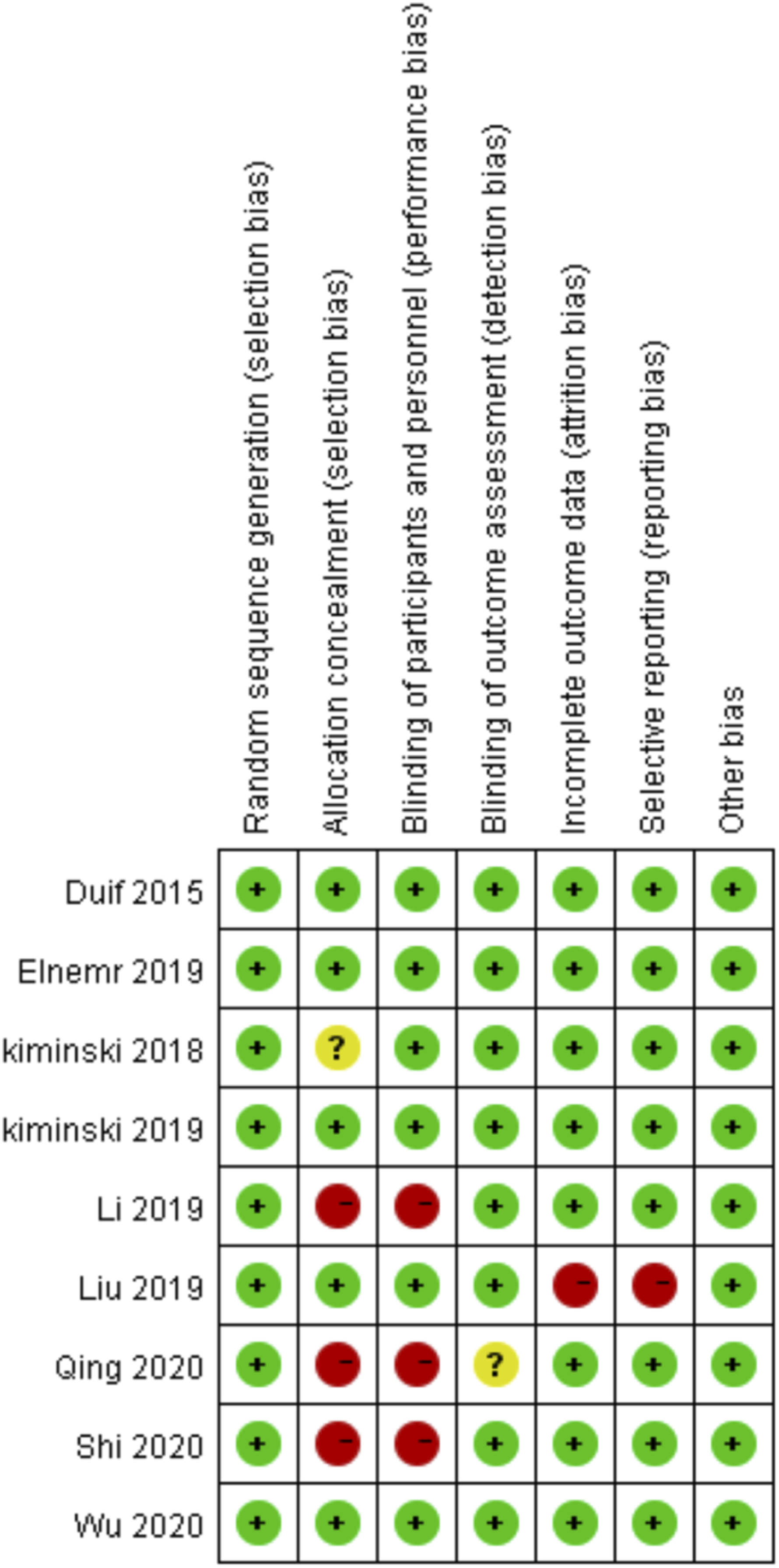
Risk of bias
Figures 2 and 3 present the results of the risk of bias graph of all RCTs. In the 3 studies,19,29–30 the surgeons knew the grouping of participants, and performance bias was a high risk. Among all the studies, only study by Liu et al.
18
showed the high risk of attrition bias and reporting bias. The meta-analysis included in the number of studies is very small, and we could not use funnel plots to assess publication bias. Therefore, publication bias could not be completely eliminated. Performance of each type of bias in all studies. Assessment of the risk of bias for included studies. +, low bias risk; -, high bias risk; ?, unclear bias risk.
The pain scores
A pooled analysis of 8 articles had evaluated The pain scores after treatment (Figure 4). As shown in the Figures 4(a) studies19,26,28–29 had evaluated The pain scores at 1 months after treatment. Compared with the control group, the pain scores of patients receiving PRP did not significantly decrease (standard mean difference [SMD]: -0.23, p = 0.09.95% CI: -0.50–0.04, I2=41%). As shown in the Figure 4(b), 5 studies18–19,26–28 had evaluated The pain scores at 6 months after treatment. Compared with the control group, the pain scores of patients receiving PRP significantly decrease (SMD: -0.95, p<0.00001,95% CI: -1.22 to -0.69, I2 = 42%). As shown in the Figure 4(c), only 3 studies17,21,26 had evaluated The pain scores beyond 12 months after treatment. Compared with the control group, the pain scores of patients receiving PRP significantly decrease (mean difference [MD]: -0.09, p = 0.14.95% CI: -0.24–0.06, I2 = 28%). Forest plot for pain scores. PRP, platelet-rich plasma.
The knee joint function scores
A pooled analysis of 8 studies had evaluated knee joint function scores (Figure 5). As shown in the Figure 5(a), 4 studies19,26,28,30 had evaluated The pain scores at 1 months after treatment. Compared with the control group, the pain scores of patients receiving PRP did not significantly improve (SMD: 0.13, p = 0.49.95% CI: -0.24–0.51, I2 = 46%). As shown in the Figure 5(b), 5 studies19,26,28,30 had evaluated The pain scores at 6 months after treatment. Compared with the control group, the pain scores of patients receiving PRP significantly improve (SMD: 1.00, p = 0.01.95% CI: 0.22 to 1.79, I2 = 89%). As shown in the Figure 5(c), 3 studies17,21,26 had evaluated The pain scores beyond 6 months after treatment. Compared with the control group, the pain scores of patients receiving PRP significantly improve (SMD: 1.27, p = 0.19.95% CI: -0.62–13.15, I2 = 96%). Forest plot for knee joint function. PRP, platelet-rich plasma.
Therefore, taking into account the obvious heterogeneity of knee joint function scores at 6months, we conducted a sensitivity analysis that excluded every trial in turn, and the analysis revealed that the heterogeneity originated from the study of Liu et al.
18
After excluding this study, 4 studies19,26,28,30 were re-incorporated (Figure 6). There was no significant heterogeneity in all studies (I2=0%), so the fixed effects model was used. The statistical difference remained the same as before the sensitivity analysis, which was conducive to PRP enhancement (SMD: 0.59, p < 0.0001, 95% CI: 0.31–0.87). Forest plot of the sensitivity analysis for knee joint function at 6 months after treatment. PRP, platelet-rich plasma.
Healing rate
In a pooled analysis of 4 studies17–18,21,30 that had evaluated the healing rate at 24–33 weeks’ follow-up (Figure 7), there were 238 patients in all, with 120 in the PRP group and 118 in the control group. In the 24–33 weeks of follow-up, compared with the control group, the PRP enhancement technique showed benefit in improving the cure rate of meniscus repair (RR:1.44; p < 0.0001, 95%CI: 1.20–1.73). Forest plot of the subgroup analysis for healing rate. PRP, platelet-rich plasma.
Adverse reactions
Adverse events were reported in only one study. 19 During the treatment, two patients developed mild postoperative joint swelling and pain and limited mobility, which were eliminated within 3 days after local ice compress, restricted mobility, and oral analgesics. Unfortunately, this study did not indicate how these adverse events were determined.
Discussion
Based on 9 RCTs, the application of PRP in meniscus repair might have a positive effect on patients’ pain score and knee joint function scores at 6 months, and the healing rate at follow-up. However, we don’t find significant improvement on patients’ pain score and knee joint function scores at 1 months and beyond 12 months. Therefore, these results should be interpreted carefully. We only included randomized, placebo-controlled trials. Most studies had explained their randomization methods, and a few studies had explained allocation concealment methods. The experiments reported varying results. Most studies reported were adequate. There were a small number of cases where there was no standard deviation or graphic representation. There was significant heterogeneity in a part of our analysis, and we therefore used sensitivity analysis to address this concern.
The results of pain score at 1 months and beyond 12 months after treatment show that the meniscus repair surgery combined with PRP group has no advantage in relieving postoperative pain. This is consistent with the research results of Dai et al., 15 Hak et al. 31 and Kaminski et al.17,21 These study concluded that PRP has no significant effect on postoperative pain improvement. The reason may be related to the high content of tumor necrosis factor-α (TNF-α) in conventional PRP preparations. 32 The high concentration of TNF-α causes the postoperative inflammatory pain index to increase, which is contrary to the positive effect of PRP. However, the results of pain score at 6 months after treatment show that the meniscus repair surgery combined with PRP group has advantage in relieving postoperative pain. This is consistent with the research of Li et al. 19 and Wang - Saegusa et al. 33 Our findings suggest that PRP may only have mid-term effect in relieving pain.
These study concluded that PRP has no significant effect on postoperative knee joint function improvement at short-term or long-term follow-up. This is consistent with the previous studies.34–35 However, Our findings suggest that PRP have mid-term effect in improving knee joint function. There was significant heterogeneity in the knee joint function at 6months; the pooled analysis, after excluding a study, 18 still suggested that PRP could improve knee joint function after meniscus repair at 6months. Analysis of the full text of Liu et al.’s study 18 showed that the mean age is 34.7 years, which is much smaller than other included studies, which might be the reason for the high heterogeneity between this and other studies. Different types of meniscus tears and age-related causes of meniscus tears can lead to different healing abilities. In general, PRP can further improve the healing rate of patients after meniscus repair and a few trials have adverse events and found that there may be no difference in the incidence of adverse events between participants receiving and not receiving PRP treatment.
Many potentially confounding variables such as the age, sex, cause and of meniscus injury, categories of meniscus tear, surgical approach, and platelet content in PRP had not been reported in detail. Routine postoperative treatment varies by research included active/passive activities and weight training, among others.
The cause of meniscus tear is often related to the patient’s age. The most common cause of meniscus tear and/or deterioration in young and elderly patients is typically related to acute trauma to the joints and degenerative changes, respectively. 36 Due to the uniqueness of the meniscus structure, there are two different mechanisms for the healing of injuries. In the red area of the meniscus (vascular area), the abundant blood supply provides nutrients for mesenchymal cells to induce healing. 37 In the white area (avascular area), the healing of the meniscus depends on its own tissue repairability, which leads to difficult healing or even non-healing. 38 Meniscus repair is effective in treating meniscus injuries in the red area, with a healing rate as high as 90%, but it has a poor effect on injuries in the white area. 39 In young men with meniscus injuries, there are often simultaneous tears in two areas.
At the same time, different meniscus repair methods have different repair capabilities. The all-inside meniscus repair systems are safer, faster, and more convenient and hence, more popular than other meniscus repair systems such as outside-in and in-outside meniscus repair. 40 Among all-inside meniscus repair, the Fast-Fix technique has a better suture effect. It adopt the conventional arthroscopic approach, no auxiliary incision is required, and the damage between the tissues is fully reduced. The operation time is short, the suture is firm, and the operation is simple. In addition, different repair methods result in different movement of meniscus and sizes of popliteal hiatus, which further leads to different biomechanics and kinematics of the lateral knee joint compartment. However, the research included in this meta-analysis did not directly mention the age stratification of the participants, area of the meniscus where the tear was located, and the type of meniscus tear; furthermore, the meniscus repair methods used were also different. Therefore, more extensive subgroup analysis could not be performed to clarify the enhancement ability of PRP on different meniscus repair procedures; moreover, it was also not possible to judge whether PRP is age-related or tear type-related for enhanced meniscus repair procedures.
PRP is a platelet concentration obtained after centrifugation of peripheral blood, and its role in the repair of cartilage damage has gradually attracted attention in recent years. PRP mainly includes platelet-related leukocyte aggregates, high-density fibrous network structure, platelet-derived growth factor, transforming growth factor-β, insulin-like growth factor, epidermal growth factor, and vascular endothelial growth factor.41–42 PRP can release a large number of anti-inflammatory factors to reduce local inflammation and can release a variety of growth factors to promote cell proliferation and regulate cell behavior. 43 In vitro studies have shown that chondrocytes and PRP exhibit a significant dose- and time-dependent increase in cell number and metabolic cell activity. 44 It has already been shown that even small variation in centrifugation settings can alter the content of the PRP product, which underlines the importance to describe the ingredients before applying PRP product. 45
All included studies only used LP-PRP. Those clinical effect of LP-PRP versus leukocyte-pich platelet-rich plasma (LR-PRP) remains controversial, as leukocytes need been argued due to their positive as well as negative properties. Leukocytes not only play a role in pro-inflammatory activity, but they also interact with platelets and other cell types to drive the regression phase of the healing cascade.46–47 However, only two studies17, 21 verified the PRP contents by using enzyme-linked immunosorbent assays and blood analyzers. The PRP content used in various studies is inconsistent, which may be an important reason for the inconsistent clinical results. Thus, future studies should not only be carried out in a randomized placebo-controlled fashion but also characterize the applied PRP product to compare results revealed in different studies.
This study has several limitations. First, only 9 RCTs were found to be suitable for analysis. Secondly, the age range of the participants in some studies is large, which makes the meniscus damage different between the participants, which affects the surgical method and results. It is recommended that more studies be included in the future to further group research and discuss the different degrees of meniscus damage. Finally, there is a lack of intuitive and effective research evidence for evaluating PRP enhanced meniscus repair, such as MRI and re-arthroscopy.
Future trials of PRP enhanced meniscus repair need to establish standardized protocols and report in detail the application of randomization, allocation concealment procedures, and blinding. The basic characteristics of the participant, cause of the meniscus injury, types of meniscus tears, meniscus repair methods, and the PRP preparation method should also be listed in detail. Platelet content in PRP should also be tested. Conventional treatment regimens should be specified in each group. Outcome measurements should include MRI data of the meniscus and serious adverse reactions.
Conclusion
As an enhancement program for meniscus repair, PRP is worthy of further consideration in improving the function and pain of patients during the mid-term follow-up after surgery, and PRP can further improve the healing rate of meniscus repair. However, due to the limited data analyzed in this paper and poor methodological quality, the results should be interpreted with caution. Therefore, future trials should be designed as high-quality RCTs with longer follow-up time and clearly defined outcomes to confirm the use and efficacy of PRP in meniscus tears.
Footnotes
Declaration of conflicting interests
The author(s) declared no potential conflicts of interest with respect to the research, authorship, and/or publication of this article.
Funding
The author(s) disclosed receipt of the following financial support for the research, authorship, and/or publication of this article: This work was supported by Sichuan Medical Research Project Plan (Q18038) and Development Project of Affiliated Hospital of North Sichuan Medical College (2021JC041, 2021ZD014).