
Abstract
Let7 microRNA implicated in many cellular processes and participated in the progress of various tumors. Similarly, Wnt signaling pathway plays an important role in morphogenesis, differentiation, cell survival, and proliferation. However, there is little research focusing on the relevance between Let7b and Wnt/β-catenin signaling pathway, especially in liver cancer cell. To study this, human liver cancer cells HUH7 and MHCC97H were cultured, enhanced, or inhibited the expression of Let7b in two cell lines. Western blotting was used to measure the expression of Wnt signaling–related protein β-catenin and Frizzled family receptor. CD24+133+ was used as a cancer stem cell marker, and the proportion of CD24+133+ in liver cancer cell lines was observed by flow cytometry. The proliferation, invasiveness, and migration of liver cancer cells were assessed by 3-(4,5-dimethylthiazol-2-yl)-2,5-diphenyltetrazolium bromide, transwell, and wound healing assays. The research revealed that enhanced expression of Let7b decreased the expression of Frizzled4, while inhibited Let7b expression increased Frizzled4 expression. Enhanced Let7b expression reduced the proportion of cancer stem cell in liver cancer cell; meanwhile, Let7b inhibition increased the proportion of cancer stem cell. Upregulated Let7b expression repressed the proliferation, invasion, and migration of liver cancer cell. This study showed that Let7b modulates the proliferation, invasiveness, and migration of liver cancer cell and reduces the proportion of cancer stem cells in liver cancer cell by inhibiting Wnt/β-catenin signaling pathway via downregulated Frizzled4.
Introduction
Liver cancer is one of the most common malignancies worldwide. 1 More than 600,000 people die from liver cancer each year. 2 As a regulatory role in the stem cells’/cancer stem cells’ self-renewal, development, differentiation, metastasis, and apoptosis, 3 microRNA (miRNA) binds to the 3′ terminal untranslated region (UTR) of target messenger RNAs (mRNAs) through a post-transcriptional regulation and induces either translation repression or mRNA degradation. Let7 miRNA family is the first known human miRNA, consisting of Let7a/b/c/d/e/f/g/i and miR-98.4,5 Besides a pivotal role for the Let7 miRNA family in embryonic development and cell maturation,6–9 the Let7 miRNA family was found to play an important role in human liver development 10 and liver disease such as liver cancer.11,12
Wnt/β-catenin signaling pathway plays an important role in cell differentiation, morphogenesis, cell survival, and the capacity of cancer cells in the proliferation and migration.13–15 The major components of Wnt pathway include the following: Wnt signal protein, Frizzled (FZD) membrane receptor family, intracytoplasmic proteins, β-catenin, Dsh, APC, GSK3, and transcription factors, T-cell factor/lymphoid enhancer factor (LEF/TCF), in the nuclei. The pathway is activated when a Wnt ligand binds to an FZD receptor at the cell membrane. β-catenin protein is associated with non-canonical and canonical pathways in Wnt signaling cascades. 16
Some result reveals that many miRNAs can affect the Wnt pathway via the targeting of Wnt components. 17 However, the specific way in this process was still not fully elucidated. Recently, research showed the inhibitory effect of Let7b on Wnt signaling pathway by regulating FZD membrane receptor family during megakaryocyte development. 18 Our preliminary study showed that the expression of Let7b was markedly lowered in the MHCC97H and HUH7 cells. We intend to demonstrate whether Let7b could modulate Wnt pathway by regulating FZD membrane receptor family in liver cancer cells.
Materials and methods
Cell cultures
Human liver cancer cells, MHCC97H and HUH7, were purchased from the Cell Bank of the Chinese Academyof Sciences (Shanghai, China) and cultured in Dulbecco’s Modified Eagle’s Medium (DMEM)/High glucose (HyClone, GE Healthcare Life Sciences, Marlborough, MA, USA) with 10% fetal bovine serum (FBS) (HyClone) at 37°C in an atmosphere of 5% CO2.
Nucleofection
Let7b NC, Let7b mimic, or Let7b inhibitor was transfected in HUH7 and MHCC97H using Lipofectamine RNAiMAX (Invitrogen Inc., Carlsbad, CA, USA). Cells were divided into six groups: HUH7/control, HUH7/Let7b NC, HUH7/Let7b inhibitor, MHCC97H/control, MHCC97H/Let7b NC, and MHCC97H/Let7b mimic, for 24–48 h. Green fluorescent protein (GPF)–labeled Anti-miR Control is used for observing transfection efficiency during transfection. Live cell number was counted using 0.4% trypan blue. After 48 h, the transfection efficiency attained approximately 70%, and the transfected cells were prepared for the following studies.
Quantitative polymerase chain reaction
Total RNA was isolated from each of the six groups’ cells using RNAiso Reagent (Takara Biotechnology Inc., Dalian, China). Complementary DNA (cDNA) was reverse transcribed using a high-capacity RNA-cDNA kit (Applied Biosystems Inc., Foster City, CA, USA) cDNA was quantified on a Roche LightCycler 480 (Roche Diagnostics, Indianapolis, IN, USA). Polymerase chain reaction (PCR) was performed using SYBR Premix Ex Taq (Takara). β-actin was used as an internal control for Let7b. Primer sequences were as follows: Pre-Let7 (forward: TGAGGT AGTAGGTTGTGTGGT, reverse: GGAAGGCAGTAGG TTGTATAG); actin (forward: AAGGTGAAGGTCGGA GTCAAC, reverse: GGGGTCATTGATGGCAACAAT A). 19
Western blotting
Total protein was extracted from each of the six groups’ cells with radioimmunoprecipitation assay (RIPA). Following centrifugation, the protein concentration was determined by bicinchoninic acid (BCA) protein assay kit (Beyotime Biotechnology, Jiangsu, China). Protein samples were separated by 10% sodium dodecyl sulfate–polyacrylamide gel electrophoresis (SDS-PAGE) under denaturing conditions and transferred to 0.22-µm polyvinylidene difluoride (PVDF) membranes (Millipore Inc., Billerica, MA, USA). Membranes were blocked in 5% milk powder for 1 h. After washing the membranes thrice with tris-buffered saline with Tween 20 (TBST), antibodies FZD4, FZD5, FZD7, c-myc, and GAPDH (Sigma, St. Louis, MO, USA), they were incubated overnight. Goat anti-rabbit IgG (Zhongshan Golden Bridge Biotechnology, Beijing, China) and Goat anti-mouse IgG (Zhongshan Golden Bridge Biotechnology) were used as secondary antibodies. Target proteins were detected with the Immobilon Western (Millipore). Each experiment was conducted in triplicate and repeated three times. Densitometric analyses were performed using ImageJ software (National Institutes of Health (NIH)).
Transwell invasion assay
Each of the six groups’ cells was cultured in the top Matrigel-coated chambers of 24-well transwell plates (8-µm pore size; Corning, NY, USA) at a density of cells at 5 × 10⁵ per well. The bottom chambers were filled with 0.6 mL of medium containing 20% FBS. After 24 or 48 h of culture, the upper surface of the filter cells was removed with a cotton swab. The migrated cells on the bottom surface were fixed with 95% alcohol, stained with 0.5% crystal violet, and counted under a microscope (magnification: ×200) in six random fields.
Proliferation assay
We cultured HUH7/control, HUH7/Let7b NC, HUH7/Let7b inhibitor, MHCC97H/control, MHCC97H/Let7b NC, and MHCC97H/Let7b mimic in 96-well plates (1 × 10⁵ cells/well) for 24 or 48 h. The cell viability was measured by 0.4% trypan blue staining. The number of live and dead cells was counted under a microscope (magnification: × 200) in six random fields.
Wound healing assay
The migration of each groups’ cells was determined by wound healing assay. Each of the six groups’ cells was cultured at a density of 5 × 10⁵ cells per well in triplicate in 24-well plates. When nearly 90% confluence was reached, the cells were scratched across the wells with tip. The cells were continually cultured for 24 or 48 h and imaged. The migration distance of cells was measured using ImageJ software (NIH).
Flow cytometry analysis
HUH7 and MHCC97H cells were cultured in 24-well plates (1 × 10⁵ cells/well) 24 h and transfected with or without Let7b mimic or inhibitor and Let7b NC using lipofectamine (Thermo Fisher Scientific Inc., Waltham, MA, USA) for 24 or 48 h. The remaining cells were stained with FITC-anti-CD24/CD133, and the percentages of CD24+133+ cancer stem cells (CSCs) in each group of cells were determined by flow cytometry.
Sphere-forming assay
HUH7/control, HUH7/Let7b inhibitor, MHCC97H/control, and MHCC97H/Let7b mimic cells were incubated with 0.25% trypsin-digested monolayer of 0.02% ethylenediaminetetraacetic acid (EDTA) and 1 × 10⁵ cells/dish were seeded in low-adhesion six-well culture plate (Corning). Serum-free DMEM containing B27 was mixed with F12 in a 1:1 ratio. Cells were cultured at 37°C, 5% CO2 for 21 days, and the formation of cell spheres was observed.
Statistical analysis
The data were expressed as the mean ± standard deviation (SD). Significance of differences was estimated by t test. All statistical analyses were performed using the SPSS 17.0. p < 0.05 was considered significant.
Results
The expression of Let7b negatively correlated with the proportion of liver CSCs in liver cancer cell lines
To research the function of Let7b in liver cancer cell, cell lines were transfected with, or without, Let7b NC, Let7b mimic, or Let7b inhibitor for 24–48 h and then divided into the following groups: HUH7/control, HUH7/Let7b NC, HUH7/Let7b inhibitor, MHCC97H/control, MHCC97H/Let7b NC, and MHCC97H/Let7b mimic. Quantitative PCR revealed that the expression of Let7b in the group of HUH7/Let7b inhibitor was significantly decreased, while in the group of MHCC97H/Let7b mimic was significantly increased (Figure 1(a)). Flow cytometry analysis revealed that the proportion of CD24+133+ CSCs in the HUH7/Let7b inhibitor cells significantly reduced compared with the control groups. In contrast, the proportion of CD24+133+CSC in the MHCC97H/Let7b mimic cells significantly reduced (Figure 1(b)). Sphere-forming assay results showed that the number of cell spheres in HUH7/Let7b inhibitor was obviously more than in HUH7/control. In MHCC97H cell line, the number of spheres in mimic group was lower than control group (Figure 1(c)). These results signified that the expression of Let7b negatively correlated with the proportion of CSCs in liver cancer cell lines.

Let7b expression affects the proportion of liver CSC in liver cancer cell lines. HUH7 and MHCC97H cells were divided into the following groups: HUH7/control, HUH7/Let7b NC, HUH7/Let7b inhibitor, MHCC97H/control, MHCC97H/Let7b NC, and MHCC97H/Let7b mimic for 24 or 48 h. (a) The expression of Let7b in different groups was determined by quantitative RT-PCR. (b) The frequency of CD24+CD133+ CSCs was determined by flow cytometry. (c) The results of sphere-forming assay. Data are representative FACS charts and expressed as the mean ± SD of each group of cells from three separate experiments. *p < 0.05.
Let7b modulates Wnt/β-catenin signaling via inhibiting FZD4 expression
Western blot analyses revealed that the expressions of FZD4, β-catenin, and c-myc in MHCC97H/Let7b mimic were significantly lower than that in both negative control (NC) and control groups. To the contrary, FZD4, β-catenin, and c-myc in HUH7/Let7b inhibitor were significantly higher than that in the NC and control groups (Figure 2), while the expressions of FZD5 and FZD7 were not related to Let7b.

Let7b inhibits FZD4 expression by targeting the Wnt/β-catenin signaling. HUH7 and MHCC97H cells were transfected with, or without, Let7b NC, Let7b inhibitor, or Let7b mimic for 24 or 48 h. The relative levels of Frizzled4, Frizzled5, Frizzled7, c-myc, and β-catenin to control GAPDH were determined by western blot. Data are representative images and expressed as the mean ± SD of each group of cells from three separate experiments. *p < 0.05.
Let7b inhibits liver cancer cell proliferation
The proliferation rates of HUH7/Let7b inhibitor were significantly higher than that of the HUH7/control and HUH7/Let7b NC groups. In contrast, the proliferation rates of MHCC97H/Let7b mimic were significantly lower than that of the MHCC97H/control and MHCC97H/Let7b NC groups (Figure 3).
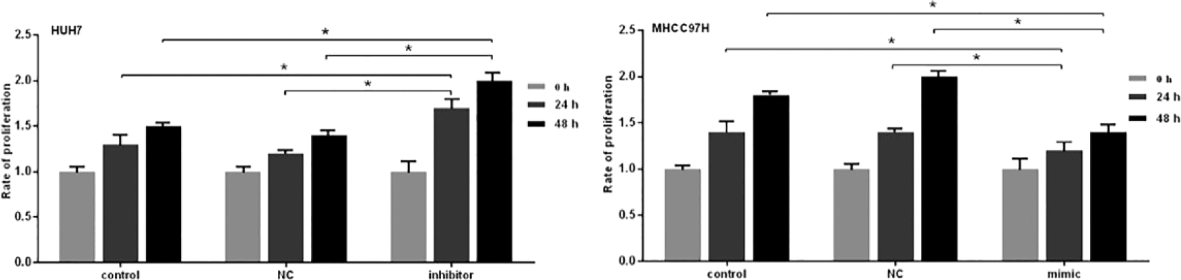
Let7b inhibits liver cancer cells’ proliferation. HUH7 and MHCC97H cells were transfected with, or without, Let7b NC, Let7b inhibitor, or Let7b mimic for 24 or 48 h. Their proliferation was determined by 0.4% trypan blue staining. Data are expressed as the mean rates ± SD of each group of cells from three separate experiments. *p < 0.05.
Let7b inhibits liver cancer cell invasion
Transwell assay results indicated that there was no significant difference in the invaded cells between the HUH7/control and HUH7/Let7b NC and MHCC97H/control and MHCC97H/Let7b NC. Besides, the numbers of invaded HUH7/Let7b inhibitor increased, and the numbers of invaded MHCC97H/Let7b mimic decreased, compared with the HUH7/Let7b NC and MHCC97H/Let7b NC (Figure 4).

Let7b inhibits the invasion of liver cancer cells. HUH7 and MHCC97H cells were transfected with, or without, Let7b NC, Let7b inhibitor, or Let7b mimic. After 24 or 48 h, the invasion of different groups was determined by transwell assay. Data are representative images and expressed as the mean ± SD of each group of cells from three separate experiments. *p < 0.05.
Let7b inhibits liver cancer cell migration
Wound healing assay results revealed that the migration of HUH7/Let7b inhibitor was significantly faster compared with HUH7/Let7b NC, while the migration of MHCC97H/Let7b mimic was significant slower than MHCC97H/Let7b NC (Figure 5).

Let7b inhibits the migration of liver cancer cells. HUH7 and MHCC97H cells were transfected with, or without, Let7b NC, Let7b inhibitor, or Let7b mimic. After 24 or 48 h, the wound healing of different groups of cells was tested. Data are representative images and expressed as the mean ± SD of each group of cells from three separate experiments. *p < 0.05.
Discussion
Total expression of Let7 family was decreased in a considerable proportion of primary liver cancer tissues and liver cancer cell lines, and also, the alterations in Let7 family members have been observed in lung and breast cancers else. 20 Let7 miRNA family plays important roles in both normal development and tumorigenesis. 21 Let7 can downregulate “stemness” by decreasing self-renewal and promoting differentiation in stem cells. 22 Among all human cancer–related miRNAs, Let7 family raised most interests since its family members have been noted to express aberrantly in human cancers.23–27
Members of the FZD family serve as receptors for the Wnt signaling glycoproteins, and the activation of the Wnt/FZD signaling plays crucial roles during development of most neoplasms, such as liver cancer. 28 Upregulation of some FZDs in cancer cell lines is associated with nuclear accumulation of β-catenin from the Wnt/β-catenin pathway which is frequently activated in tumors.
Our previous study found markedly low expression of Let7b in HUH7 and MHCC97H liver cancer cells. Because of the inhibitory effect of Let7b on Wnt signaling pathway by regulating FZD membrane receptor family during megakaryocyte development, 18 we assume that Let7b might also regulate FZD in liver cancer cells. Our study results have shown that transfection with Let7b mimic obviously reduced the FZD4, but not FZD5 and FZD7 in MHCC97 cell. Furthermore, enhanced Let7b overexpression significantly downregulated the relative content of nuclear to cytosol β-catenin and the expression of downstream gene c-myc. In contrast, inhibited Let7b expression upregulated the FZD4 expression and increased the β-catenin and c-myc in HUH7 cell. These evidence definitely showed that Let7b inhibited the FZD4 and the Wnt/β-catenin signaling in liver cancer cells. Thus, we speculated that Let7b inhibits the Wnt/β-catenin signaling by downregulating the upstream Wnt co-receptor and regulator expression in liver cancer cells. Enhanced Let7b expression significantly inhibited the proliferation, invasion, and migration of liver cancer, while Let7b inhibition significantly enhanced the proliferation, invasion, and migration. And, enhanced Let7b also inhibited the ratios of CD24+133+ CSCs in liver cancer cells. Similar results appear in sphere-forming assay. To sum up, we consider that Let7b inhibited Wnt/β-catenin signaling by downregulating the expression of FZD4 and then reduced the ratios of CSCs in liver cancer cells and repressed the proliferation, invasion, and migration of liver cancer cells. Moreover, overexpression of Let7b might be a new therapeutic option in treating liver cancers.
There are some shortcomings in this study. Because of the shortage of funds, we did not carry out in vitro experiments and gene sequencing. So, we have a lack of research on the specific molecular mechanisms. In the follow-up study, we will continue to further study its mechanism.
Footnotes
Acknowledgements
The authors thank the Central Laboratory and Biomedical transformation center of Gansu Provincial Hospital.
Declaration of conflicting interests
The author(s) declared no potential conflicts of interest with respect to the research, authorship, and/or publication of this article.
Funding
The author(s) disclosed receipt of the following financial support for the research, authorship, and/or publication of this article: This study was supported by the Longyuan Youth Innovative Talent Support Project, Gansu Province Natural Science Fund (145RJZA117), Gansu Province Health Industry Research Project (GSWSKY-2015-11), Postdoctoral Research Projects in Gansu Province (22097801), Gansu Province Health Industry Research Project Management Project(GWGL2013-47).