
Abstract
Most primarily cultured laryngeal squamous cell carcinoma cells are difficult to propagate in vitro and have a low survival rate. However, in our previous work to establish a laryngeal squamous cell carcinoma cell line, we found that laryngeal cancer-associated fibroblasts appeared to strongly inhibit the apoptosis of primarily cultured laryngeal squamous cell carcinoma cells in vitro. In this study, we investigated whether paired laryngeal cancer-associated fibroblasts alone can effectively support the growth of primarily cultured laryngeal squamous cell carcinoma cells in vitro. In all, 29 laryngeal squamous cell carcinoma specimens were collected and primarily cultured. The laryngeal squamous cell carcinoma cells were separated from cancer-associated fibroblasts by differential trypsinization and continuously subcultured. Morphological changes of the cultured laryngeal squamous cell carcinoma cells were observed. Immunocytofluorescence was used to authenticate the identity of the cancer-associated fibroblasts and laryngeal squamous cell carcinoma cells. Flow cytometry was used to quantify the proportion of apoptotic cells. Western blot was used to detect the protein levels of caspase-3. Enzyme-linked immunosorbent assay was used to detect the levels of chemokine (C-X-C motif) ligand 12, chemokine (C-X-C motif) ligand 7, hepatocyte growth factor, and fibroblast growth factor 1 in the supernatants of the laryngeal squamous cell carcinoma and control cells. AMD3100 (a chemokine (C-X-C motif) receptor 4 antagonist) and an anti–chemokine (C-X-C motif) ligand 7 antibody were used to block the tumor-supporting capacity of cancer-associated fibroblasts. Significant apoptotic changes were detected in the morphology of laryngeal squamous cell carcinoma cells detached from cancer-associated fibroblasts. The percentage of apoptotic laryngeal squamous cell carcinoma cells and the protein levels of caspase-3 increased gradually in subsequent subcultures. In contrast, no significant differences in the proliferation capacity of laryngeal squamous cell carcinoma cells cocultured with cancer-associated fibroblasts were detected during subculturing. High level of chemokine (C-X-C motif) ligand 12 was detected in the culture supernatant of cancer-associated fibroblasts. The tumor-supporting effect of cancer-associated fibroblasts was significantly inhibited by AMD3100. Our findings demonstrate that the paired laryngeal cancer-associated fibroblasts alone are sufficient to support the primary growth of laryngeal squamous cell carcinoma cells in vitro and that the chemokine (C-X-C motif) ligand 12/chemokine (C-X-C motif) receptor 4 axis is one of the major contributors.
Keywords
Introduction
Most of the established head and neck squamous cell carcinoma (HNSCC) cell lines propagate vigorously in vitro without demonstrating a decline in proliferation capacity during subsequent subculturing. However, each of these cancer cell lines has been successfully screened for via in vitro culturing. Establishing carcinoma cell lines has proven more challenging since most cultured cancer cells perish shortly after primary or secondary culture procedures when dissociated from stromal cells in the tumor microenvironment. Giard et al. 1 attempted to establish cell lines from 200 solid human tumors and succeeded in establishing 13 cell lines; the establishment rate of carcinoma cell lines was only approximately 6%. White et al. 2 generated 52 stable HNSCC cell lines from a series of 199 tumors collected between 1992 and 1997 at the University of Pittsburgh Medical Center; the overall rate of establishment was about 26%. Meanwhile, Kim et al. 3 attempted 79 cultures from 65 HNSCC patients and developed only nine cell lines, with an establishment rate of 11.3%.
The explanation of this phenomenon remains elusive, although we may speculate that some of the critical factors in the tumor microenvironment that support epithelial cancer growth are missing in primary and secondary in vitro cultures. The tumor microenvironment is composed of complex components, including cancer-associated fibroblasts (CAFs), tumor-associated macrophages, vascular endothelial cells, bone marrow–derived cells, and immunocytes, as well as various cell factors such as inflammatory mediators and chemokines.4–7 In addition, the acidic and hypoxic tumor microenvironment also seems to be important for cancer cell growth.8–10 However, it is still unknown which of the abovementioned factors is the most influential. Although CAFs secrete various cytokines, such as chemokine (C-X-C motif) ligand 12 (CXCL12), 11 chemokine (C-X-C motif) ligand 7 (CCL7), 12 hepatocyte growth factor (HGF), 13 and fibroblast growth factor 1 (FGF1), 14 and exert particular pro-tumorigenic influence on adjacent tumor cells in several solid tumors, the mechanism of tumor-supporting capacity of CAFs in laryngeal squamous cell carcinoma (LSCC) is unclear, especially in the primarily cultured LSCC cells.
In our previous work, we developed a permanent LSCC cell line, FD-LSC-1, from 40 laryngeal cancer specimens. 15 We found that CAFs in the tumor tissue survived more easily under in vitro conditions than LSCC cells. In addition, CAFs appeared to strongly inhibit the apoptosis of primarily cultured LSCC cells in vitro. In this study, we investigated whether laryngeal CAFs alone can effectively support the primary growth of LSCC cells by examining LSCC cells in isolation as well as their coculture with CAFs. Meanwhile, we detected several cytokines in the supernatants of LSCC cells and CAFs and assessed the tumor-supporting influence of CXCL12 and CCL7 on LSCC cells.
Materials and methods
Ethics statement
Tumor specimens were collected with the approval of the Ethics Committee of the Eye, Ear, Nose, and Throat Hospital, Fudan University, Shanghai, China. Signed informed consent was obtained from all individual participants included in the study.
Patient information
A total of 29 specimens from 29 male patients diagnosed with LSCC by biopsy and pathology were obtained immediately after surgical resection. Patient information, including sex, age, tumor site, tumor stage, and tumor differentiation, was recorded. Of these specimens, 25 were epiglottic LSCC, and 4 were glottic LSCC. Tumor staging was based on the classification guidelines, 6th edition, of the Union for International Cancer Control (UICC). From January 2015 to June 2015 and from September 2016 to November 2016, tumor specimens were collected and the study was conducted.
Primary culture of tumor specimens
The LSCC specimens obtained from fresh surgically resected tumors were immersed immediately in cold RPMI 1640 medium and then immersed in saline-diluted povidone-iodine solution (1:10, 5 min), gentamycin sulfate solution (1:8, 5 min), and lincomycin hydrochloride solution (1:8, 5 min). After being washed in phosphate-buffered saline (PBS), specimens were scissored into small fragments (1–2 mm3), followed by trypsinization in RPMI 1640 medium containing type IV collagenase (200 U/mL, Sigma, St. Louis, MO, USA) for at least 12 h at 37°C. The digested fragments and remaining cells were then rinsed in PBS and suspended in bronchial epithelial cell growth medium (BEGM; Catalog CC-3170; Lonza, Walkersville, MD, USA) supplemented with 1% penicillin/streptomycin and 10% fetal bovine serum (FBS; Gibco, Life Technologies Corporation, Grand Island, NY, USA). The suspension was seeded in 6-cm petri dishes (Corning Inc., Corning, NY, USA) in an incubator humidified with 5% CO2 at 37°C. After 3–7 days of incubation, the dishes were removed from the incubator and examined under a phase-contrast microscope. The contaminated dishes were discarded. The LSCC cells and CAFs that grew from the fragments of the 20 specimens were continuously subcultured; subsequent assays were performed unless cultures were contaminated or cells perished. Data were collected from the two specimens (collected from January 2015 to June 2015) with LSCC cells that continued proliferating after continuous subculturing until passage 4 except that the data of enzyme-linked immunosorbent assay (ELISA) assay and AMD3100 inhibition assays were collected from the 9 specimens collected from September 2016 to November 2016.
Secondary culture and morphological examination of LSCC cells and CAFs
CAFs were separated from LSCC cells by brief exposure to 0.25% trypsin-EDTA (Invitrogen, Grand Island, NY, USA); both CAFs and LSCC cells were collected for subculture. LSCC cells detached from CAFs as well as CAFs in isolation were continuously subcultured for 3–4 passages in order to evaluate any morphological changes. LSCC cells consistently cocultured with CAFs also served as controls. Representative microscope images were recorded for each passage.
Immunocytochemical staining of LSCC cells and CAFs
Immunocytochemistry was used to verify the identities of LSCC cells and CAFs. The LSCC cells and the CAFs suspended in BEGM were plated on glass coverslips and incubated at 37°C for 24 h. Cells were fixed on the coverslips with 4% paraformaldehyde (PFA) for 15 min. The coverslips were then washed in PBS and incubated in 10% normal goat serum (Boster, Wuhan, China) in either the presence or absence of 0.3% Triton X-100 to permeabilize the cells for 40 min at room temperature and to block nonspecific interactions. The coverslips were then immersed in rabbit anti-human primary antibodies of pan-cytokeratin (CK; 1:400; Abcam, Cambridge, UK), vimentin (1:200; Abcam), α-smooth muscle actin (α-SMA; 1:200; Abcam), and fibroblast activation protein (FAP; 1:250; Abcam) at 4°C overnight. Followed by incubation in the dark for 1 h at 37°C in secondary antibodies (Jackson ImmunoResearch, West Grove, PA, USA) of fluorescein isothiocyanate (FITC)-conjugated goat anti-rabbit IgG (H + L; 1:100) or CyTM3-conjugated goat anti-rabbit IgG (1:100). To stain the nuclei, 4′,6-diamidino-2-phenylindole (DAPI; Boster) was used.
Flow cytometry for the quantification of apoptotic cells
The apoptotic cells were detected by Annexin V-FITC/propidium iodide (PI) double staining using an Annexin V-FITC apoptosis detection kit (Beyotime Biotechnology, Nantong, China) according to the manufacturer’s instructions. The LSCC cells of passage 1, passage 2, and passage 3 were submitted to apoptosis detection. LSCC cells continuously cocultured with CAFs were used as controls. In brief, the culture medium (containing floating apoptotic cells) of each 6-cm dish was collected and transferred to a corresponding labeled tube. The cells attached to the dishes were washed with PBS, which was also transferred to the corresponding tube. The cells were then harvested by trypsinization, washed with PBS, and resuspended in PBS. The cell density of each tube was calculated. Approximately 5 to 10 × 104 cells suspended in PBS were centrifuged. At following, 195 µL Annexin V-FITC Binding Buffer, 5 µL Annexin V-FITC, and 10 µL PI were added. After gentle mixture, the cells were incubated for 15 min in the dark and mixed at intervals before being analyzed by the flow cytometer.
Western blot for the detection of caspase-3
The total proteins from the lysates of the LSCC cells and the controls of LSCC cells cocultured with CAFs were analyzed with electrophoresis using sodium dodecyl sulfate-polyacrylamide gel (Beyotime Biotechnology). The samples were then electrotransferred to a polyvinylidene fluoride membrane (Millipore, Billerica, MA, USA) and incubated overnight in the primary antibody of caspase-3 (1:5000; Abcam) with gentle shaking, followed by incubation in a secondary antibody of horseradish peroxidase–conjugated goat anti-rabbit IgG (H + L; 1:2000; Jackson ImmunoResearch). A BeyoECL Plus Kit (Beyotime) was used for signal detection.
Enzyme-linked immunosorbent (ELISA) assay
The purified LSCC cells, CAFs, and the controlled LSCC cells cocultured with CAFs at passage 1 from newly collected 9 specimens were seeded in six-well plates at a density of 4 × 10⁵ per well in BEGM medium supplemented with 10% FBS. After 12 h of incubation at 37°C, the media was changed to 2 mL of serum-free BEGM, followed by incubation for 48 h. Then, the supernatants of each well were collected and centrifugated (2000g, 5 min) to remove particulates prior to detection of CXCL12, CCL7, HGF, and FGF1 using ELISA kits (R&D Systems, Minneapolis, MN, USA) according to the manufacturers’ protocol. The assay was conducted in triplicate. The absorbance was recorded at a wavelength of 450 nm using an ELISA microplate reader.
Proliferation assay after AMD3100 and anti-CCL7 inhibition
The influence of AMD3100 and anti-CCL7 on the tumor-supporting capacity of CAFs was measured by proliferation assay. The LSCC cells were cocultured with the CAFs in BEGM medium supplemented with 10% FBS at 37°C for 24 h, and then, the supernatants were collected as conditioned medium (designated as CM). The purified LSCC cells of passage 1 were seeded in 96-well plates in 0.2 mL of CM at a density of 5 × 10³ per well in the presence of AMD3100 (Sigma, a chemokine (C-X-C motif) receptor 4 (CXCR4) antagonist, final concentration of 10 µg/mL) or anti-CCL7 neutralizing antibody (Sigma, final concentration of 2 µg/ml). The LSCC cells cultured in normal BEGM supernatants from the LSCC cells (24 h after seeding) and LSCC cells cultured in CM without AMD3100 or anti-CCL7 antibody served as controls. Wells that contain CM only served as blank controls. Each group was repeated in six wells. Then, the cells were cultured at 37°C for 72 h. Cell growth was analyzed at 24, 48, and 72 h using the Cell Counting Kit-8 (CCK-8; Dojindo Laboratories, Kumamoto, Japan) 3 h before performing spectrophotometric reading according to the manufacturer’ s directions. The assay was conducted in triplicate. Ultraviolet absorbance was measured at 450 nm with an ELISA plate reader.
Apoptosis assay after AMD3100 inhibition
The influence of AMD3100 on the tumor-supporting capacity of CAFs was also measured by apoptosis assay. Briefly, the purified LSCC cells of passage 2 were seeded in six-well plates in CM mentioned in the proliferation assay at a density of 2 × 10⁵ per well in the presence or absence of AMD3100 (Sigma) at the final concentration of 10 µg/mL. The LSCC cells cultured alone in normal BEGM and LSCC cells cocultured with CAFs in BEGM served as controls. The cells were cultured at 37°C for 72 h. Then, the cells in each well were harvested for apoptosis detection as mentioned above.
Statistical analysis
Data were presented as the mean ± standard deviation (SD) of three independent assays. The GraphPad Prism software version 6.00 for Windows (GraphPad Software, San Diego, CA, USA) was used to analyze the data. Statistical analysis was performed by Student’s t-test or Fisher’s exact test. Statistical significance was acceptable to a level of p < 0.05.
Results
Primary and secondary culture of LSCC cells and CAFs
Of the 29 cultured specimens, 4 were contaminated by bacteria or fungus during the primary culture. Although LSCC cells successfully grew from the remaining 25 specimens (3 from glottis and 22 from epiglottis), they stopped propagating and perished within passage 4 when dissociated from the coexisting CAFs. Among the 25 specimens, 6 stopped propagating in passage 1, 10 in passage 2, 6 in passage 3, and 3 in passage 4 (Table 1). The clinical characteristics (tumor site, stage, and differentiation) of LSCC cells were not significantly related to their in vitro proliferation capacity according to statistical analysis (Table 2, Figure 1). In addition, the CAFs proliferated vigorously in primary and secondary cultures even when detached from LSCC cells and did not show any apoptotic signs within passage 10.
Clinical characteristics and results of 25 continuous cultures of uncontaminated LSCC samples.

LSCC: laryngeal squamous cell carcinoma; W/D: well-differentiated; M/D: moderately differentiated; P/D: poorly differentiated.
Analysis of clinical characteristics in relationship to proliferation capacity of LSCC cells by Fisher’s exact test.

LSCC: laryngeal squamous cell carcinoma; W/D: well-differentiated; M/D: moderately differentiated.

Statistical analysis of clinical characteristics in relationship to proliferation capacity of LSCC cells. No significant correlations were found between the (a) tumor site, (b) stage, and (c) level of differentiation and the in vitro proliferation capacity of LSCC cells (n.s.p > 0.05).
Pathology of patients and authentication of LSCC cells and CAFs by immunocytofluorescence
The pathological characteristics of the two patients showed typical squamous cell carcinoma of the larynx (Figure 2(a) and (b)). The LSCC cells showed positive staining of pan-CK and negative staining of vimentin (Figure 2(c) and (d)), validating their epithelial origin. In contrast, the CAFs showed positive staining of vimentin and negative staining of pan-CK (Figure 2(e) and (f)), indicating their identity as fibroblasts. In addition, the CAFs showed positive staining of FAP and α-SMA, two markers found in activated fibroblasts, further verifying their identity as CAFs (Figure 2(g) and (h)).

Pathology of patients and authentication of LSCC and CAFs using immunocytochemistry. (a and b) Hematoxylin and eosin staining of the two patients. Both were typical squamous cell carcinoma and keratin pearls (white arrows) were found in patient 1. (c and d) The LSCC cells showed positive staining of pan-CK and negative staining of vimentin. (e–h) The CAFs showed negative staining of pan-CK and positive staining of vimentin, α-SMA, and FAP.
Morphological changes of subcultured LSCC cells and CAFs
After 3–7 days of incubation, LSCC cells grew from the trypsinized fragments and were surrounded by coexisting CAFs. The well-attached LSCC cells grew as a monolayer in a cobblestone pattern and tended to cluster together, exhibiting typical epithelial appearance. The CAFs also grew as a monolayer and exhibited a long spindle shape with several projections on the cell surface (Figure 3(a)–(e)). The LSCC cells dissociated from the CAFs showed an increasingly slow response to trypsin during trypsinization than the LSCC cells cocultured continuously with CAFs. Interestingly, dissociated LSCC cells exhibited obvious morphological changes from passage 1, including an increasingly enlarged and flattened outline and an increasing number of apoptotic cells floating in the medium due to their decreased capacity to attach to the flask (Figure 3(e)–(h)). In addition, huge vacuoles were observed in the cytoplasm of the apoptotic cells (Figure 3(h)). In contrast, both the CAFs and the LSCC cells consistently in the coculture system did not show significant morphological changes before passage 4 (Figure 3(a)–(d)). Furthermore, the CAFs detached from LSCC cells did not show significant morphological changes before passage 10 (Figure 3(i)–(l)).
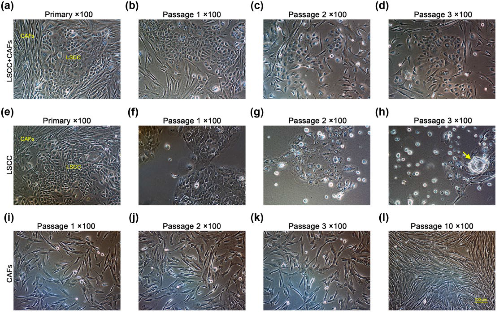
Morphological changes of LSCC and CAFs in different in vitro culture systems. (a–d) When consistently cocultured, neither CAFs nor LSCC cells showed significant morphological changes in the primary culture, passage 1, passage 2, or passage 3. (e–h) In contrast, LSCC cells detached from CAFs showed significant morphological changes in the primary culture, passage 1, passage 2, and passage 3, including increasingly flattened outline, a decreasing capacity to attach to the flask, and an increasing number of apoptotic cells floating in the medium. Notably, apoptotic cells with huge cytoplasmic vacuoles were observed (yellow arrow). (i–l) When CAFs were separated from LSCC cells, obvious morphological changes were not observable at passage 1, passage 2, passage 3, or passage 10.
Flow cytometric quantification of apoptosis
Once LSCC cells were detached from CAFs and subcultured alone, and they perished shortly after in the culture medium. An increasingly upregulated fraction of apoptotic cells was detected in passage 1, where the total proportion of apoptotic cells was 7.187% ± 1.853%, followed by a proportion of 61.967% ± 3.702%, and 78.343% ± 3.850% in passage 2 and passage 3, respectively (*p < 0.05; Figure 4(g)–(l)). In contrast, the LSCC cells continuously cocultured with CAFs did not show a large extent of apoptosis in the primary culture nor up to passage 3, in which the proportion of apoptotic cells was 2.417% ± 0.040%, 2.347% ± 0.080%, 2.393% ± 0.240%, and 2.577% ± 0.136%, respectively (n.s.p < 0.05; Figure 4(b)–(e), (k) and (l)).

Changes in apoptotic LSCC cells quantified by flow cytometry in two different in vitro culture systems. (a and f) Blank controls. (b–e) When LSCC cells were cocultured with CAFs, the flow cytometry detected consistently few apoptotic cells in the primary culture and in passage 1, passage 2, and passage 3. (g–j) In contrast, the LSCC cells separated from CAFs showed an increasingly upregulated proportion of apoptotic cells in the primary culture, passage 1, passage 2, and passage 3. (k and l) The statistical analyses of apoptotic cells in two different culture systems are shown (n.s.p > 0.05, *p < 0.05).
Elevated expression of caspase-3 protein
The apoptosis of LSCC cells was also detected by the Western blot analysis during in vitro culturing. Once detached from CAFs, the LSCC cells showed an increasingly elevated expression of caspase-3 protein beginning in passage 1, with a relative protein expression (LSCC/LSCC + CAFs) of 2.488 ± 1.102, followed by 5.068 ± 1.216 and 9.675 ± 4.285 in passage 2 and passage 3, respectively. In contrast, a consistently low expression of caspase-3 protein was detected in the LSCC cells cocultured with CAFs in the primary culture and up to passage 3 (n.s.p > 0.05, *p < 0.05; Figure 5).

Changes in caspase-3 detected by the western blot in two different in vitro culture systems. (a) The LSCC cells cocultured with CAFs showed consistently low expression of caspase-3 in the primary culture, passage 1, passage 2, and passage 3. (b) In contrast, LSCC cells separated from CAFs showed increasingly enhanced expression of caspase-3 from the primary culture through passage 3. (c and d) The statistical analyses of relative protein expression of LSCC cells in two different culture systems are shown (n.s.p > 0.05, *p < 0.05).
Quantification of CXCL12, CCL7, FGF1, and HGF by ELISA
Using ELISA, we measured secreted levels of CXCL12, CCL7, HGF, and FGF1 in the supernatants from purified LSCC cells, CAFs, and the LSCC cells cocultured with CAFs. The protein levels of CXCL12, CCL 7, HGF, and FGF1 secreted by purified LSCC cells were undetectable. By contrast, The CAFs secreted high level of CXCL12 in the culture supernatants. Elevated protein levels of CCL7, HGF, and FGF1 were also detected in the supernatants of CAFs. In addition, the protein levels of the four cytokines secreted by CAFs were increased significantly by co-culturing with LSCC cells (*p < 0.05, **p < 0.01) (Figure 6(a) and (b)).

ELISA, proliferation, and apoptosis assay. (a) Quantification of CXCL12, CCL7, HGF, and FGF1 in the supernatants of LSCC cells, CAFs, and CAFs cocultured with LSCC cells by ELISA. Significantly high level of CXCL12 was detected in the culture supernatant of CAFs. Relatively lower levels of CCL7, HGF, and FGF1 were also detected in the supernatants of CAFs. By contrast, the LSCC cells showed undetectable (u.d.) levels of these four cytokines. (b) Coculture with LSCC cells significantly promoted the production of these four cytokines by CAFs. (c) Tumor-supporting capacity of CXCL12 and CCL7 evaluated by CCK-8 assay. The AMD3100 significantly inhibited the tumor-supporting influence of the CM on the LSCC cells. By contrast, the inhibition effect of anti-CCL7 was not significant at all time points. (d and e) Apoptosis-inhibiting capacity of CXCL12 evaluated by flow cytometer. The LSCC cells cultured in normal BEGM and conditioned medium (CM) (in the presence or absence of AMD3100) or cocultured with CAFs in normal BEGM showed different percentages of apoptotic cells (n.s.p > 0.05, *p < 0.05, and **p < 0.01).
Influence of CXCL12 and CCL7 on the tumor-supporting potential of CAFs
Based on the ELISA assay, we investigated the tumor-supporting capacity of CXCL12 and CCL7 individually. At the time point of 24 h, the growth of LSCC cells cultured in CM was significantly inhibited by AMD3100 (*p < 0.05), and the inhibition effect increased further at the time point of 48 and 72 h (**p < 0.01). By contrast, the inhibition effect of anti-CCL7 on the proliferation of LSCC cells was not significant at the time points of 0, 24, 48, and 72 h (n.s.p > 0.05). Interestingly, the LSCC cells, cultured in the CM and inhibited by AMD3100, proliferated still faster than the LSCC cells cultured in BEGM at the time points of 48 and 72 h (*p < 0.05), indicating that some other cytokines in the CM can also support the growth of the LSCC cells in vitro except the dominant tumor-supporting effect of ADM3100 (Figure 6(c)).
Influence of AMD3100 on the apoptosis-inhibiting capacity of CAFs
Since the CXCL12/CXCR4 axis exerts a critical influence on the proliferation of LSCC cells, we next assessed the influence of AMD3100 on the apoptosis-inhibiting capacity of CAFs. The LSCC cells cocultured with the CAFs showed the lowest percentage of apoptotic cells (2.58% ± 0.93%), and the LSCC cells cultured in BEGM showed the highest percentage of apoptotic cells (61.80% ± 3.76%). The LSCC cells cultured in CM in the presence of AMD3100 showed significantly higher percentage of apoptotic cells (40.95% ± 4.89%) than the LSCC cells cultured in CM (11.49% ± 2.12%; **p < 0.01), indicating that the CXCL12/CXCR4 pathway exerted a crucial apoptosis-inhibiting influence on the LSCC cells. The LSCC cells cultured in CM in the presence of AMD3100 showed significantly lower percentage of apoptotic cells than the LSCC cells cultured in BEGM (*p < 0.05), indicating that some other cytokines in the CM may inhibit the apoptosis of the LSCC cells except CXCL12. Likewise, the LSCC cells cultured in CM showed higher percentage of apoptotic cells than the LSCC cells cocultured with the CAFs (*p < 0.05), indicating that the cocultured CAFs harbored higher apoptosis-inhibiting capacity than the CM (Figure 6(d) and (e)).
Discussion
CAFs are some of the most crucial mesenchymal cells in the tumor microenvironment. These cells secrete ample soluble paracrine and autocrine growth factors to autoregulate and to regulate extracellular stroma, adjacent non-neoplastic cells, and cancer cells. It is becoming increasingly clear that the mutual interaction between CAFs and cancer cells plays a critical role in the tumorigenesis, progression, and metastasis of cancer.16–19 It was recently documented that CAFs from several solid tumors harbored the ability to partially promote the proliferation, invasion, and metastasis of corresponding tumor cells.20–23 All of these studies were performed using cell lines established from solid tumors, and these cell lines clearly harbored vigorous capacities for proliferation and invasion in vitro. However, it is not clear whether CAFs are able to promote the primary growth of cancer cells in solid tumor specimens nor to what extent.
In our previous work, we successfully separated CAFs from primarily cultured human LSCC specimens and mouse xenografted laryngeal tumor model. We found that laryngeal CAFs were able to partially enhance the tumor-promoting capacity of the laryngeal cancer cell line.24,25 In addition, in our previous work to establish a LSCC cell line, we found that the coexisting CAFs in the LSCC specimens appeared to have the capacity to strongly hamper the apoptosis of LSCC cells in primary and secondary cultures. 15 Furthermore, the establishment of cancer cell lines is challenging because most cancer cells are unable to adapt to in vitro culture systems and perish shortly after their dissociation from other stromal cells in the tumor microenvironment; the exact mechanism behind this phenomenon remains to be clarified.1–3 Given this context, we were eager to determine whether laryngeal CAFs alone could effectively support the primary growth of LSCC cells. To examine this phenomenon, we detached LSCC cells from coexisting CAFs and compared these cultures with those in which LSCC cells were continuously cocultured with CAFs.
In all, 29 specimens from 29 LSCC patients were analyzed in this study. Despite our efforts in specimen selection, collection, preservation, and sterilization, 4 of the 29 specimens were contaminated with bacteria or fungus during the primary or secondary culture. We performed subsequent assays for the 25 uncontaminated samples. Of the specimens, 22 were derived from the supraglottis area (epiglottis), among which 3 specimens contained LSCC cells that stopped propagating at passage 4 (3/22). Three specimens were derived from the glottis, among which no specimen contained LSCC cells that stopped propagating at passage 4 (0/3) (Table 1). The study of histoembryology shows that the glottis, epiglottis, and subglottis are derived from different primordiums; therefore, the malignancy of tumors from different sites was also different. The epiglottis derives from the caudal half of the hypobranchial eminence, a derivative of branchial arches III and IV.26,27 Supraglottic cancers have a high propensity to disseminate into regional lymphatics compared with glottic and subglottic cancer. 28 Therefore, tumor cells from the supraglottic area and from relatively late stages of cancer (III/IV) appeared to be more likely to be subcultured up to passage 4 compared with those from the glottic area and relatively early stages (I/II). However, according to the statistical analysis, we found no significant correlation between the tumor site, tumor stage, and the in vitro proliferation capacity of LSCC cells. Similarly, no significant differences in proliferation capacity were found between moderately differentiated and well-differentiated LSCC cells (n.s.p > 0.05; Table 2, Figure 1).
We verified the identity of LSCC cells and CAFs by immunocytochemistry. LSCC cells were positively stained with pan-CK and negatively stained with vimentin (Figure 2(c) and (d)), indicating their epithelial origin. CAFs were positively stained with vimentin and negatively stained with pan-CK (Figure 2(e) and (f)), indicating their identity as fibroblasts. Positive staining was also shown for two CAFs markers, FAP and α-SMA, further authenticating the identity of CAFs (Figure 2(g) and (h)).
The morphology of cells reflects the state and survival of cells under in vitro conditions; a decreased proliferation capacity would be easily observed in the corresponding morphological changes. In this study, when LSCC cells were cocultured with CAFs, both LSCC cells and CAFs showed a vigorous, three-dimensional appearance with bright outlines, indicating the reflection of light. The LSCC cells formed a cobblestone pattern, and the CAFs exhibited long spindle shapes (Figure 3(a)–(e)). However, when detached from CAFs, the LSCC cells exhibited interesting morphological changes during initial subculturing, starting in passage 1. These changes included increasingly enlarged and flattened outlines, increasing number of apoptotic cells floating in the medium, and the presence of huge vacuoles in the cytoplasm of apoptotic cells (Figure 3(e)–(h)). In contrast, CAFs detached from LSCC cells did not show significant morphological changes before passage 10 (Figure 3(i)–(l)). These findings demonstrated that the presence of CAFs in the coculture system is sufficient for supporting the primary growth of LSCCs, yet coexistence with LSCCs was not necessary for the primary survival of CAFs.
We then used an apoptosis detection kit and flow cytometry to further quantify the apoptotic extent of LSCC cells. We found that LSCC cells cocultured with the CAFs showed consistently lower proportions of apoptotic cells in the primary culture and through passage 3 or 2.417% ± 0.040%, 2.347% ± 0.080%, 2.393% ± 0.240%, and 2.577% ± 0.136% in the primary and successive subcultures, respectively (n.s.p < 0.05; Figure 4(b)–(e), (k) and (l)). However, when dissociated from CAFs, LSCC cells showed increasingly elevated fractions of apoptotic cells starting in passage 1 (*p < 0.05; Figure 4(g)–(l)). These results also revealed that coexisting CAFs strongly inhibit the apoptosis of LSCC cells.
Caspases are crucial mediators involved in the initiation and execution of programmed cell death. In particular, caspase-3 is a frequently activated death protease responsible for the specific cleavage of many key cellular proteins. The pro-form of caspase-3 exists as an inactive 32-kDa proenzyme. It is activated by proteolytic cleavage into a 17-kDa enzyme capable of disassembling cells once apoptosis is initiated.29,30 In this study, we used the 17-kDa active antibody to detect changes in the caspase-3 protein by western blot analysis. Similar to the results of flow cytometry, we found that LSCC cells showed consistently low expression of caspase-3 when cocultured with CAFs in the primary culture and up to passage 3 (n.s.p > 0.05, *p < 0.05). Interestingly, when dissociated from CAFs, LSCC cells showed increasingly upregulated expression of caspase-3 beginning in passage 1 (Figure 5). These results further strengthened the hypothesis that coexistence with CAFs alone is sufficient for supporting the primary growth of LSCC cells.
According to the present reports, CAFs may exert the tumor-promoting effect on the tumor cells through paracrine-mediated CAF–carcinoma cell interaction, CAF–endothelial cell interactions, CAF-modulated extracellular matrix remodeling, CAF-induced inflammation and immune escape, and CAF-mediated anti-immune responses. 19 However, in our study, the CAFs most likely exert the tumor-supporting effect through the paracrine effect because CAFs have been shown to be a source of various cytokines in the tumor microenvironment. Some CAF-derived cytokines have been reported to exert particular paracrine effects via interacting with their receptors expressed in or on adjacent tumor cells, thus promoting tumor progression. CCL12, 11 CCL7, 12 HGF, 13 and FGF1 14 are reported cytokines to harbor such tumor-promoting capacity in solid tumors. Therefore, we detected the four cytokines in the supernatants from purified LSCC cells, CAFs, and the LSCC cells cocultured with CAFs by ELISA. We found that the protein levels of CXCL12, CCL 7, HGF, and FGF1 secreted by purified LSCC cells were undetectable. By contrast, the CAFs secreted high level of CXCL12 in the culture supernatants, followed by CCL7. Elevated protein levels of HGF and FGF1 were also detected. In addition, the protein levels of the four cytokines secreted by CAFs were increased significantly by co-culturing with LSCC cells (*p < 0.05, **p < 0.01; Figure 6(a) and (b)), indicating that the reciprocal interaction between CAFs and the LSCC cells enhanced the paracrine function of CAFs.
We next evaluated the tumor-supporting capacity of CXCL12 and CCL 7 using AMD3100 (a CXCR4 antagonist) and anti-CCL7 neutralizing antibody. We found that AMD3100 could inhibit the proliferation and promote the apoptosis of LSCC cells cultured in the CAF-derived CM significantly. The influence of CCL7 on the LSCC cells was not significant (Figure 6(c)–(e)). These findings indicated that the CXCL12/CXCR4 axis exerted a crucial influence on the survival of primarily cultured LSCC cells, consistent with the previous reports. 31 However, in the proliferation assay, the LSCC cells cultured in the CM and inhibited by AMD3100 proliferated still faster than the LSCC cells cultured in BEGM at the time points of 48 and 72 h (*p < 0.05). Likewise, in the apoptosis assay, the LSCC cells cultured in the CM and inhibited by AMD3100 showed lower percentage of apoptotic cells than the LSCC cells cultured in BEGM; the LSCC cells cultured in CM showed higher percentage of apoptotic cells than the LSCC cells cocultured with the CAFs (*p < 0.05). These observations revealed that some other cytokines except CCL12 in the CM can also support the growth of the LSCC cells to some extent and that the cocultured CAFs harbored higher apoptosis-inhibiting capacity than the CM alone. Some other mechanisms, such as the direct physical contact between the CAFs and the tumor cells 32 and change in cytokine production by tumor–stromal interaction, 33 are needed to be further explored.
In conclusion, for the first time, we proposed the hypothesis that CAFs in LSCC tumor specimens may strongly support the primary growth of LSCC cells, which is based on our previous work. In this study, we validated our hypothesis by obtaining LSCC specimens and performing a primary culture. We found that LSCC cells cocultured with CAFs hardly underwent apoptosis, while LSCC cells separated from CAFs perished soon afterwards. Our findings firmly verified that the CAFs coexisting with LSCC cells in tumor specimens effectively inhibited the apoptosis of LSCC cells when primarily cultured in vitro. The cytokine of CXCL12 exerts a critical influence on the tumor-supporting capacity of CAFs. However, some other factors seem to also exert the tumor-supporting influence on the LSCC cells to some extent, which needs to be explored in future investigation.
Footnotes
Acknowledgements
The authors thank the editors Xiu-Feng Zhang and Ling Shen from the Fudan University Journal of Medical Sciences for their great help in preparing this manuscript. M.W. and C.W. contributed equally to this work.
Declaration of conflicting interests
The author(s) declared no potential conflicts of interest with respect to the research, authorship, and/or publication of this article.
Funding
The author(s) disclosed receipt of the following financial support for the research, authorship, and/or publication of this article: This study was partly funded by the Shanghai Science and Technology Foundation of China (15ZR1406000).