
Abstract
Circular RNAs are new type of endogenous RNAs, which play an important role in the regulation of gene expression and indicate a great capacity in clinical diagnosis and treatments of diseases. However, the role of circular RNAs in gastric cancer remains unknown. In this study, we chose hsa_circ_0006633 as the target circular RNA and measured its levels in human gastric cancer tissues, plasma, and gastric cell lines by real-time quantitative reverse transcription polymerase chain reaction. Hsa_circ_0006633 levels at multiple stages of gastric tumorigenesis were then explored, and its relationships with clinicopathological features were analyzed as well. We found that the expression levels of hsa_circ_0006633 in four gastric cancer cell lines, HGC-27, SGC-7901, MGC-803, and AGS, were downregulated than those in normal gastric mucosal epithelial cell line GES-1. Then, we further detected that it was downregulated in 79.2% (76/96) gastric cancer tissues compared with the adjacent non-tumorous tissues. The lower expression of hsa_circ_0006633 was associated with cancer distal metastasis (p = 0.037) and tissue carcinoembryonic antigen level (p = 0.041). In addition, hsa_circ_0006633 expression was significantly decreased in gastritis and dysplasia tissues comparing with the healthy control. Moreover, plasma hsa_circ_0006633 levels were significantly increased in gastric cancer compared with healthy control. Our data imply that hsa_circ_0006633 may play an important role in gastric carcinogenesis and is also a potential biomarker for screening gastric cancer.
Introduction
Gastric cancer is one of the most common malignant tumors worldwide and the second leading cause of global cancer deaths in the world, especially in Eastern Asian countries.1,2 As gastric cancer often remains symptomless, it may be detected in late stage, and the 5-year survival rate remains 4%–27% from developing to developed countries, respectively. 3 While the 5-year survival rate can be more than 90% after the medical measures if detected in the early stage of gastric cancer. 4 Improved outcomes can be achieved with the early recognition and aggressive managements if detailed molecular characterization of tumors can be discovered. 5 Therefore, it is necessary to improve the diagnosis, preferably non-invasive and cost-effective methods.
Nowadays, with the development of RNA sequencing (RNA-seq) and bioinformatic analysis, circular RNA (circRNA) has become a hot spot in the RNA field as a novel kind of RNA. 6 Some characteristics such as diversity, abundance, and conservation indicate that circRNAs have biological functions. 7 With their dynamic and cell-type-specific expression patterns during development, a variety of functions have been proposed for circRNAs.6,7 CircRNAs also play specific roles in a wide variety of cancers and in regulating the biological behavior of cancers, providing a novel potential perspective in cancer treatment. 8
Based on our previous microarray screening results of the differential expression of circRNAs in gastric cancer tissues comparing with adjacent non-tumorous tissues (GEO No. GSE89143; https://www.ncbi.nlm.nih.gov/geo/query/acc.cgi?acc=GSE89143), here we choose hsa_circ_0006633 as the target in the study. Among the dysregulated circRNAs, hsa_circ_0006633 is one of the most downregulated circRNAs in gastric cancer tissues. Its gene is located at chromosome 1: 59805629-59844509, and the spliced sequence length is 353 nt. Our data here showed that hsa_circ_0006633 may play an important role in the tumorigenesis of gastric cancer.
Materials and methods
Specimens and clinical data collection
All 338 samples were collected from the Affiliated Hospital of Ningbo University School of Medicine, China, from November 2012 to June 2016. The 96 gastric cancer tissues and their adjacent non-tumorous tissues 5 cm from the edge of the tumor were obtained from surgical operations. Another 35 human healthy gastric mucosa, 51 gastritis mucosa, and 20 gastric dysplasia tissues were gained from biopsy specimens. All tissues were preserved in RNA-fixer Reagent (Bioteke, Beijing, China) at −80°C until use. Plasma samples were obtained from 20 healthy volunteers and 20 gastric cancer patients and stored at −80°C until RNA extraction. No patient or healthy volunteer received local or systemic treatment before the upper gastrointestinal endoscopy examination or surgical excision. The final diagnosis was histopathologically confirmed. Tumors were staged according to the tumor–node–metastasis (TNM) stage system of the international Union Against Cancer (7th edition). Histological grade was assessed following the National Comprehensive Cancer Network (NCCN) clinical practice guideline of oncology (V.1, 2011). The experimental operators were blind to the clinical data, which were collected by doctors. The study was approved by the Human Research Ethics Committee of Ningbo University, and all patients agreed to participate in the study and written informed consent was obtained from all subjects.
Cell cultures
Human gastric epithelial cell line GES-1 and four gastric cancer cell lines, HGC-27, SGC-7901, MGC-803, and AGS, were obtained from Shanghai Institute of Biochemistry and Cell Biology, Chinese Academy of Sciences (Shanghai, China). Cells were cultured with RPMI 1640 Medium (Invitrogen, Grand Island, NY, USA) consists of 50 U/mL penicillin, 10% fetal bovine serum, and 50 U/mL streptomycin in culture flasks at 37°C in a humidified atmosphere of 5% CO2 as previous report. 9
Total RNA preparation and qRT-PCR detection
We first extracted total RNA from specimens by TRIzol reagent (Ambion, Carlsbad, CA, USA) and then reverse transcribed it into cDNA by GoScript Reverse Transcription (RT) System (Promega, Madison, WI, USA) following the manufacturer’s instructions. Real-time quantitative reverse transcription polymerase chain reaction (qRT-PCR) was achieved using GoTaq qPCR Master Mix (Promega) on an Mx3005P real-time PCR System (Stratagene, La Jolla, CA, USA). Divergent primers rather than the more commonly used convergent primers of hsa_circ_0006633 were designed. 10 Primers for hsa_circ_0006633 and glyceraldehyde 3-phosphate dehydrogenase (GAPDH) were synthesized by Sangon Biotech (Shanghai, China). The sequences of the PCR primers for GAPDH and hsa_circ_0006633 were as follows: 5′-TCGACAGTCAGCCGCATCTTCTTT-3′ and 5′-ACCAAATCCGTTGACTCCGACCTT-3′ for GAPDH; 5′-CTCCCGGACTTCTTATCGTGG-3′ and 5′-CGATGGTCCAGCCACATGAT-3′ for hsa_circ_0006633. The data were analyzed through the ΔCt method, 11 and all the results were expressed as mean ± standard deviation (SD) of three independent experiments. Higher ΔCt means lower expression of hsa_circ_0006633. The conditions of thermal cycling were as follows: 95°C at 5 min for a hot-start, then 40 cycles at 94°C for 15 s, 56°C for 30 s, and 72°C for 30 s. The cycle threshold (Ct) values were recorded for both hsa_circ_0006633 and GAPDH, which is used as a control.
Immunohistochemical analysis of tissue carcinoembryonic antigen and CA19-9
We incubated the paraffin tissue sections in primary anti-carcinoembryonic antigen (CEA) or anti-carbohydrate antigen 19-9 (CA19-9) (Dako, Glostrup, Denmark) for 1 h at room temperature, and then, the tissues were incubated in diaminobenzidine (DAB; Dako) for color development after incubation with broad-spectrum second antibody K5007 (Dako). 12 The standard for determination of the results was according to the 2010 American Society of Clinical Oncology (ASCO)/the College of American Pathologists (CAP) guideline. 12
Statistical analysis
All statistical analyses were performed by Statistical Program for Social Sciences (SPSS) 20.0 software (SPSS, Chicago, IL, USA), GraphPad Prism 5.0 (GraphPad Software, La Jolla, CA, USA), and SigmaPlot 10.0 (SigmaPlot Software, La Jolla, CA, USA). We used Student’s t test, one-way analysis of variance (ANOVA) test, and rank-sum test as appropriate. Statistical significance was accepted at p < 0.05.
Results
Expression of hsa_circ_0006633 in gastric cancer cell lines and tissues
We first sequenced the qRT-PCR product, which was coincident with that in circBase (http://circbase.org/; Figure 1). Then, we found that the expression levels of hsa_circ_0006633 in four gastric cancer cell lines, HGC-27, SGC-7901, MGC-803, and AGS, were significantly downregulated than those in normal gastric epithelial cell line GES-1 (Figure 2).
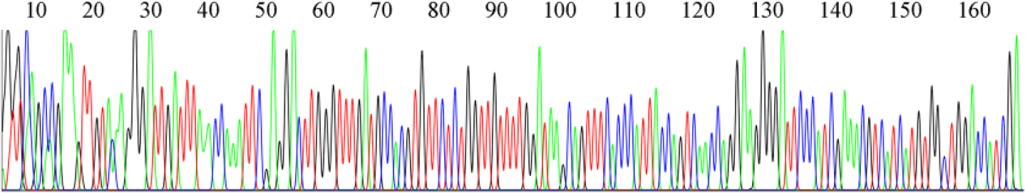
DNA sequencing results of hsa_circ_0006633 in gastric tissues. The qRT-PCR products of tissue hsa_circ_0006633 were sequenced by Qingkezixi Biotech (Hangzhou) Co., Ltd.

Relative expression of hsa_circ_0006633 in gastric cancer cell lines and normal gastric epithelial cell line. Hsa_circ_0006633 expression levels in four gastric cancer cell lines, HGC-27, SGC-7901, MGC-803, and AGS, and normal gastric epithelial cell line GES-1 were determined by qRT-PCR. Data are expressed as means ± SD of three independent experiments. Asterisks indicate p values that are significantly different comparing with those in GES-1 (*p < 0.05, **p < 0.01).
We then explored hsa_circ_0006633 expression levels in gastric cancer tissues. Its level in gastric cancer tissues was significantly lower than that in corresponding non-tumorous tissues (p < 0.001, Figure 3). Its expression levels were significantly downregulated in 79.2% (76/96) gastric cancer tissues compared with the adjacent normal tissues (Figure 3(c)). To explore its diagnostic values, a receiver operating characteristic (ROC) curve was established. The area under the ROC curve was 0.741 (Figure 3(d)). When the cutoff value was 8.17, its sensitivity and specificity were 0.60 and 0.81, respectively.

Hsa_circ_0006633 expression levels in gastric cancer tissues. (a) The expression levels of hsa_circ_0006633 in cancer tissues (n = 96) and adjacent normal tissues (n = 96). Higher ΔCt value indicates lower expression (***p < 0.001). (b) The expression levels of hsa_circ_0006633 are significantly lower than those in corresponding non-tumorous tissues. (c) The expression level of hsa_circ_0006633 was significantly downregulated in 79.2% (76/96) gastric cancer tissues compared with the adjacent normal tissues. (d) ROC curve of hsa_circ_0006633 in differentiating gastric cancer tissues from controls. The area under the curve was up to 0.741.
We further detected the hsa_circ_0006633 expression levels in different stages of gastric cancer development. As shown in the Figure 4, hsa_circ_0006633 expression levels were significantly decreased in gastritis, dysplasia, and cancer tissues comparing with healthy control; meanwhile, it was significantly downregulated in gastritis tissues comparing the cancer tissues (Figure 4). There were no significant differences of hsa_circ_0006633 expression levels between dysplasia and cancer tissues.

Differences of hsa_circ_0006633 expression levels among different stages of gastric mucosa. The hsa_circ_0006633 expression levels in healthy gastric mucosa (n = 35), gastritis (n = 51), dysplasia (n = 20), and gastric cancer (n = 96) tissues were determined by qRT-PCR. Higher ΔCt value indicates lower expression (**p < 0.01, ***p < 0.001).
Relationships between hsa_circ_0006633 expression levels and clinicopathological factors
Based on the above findings, we analyzed the association between hsa_circ_0006633 expression levels and clinicopathological characteristics of patients with gastric cancer. As shown in Table 1, low expression of hsa_circ_0006633 in human gastric cancer tissues was associated with distal metastasis (p = 0.037) and tissue CEA level (p = 0.041). However, its levels were not significantly associated with other clinicopathological characteristics such as age, gender, diameter, stage, pathological diagnosis, invasion, and CA19-9 levels.
Relationship of Hsa_circ_0006633 expression levels (ΔCt) in cancer tissues with clinicopathological factors of patients with gastric cancer..

CEA: carcinoembryonic antigen; SD: standard deviation.
Detection of hsa_circ_0006633 level in human plasma
As human plasma is the main material used in the screening of cancers, we wondered whether plasma hsa_circ_0006633 levels were also altered in gastric cancer patients. We detected plasma hsa_circ_0006633 levels in 20 healthy volunteers and 20 gastric cancer patients by qRT-PCR method. Our data showed that hsa_circ_0006633 levels were significantly increased in gastric cancer patients’ plasma compared with healthy control (Figure 5).

Detection of hsa_circ_0006633 level in human plasma. Plasma hsa_circ_0006633 levels in healthy volunteers (n = 20) and gastric cancer patients (n = 20) were detected by qRT-PCR method. Lower ΔCt value indicates higher level (*p < 0.05).
Discussion
CircRNAs were once thought to be the byproducts of splicing errors caused by low abundance and the technological limitations, while they have been received great attention recent years with the development of RNA-seq and bioinformatic analysis. 13 Unlike linear RNA, circRNA forms covalently closed-loop structure by jointing 3′ and 5′ ends together via exon circularization or intron circularization. 13 CircRNAs are abundant, multiple, and stable in creatures.7,14 Many researchers have found that circRNAs play an important part in the regulation of gene expression. 6 It is widely accepted that some RNAs regulate with microRNA (miRNA) response elements (MREs) and works as “competing endogenous RNA (ceRNA)” hypothesis.15,16 Recent studies have showed that circRNAs can work as a miRNA sponge. For example, ciRS-7 (circRNA sponge for miR-7) containing approximately 70 binding sites acts as a miR-7 sponge, which can suppress the miR-7 activity and thus increase levels of miR-7 targets; sex-determining region Y (Sry) works as a miR-138 sponge. 17
In addition, circRNAs have been implicated in many diseases such as Parkinson disease, Alzheimer’s disease, atherosclerosis risk, and prion diseases.18–21 Increasing studies have showed that circRNAs play an important role in tumorigenesis and tumor progression. 22 In colorectal cancer, there exists a negative correlation of global circRNA abundance and proliferation. 23 Hsa_circ_0001649 was downregulated in hepatocellular carcinoma. 24 Hsa_circ_100855 and hsa_circ_104912 were dysregulated in laryngeal squamous cell cancer (LSCC) tissues and associated with some clinicopathological factors. 25 In gastric cancer, lower expression of hsa_circ_002059 expression was first found to be significantly correlated with distal metastasis, TNM stage, gender, and age. 26 In this study, we found that expression levels of hsa_circ_0006633 in four gastric cancer cell lines, AGS, HGC-27, MGC-803, and SGC-7901, were significantly downregulated than those in normal gastric epithelial GES-1 cells (Figure 2). Moreover, it was also significantly downregulated in gastric cancer tissues than those in paired adjacent non-tumorous tissues (Figure 3). The data realized that hsa_circ_0006633 may play an important role in the carcinogenesis of gastric cancer.
The occurrence of gastric cancer is the result of multi-stage and multi-factors.2,27 It is a progressive process of the gradual emergency of gastritis, gastric atrophy, intestinal metaplasia, and dysplasia, eventually leading to the occurrence of gastric cancer. 28 The progression of gastric cancer is mainly caused by microbial pathogens and is closely related to host inflammatory factors; during this process, the altered microenvironment may cause random mutations to occur in gastric cells, which inevitably leads to carcinoma.2,29 Increasing evidence has revealed that several proinflammatory cytokinesgenes, such as those for tumor necrosis factor (TNF) and interleukin-1 (IL-1), may increase the risk of gastric cancer. 30 In this study, we made further explorations of hsa_circ_0006633 expression in multiple stages of tissues during the gastric cancer progression. We discovered that hsa_circ_0006633 expression was significantly decreased in gastritis, dysplasia, and gastric cancer tissues (Figure 4). Since mucosal inflammation is an initiation factor eventually resulting in gastric cancer,2,28 we found that the expression of hsa_circ_0006633 in gastritis was significantly lower than that in gastric cancer tissues (Figure 4), indicating that hsa_circ_0006633 may be a dysregulative molecule underlying gastric benign lesions.
Distal metastasis is a crucial factor in evaluating the prognosis of gastric cancer. A study indicated that patients with both serum CEA and CA19-9 positive significantly correlated higher frequencies of lymph node metastasis, deeper invasion by the tumor, and higher rates of hepatic metastasis. 31 Our research found that the lower expression of hsa_circ_0006633 was significantly correlated with distal metastasis and tissue CEA level (Table 1). Moreover, hsa_circ_0006633 can exist in human plasma. Plasma hsa_circ_0006633 levels were significantly increased in gastric cancer patients compared with healthy control. This means that hsa_circ_0006633 is also a potential biomarker for screening gastric cancer.
In conclusion, our data implied that hsa_circ_0006633 expression was not only significantly decreased in gastric cancer tissues and its cell lines but also decreased in dysplasia. The downregulation expression of hsa_circ_0006633 was associated with distal metastasis and tissue CEA level. Moreover, compared with healthy control, plasma hsa_circ_0006633 levels were significantly increased in gastric cancer patients. Our findings suggest that hsa_circ_0006633 may play a crucial role during gastric cancer occurrence and is also a potential biomarker for screening gastric cancer.
Footnotes
Declaration of conflicting interests
The author(s) declared no potential conflicts of interest with respect to the research, authorship, and/or publication of this article.
Ethical approval
This study was approved by the Human Research Ethics Committee of Ningbo University School of Medicine.
Funding
The author(s) disclosed receipt of the following financial support for the research, authorship, and/or publication of this article: This study was supported by grants from the Social Development Research Project of Ningbo (No. 2016C51015), the Medical Research Project of the Affiliated Hospital of Ningbo University School of Medicine (No. XYY16022), the Foundation of Zhejiang Key Laboratory of Pathophysiology (No. 201601), the Medical and Health Research Project of Zhejiang Province (No. 2017KY598), Applied Research Project on Nonprofit Technology of Zhejiang Province (Nos 2016C33177 and 2017C35004), and the K. C. Wong Magna Fund in Ningbo University.