
Abstract
DCAMKL1 (doublecortin and CaM kinase-like 1) has been found to be overexpressed and function as an oncogene in several types of cancer, but there are limited reports on the role of DCAMKL1 in bladder cancer. The messenger RNA and protein expression of DCAMKL1 in bladder cancer tissues and cell lines was measured by quantitative reverse transcription polymerase chain reaction, western blotting, or immunohistochemistry. The correlation between DCAMKL1 protein expression and clinicopathological characteristics was analyzed. Univariate and multivariate Cox regression models were adopted to evaluate prognostic significance of DCAMKL1 in bladder cancer patients. In our results, DCAMKL1 messenger RNA and protein were overexpressed in bladder cancer tissues compared with adjacent normal tissues. DCAMKL1 protein overexpression was positively associated with clinical stage, muscularis invasion, lymph node metastasis, and distant metastasis. The univariate and multivariate analyses suggested DCAMKL1 protein overexpression was an unfavorable prognostic factor in bladder cancer patients. In conclusion, DCAMKL1 is an independent poor prognostic factor for bladder cancer patients.
Introduction
Bladder cancer is the most common urinary tract malignant tumor in the developed world, while bladder cancer is the first most common urological cancer in China. 1 About 80,500 newly diagnosed bladder cancer cases and 32,900 bladder cancer deaths were appeared in China in 2015. 2 According to American Cancer Statistics 2017, bladder cancer is the sixth most common cancer with an estimated 79,030 new cases and 16,870 deaths in the United States in 2017. 3 Although surgical resection and chemotherapy are widely used for bladder cancer treatment, the recurrence rate is still high, as 50%–70% of bladder cancer cases recur within 5 years.4,5 The 5-year survival rate for bladder cancer patients remains at 50%–60%.6,7 Up to now, the management of bladder cancer relies on the patient’s clinicopathological characteristics including tumor–node–metastasis (TNM) staging and histological grade, as indicators of favorable/unfavorable prognosis. However, these characteristics are not enough to predict bladder cancer patient’s outcome. Thus, it is urgent to identify biomarkers which provide accurate prognosis prediction and development of targeted molecular therapies for bladder cancer patients.
Doublecortin and CaM kinase-like 1 (DCAMKL1) is a microtubule-binding member of the calmodulin-dependent kinase that plays important roles in regulating cell differentiation, migration, apoptosis, and epithelial–mesenchymal transition (EMT) and has been identified as a marker for gastrointestinal stem cell and cancer stem cell.8–12 DCAMKL1 has been found to be overexpressed and functions as an oncogene in several types of cancer such as pancreatic cancer, 10 colorectal cancer, 13 hepatocellular carcinoma, 14 and gastric cancer. 15 However, there are limited reports on the role of DCAMKL1 in bladder cancer.
In order to clarify the role of DCAMKL1 in the pathogenesis of bladder cancer, we explored the association between DCAMKL1 expression and clinicopathological features of bladder cancer patients. Meanwhile, the prognostic significance of DCAMKL1 in bladder cancer patients was estimated through univariate and multivariate Cox regression analyses.
Materials and methods
Ethical statement
This study was approved by the Ethical Committees of Jining No. 1 People’s Hospital. An informed consent was obtained from all the participants before enrollment in the study. The entire study was performed based on the Declaration of Helsinki.
Tissue samples collection
A total of 118 bladder cancer tissues and 40 matched adjacent non-tumor normal tissues were obtained from Jining No. 1 People’s Hospital from January 2005 to December 2015. Before surgical therapy, none of the bladder cancer patients in this study had received neoadjuvant treatment. Tissues were respectively stored in liquid nitrogen and formaldehyde solution after surgical resection. All fresh tissues were stored at −80°C for reverse transcription polymerase chain reaction (RT-PCR) and western blot experiments. The diagnosis and system treatment was according to Chinese guidelines for bladder cancer.
RT-PCR
Total RNA was obtained from tissues by one-step extraction method using TRIzol reagent (Invitrogen, USA) and then reverse-transcribed to complementary DNAs (cDNAs) using the PrimeScript RT Master Mix (TaKaRa, Japan), according to the manufacturer’s instructions. The LightCycler (Roche Diagnostics, USA) was selected to conduct the amplification of cDNAs using SYBR Premix Ex Taq™ II (TaKaRa). The primers used for RT-PCR were purchased from TaKaRa. Primers for the human DCAMKL1 are as follows—forward primer: 5′-TGAAGGGTACGCTCCTCAGT-3′ and reverse primer: 5′-GCTACACTCTGACCGCATGA-3′. Primers for the human β-actin are as follows: forward primer: 5′-GGACTTCGAGCAAGAGATGG-3′ and reverse primer: 5′-AGCACTGTGTTGGCGTACAG-3′. Relative expression was calculated via the comparative cycle threshold method and was normalized to the expression of β-actin.
Western blotting
Total protein was extracted using cell lysis buffer (Beyotime, China) for western blotting. Then the proteins in samples were separated through sodium dodecyl sulfate–polyacrylamide gel electrophoresis (SDS-PAGE) and subsequently transferred to a polyvinylidene fluoride (PVDF) membrane. After the membrane was blocked for 2 h using 5% skim milk, it was incubated with primary antibodies against DCAMKL1 (Cell Signaling Technology, USA) and β-actin (CWBIO, China) for 2 h. Finally, the membrane was incubated with horseradish peroxidase (HRP)-conjugated secondary antibodies for another 2 h. Band signals were visualized using enhanced chemiluminescence (ECL; CWBIO). Quantity One Software (Bio-Rad, USA) was used to analyze the intensity of blots.
Immunohistochemistry
Immunohistochemical analysis was performed to measure DCAMKL1 protein expression in 118 primary bladder cancer tissues and 40 adjacent non-cancerous tissues. In brief, slides were baked at 60°C for 1 h, followed by deparaffinization with xylene, and rehydrated. The sections were submerged in ethylenediaminetetraacetic acid (EDTA) antigenic retrieval buffer and microwaved for antigen retrieval. They were then treated with 3% hydrogen peroxide in methanol to quench endogenous peroxidase activity, followed by incubation with 5% bovine serum albumin to block non-specific binding. Sections were incubated with anti-DCAMKL1 (Cell Signaling Technology) overnight at 4°C. After washing, tissue sections were treated with secondary antibody, followed by incubation with HRP-conjugated streptavidin. Tissue sections were then counterstained with hematoxylin, dehydrated, and mounted. Finally, the sections were viewed under a bright-field microscope. The tissue sections stained immunohistochemically for DCAMKL1 were reviewed and scored separately by two pathologists blinded to the clinical parameters. For DCAMKL1 assessment, extent of staining was scored as 0: <5%, 1: 5%–25%, 2: 26%–50%, 3: 51%–75%, or 4: >75% positive cells, and the staining intensity was scored as 0: negative, 1: weak, 2: moderate, or 3: strong. The final score was determined by the combined staining score and proportion score (immunoreactive score = intensity score × proportion score), which generated scores of 0, 1, 2, 3, 4, 6, 8, 9, and 12. Low expression of DCAMKL1 was defined as 0–3 scores; high expression of DCAMKL1 was defined as >4 scores (including 4 scores). 13
Database analysis
The OncoLnc database (http://www.oncolnc.org/) was used to analyze the significance of DCAMKL1 in bladder cancer patients. The bladder cancer patient’s cohort from OncoLnc database included 402 cases. DCAMKL1 (DCLK1) was entered as the gene symbol, and the median value of DCAMKL1 expression was selected as the cutoff of the high and low DCAMKL1 groups.
Statistical analysis
SPSS 17.0 software (SPSS Inc., USA) was used to perform statistical analysis. The Wilcoxon signed-rank test was applied to test the expression of DCAMKL1 in cancer tissues compared to paired adjacent normal tissues. The association of clinicpathological characteristics with DCAMKL1 expression was determined using χ2 tests. Survival curves were plotted using the Kaplan–Meier method and the log-rank test. Univariate and multivariate Cox regression models were adopted to evaluate prognostic significance. p values in all experiments were considered to be statistically significant at less than or equal to 0.05.
Results
DCAMKL1 messenger RNA and protein are overexpressed in bladder cancer tissues
In order to explore the status of DCAMKL1 in bladder cancer tissues, we conducted quantitative RT-PCR (qRT-PCR), western blotting, and immunohistochemistry to determine DCAMKL1 messenger RNA (mRNA) and protein levels in bladder cancer tissue samples and adjacent normal tissue samples. Compared with adjacent normal tissues, DCAMKL1 mRNA was overexpressed in bladder cancer tissues (p < 0.001, Figure 1(a)). Meanwhile, the result of western blotting showed that DCAMKL1 protein expression was overexpressed in bladder cancer tissues compared with adjacent normal tissues (Figure 1(b)). Furthermore, DCAMKL1 protein expression in 118 bladder cancer tissues and 40 adjacent normal tissues was detected by immunohistochemical staining (Figure 2(a)–(h)). We observed that DCAMKL1 protein was overexpressed in bladder cancer tissues in 55.1% (65/118). In comparison, only 12.5% (5/40) of adjacent normal tissues suggested overexpression of DCAMKL1 protein, which was significantly lower than that in the bladder cancer tissues (p < 0.001, Table 1).

DCAMKL1 mRNA and protein levels are overexpressed in bladder cancer tissues. (a) Expression of DCAMKL1 mRNA was increased in bladder cancer tissues compared with adjacent normal tissues. (b) DCAMKL1 protein expression was elevated in bladder cancer tissues compared with adjacent normal tissues.

Immunohistochemical staining of DCAMKL1 in bladder cancer tissues and adjacent normal tissues (the label = 100 µm). (a) Negative expression of DCAMKL1 in bladder cancer tissue (staining intensity score = 0). (b) Weak expression of DCAMKL1 in bladder cancer tissue (staining intensity score = 1). (c) Moderate expression of DCAMKL1 in bladder cancer tissue (staining intensity score = 2). (d) Strong expression of DCAMKL1 in bladder cancer tissue (staining intensity score = 3). (e) Negative expression of DCAMKL1 in normal bladder tissues (staining intensity score = 0). (f) Weak expression of DCAMKL1 in normal bladder tissues (staining intensity score = 1). (g) Moderate expression of DCAMKL1 in normal bladder tissues (staining intensity score = 2). (h) Strong expression of DCAMKL1 in normal bladder tissues (staining intensity score = 3).
DCAMKL1 protein expression is increased in bladder cancer.

DCAMKL1: doublecortin and CaM kinase-like 1.
DCAMKL1 protein expression is associated with malignant status of bladder cancer patients
The clinical significance of DCAMKL1 protein expression in bladder cancer patients was explored in 118 bladder cancer tissue samples through immunohistochemistry. The association between clinicopathological features and DCAMKL1 protein expression in bladder cancer patients was summarized in Table 2. We observed that DCAMKL1 protein overexpression was positively associated with clinical stage (I–II vs III–IV, p = 0.001), muscularis invasion (absent vs present, p < 0.001), lymph node metastasis (absent vs present, p = 0.001), and distant metastasis (absent vs present, p = 0.012). However, there were no significant relationships between DCAMKL1 protein expression and age (<60 years vs ≥60 years, p = 0.912), gender (female vs male, p = 0.849), tumor size (<3 cm vs ≥3 cm, p = 0.527), histological grade (high grade vs low grade, p = 0.107), and mutiplicity (single vs multiple, p = 0.675).
Association between DCAMKL1 protein expression and clinicopathological characteristics in bladder cancer patients.

DCAMKL1: doublecortin and CaM kinase-like-1.
DCAMKL1 protein overexpression is an unfavorable prognostic factor in bladder cancer patients
The prognostic value of DCAMKL1 protein expression in bladder cancer patients was also explored in 118 bladder cancer tissue samples through immunohistochemistry. Kaplan–Meier survival analysis showed that bladder cancer patients who expressed high level of DCAMKL1 protein had lower overall survival compared to patients with low level of DCAMKL1 protein expression (p < 0.001, Figure 3(a)). Similar to our result, we also found bladder cancer patients with DCAMKL1 high expression had a shorter survival time than those with DCAMKL1 low expression (p = 0.008, Figure 3(b)) through analyzing OncoLnc database. Moreover, we conducted univariate analysis and identified six prognostic parameters: clinical stage, tumor size, muscularis invasion, lymph node metastasis, distant metastasis, and DCAMKL1 protein expression. Finally, we found that DCAMKL1 protein overexpression was an independent poor prognostic factor for bladder cancer patients through multivariate analysis (hazard ratio (HR) = 1.841, 95% confidence interval (CI) = 1.026–3.305, p = 0.041, Table 3).

DCAMKL1 high expression predicts an unfavorable prognosis in bladder cancer patients. (a) Survival analysis was performed in 118 bladder cancer patients from our study. (b) Survival analysis was performed in 402 bladder cancer patients from OncoLnc database.
Summary of univariate and multivariate Cox regression analysis of overall survival duration in bladder cancer patients.
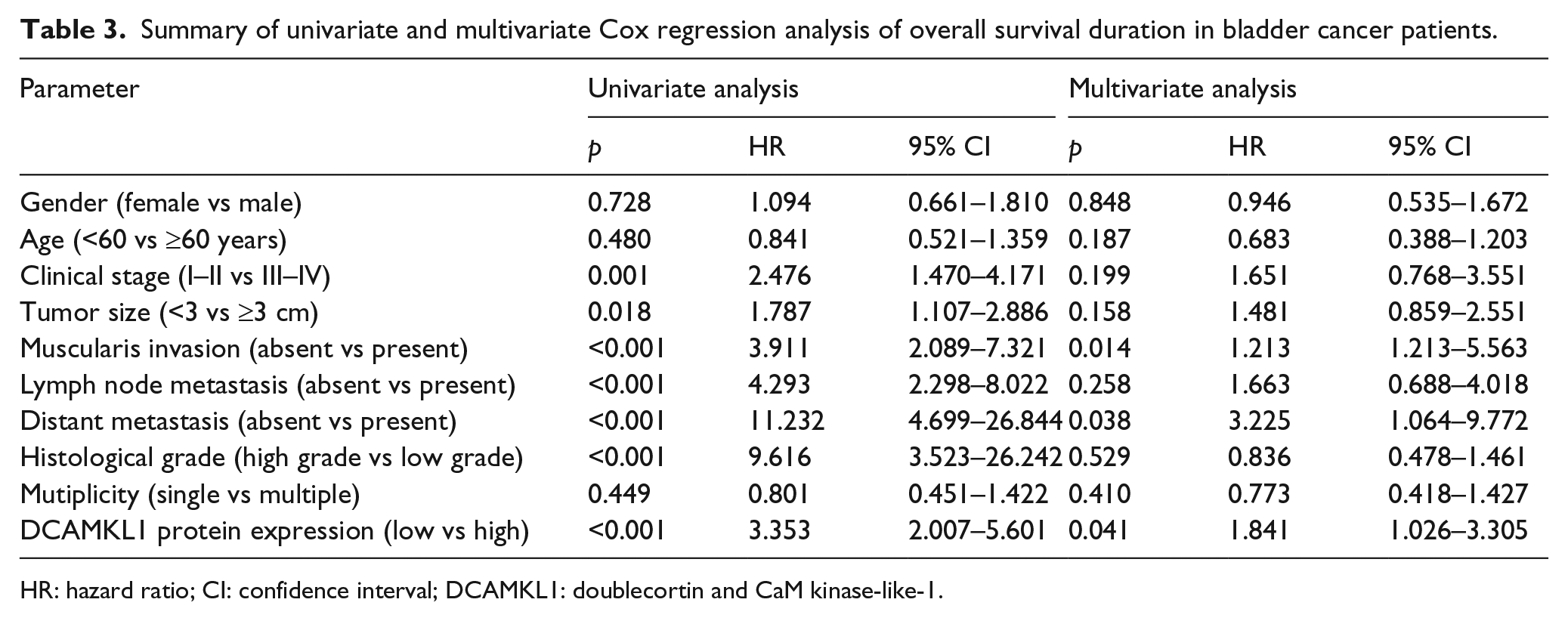
HR: hazard ratio; CI: confidence interval; DCAMKL1: doublecortin and CaM kinase-like-1.
Discussion
DCAMKL1 is a serine/threonine kinase that belongs to the family of microtubule-associated proteins. 16 The C-terminal region, which includes a serine/threonine kinase domain, is highly related to a Ca2+/calmodulin-dependent protein 1 (CaMKI) kinase domain, while the N-terminal region of DCAMKL1 harbors a tandem doublecortin domain that drives the microtubule-associating function. 17 DCAMKL1 was originally identified in neurogenesis, but DCAMKL1 has recently been shown to regulate biological processes including cell differentiation, migration, apoptosis, and EMT.18,19 In clear cell renal carcinoma, Weygant et al. 20 found that knockdown of DCAMKL1 could inhibit EMT biomarkers and pluripotency factor expression and markedly suppressed tumor cell migration, invasion, focal adhesion, clonogenic capacity, and drug-resistance. In colorectal cancer, Gao et al. 13 reported that DCAMKL1 overexpression significantly accelerated tumor cell migration and invasion, upregulated the mesenchymal markers vimentin and zinc finger E-box-binding homeobox 1 (ZEB1) expression, and downregulated the epithelial marker E-cadherin expression. Moreover, DCAMKL1 has been suggested to serve as a functional target for microRNAs involving tumorigenicity.14,21–24 Wang et al. 21 showed that microRNA 613 (miR-613) suppressed hepatocellular carcinoma cell growth and invasiveness through targeting DCAMKL1. Sureban et al. 22 suggested that knockdown of DCAMKL1 in human pancreatic cancer cells regulated miR-200a, let-7a, and miR-144 to control cell signaling pathways. Interestingly, several biological experimentations indicated that DCAMKL1 was an important target for anti-tumor drugs. DCAMKL1 has been confirmed as a target for XMD8-92 (a kinase inhibitor with anti-cancer activity) to modulate its downstream oncogenic pathways (EMT, pluripotency, angiogenesis, and anti-apoptotic). 25 Meanwhile, LRRK2-IN-1 (small molecule inhibitors of DCAMKL1 kinase) obviously inhibited cancer cell proliferation, migration, and invasion as well as induction of apoptosis and cell cycle arrest in colorectal and pancreatic cancer. 26 These biological studies consistently suggested that DCAMKL1 acted as an oncogene in human cancer.
The clinical value of DCAMKL1 in human cancer is still deficiently understood. In colorectal cancer patients, DCAMKL1 mRNA and protein expression levels were both increased in colorectal cancer clinical tissue samples, and DCAMKL expression was positively correlated to TNM stage, tumor grade, and neoadjuvant chemoradiotherapy. 27 In renal clear cell carcinoma patients, DCAMKL was overexpressed and dysregulated on the mRNA and epigenetic level in more than 93% of renal clear cell carcinoma compared with adjacent normal tissue through analyzing Cancer Genome Atlas dataset, and DCAMKL overexpression was demonstrated in stage II–III tumors compared to normal kidney and stage I tumors. 20 Meng et al. 28 reported that DCAMKL expression was significantly upregulated in gastric cancer and strongly associated with pN stage and lymphovascular invasion. Up to now, the expression status of DCAMKL in bladder cancer is unknown. Our study first showed DCAMKL1 mRNA and protein were overexpressed in bladder cancer tissues compared with adjacent normal tissues. Furthermore, we observed that DCAMKL1 protein overexpression was positively associated with clinical stage, muscularis invasion, lymph node metastasis, and distant metastasis. These studies consistently implied that DCAMKL1 overexpression may serve as an unfavorable factor in predicting human cancer prognosis.
In recent years, DCAMKL1 overexpression has been shown to be a prognostic factor in several types of tumors, which has a favorable or unfavorable prognostic significance depending on tumor types. In breast cancer patients, DCAMKL1 overexpression was significantly associated with better overall survival and disease-free survival. 29 Conversely, there was more evidence which showed that DCAMKL1 overexpression in tumor tissues was an unfavorable prognosis biomarker in gastric cancer, 28 lung cancer, 30 colorectal cancer,13,31 and salivary gland cancer. 32 Meng et al. 28 demonstrated that gastric cancer patients with DCAMKL1 high expression had a significantly shorter overall survival and disease-free survival, and univariate and multivariate analyses showed that DCAMKL1 expression was an independent factor for predicting overall survival and disease-free survival based on univariate and multivariate analyses. In colorectal cancer patients, DCAMKL1 expression was inversely correlated with overall survival. 13 Interestingly, we analyzed OncoLnc database and found that bladder cancer patients with DCAMKL1 high expression had a shorter survival time that those with low DCAMKL1 expression. Similarly, our study suggested that bladder cancer patients who expressed high level of DCAMKL1 protein had lower overall survival compared to patients with low level of DCAMKL1 protein expression, and DCAMKL1 protein overexpression was an independent poor prognostic factor for bladder cancer patients through univariate and multivariate analyses.
In conclusion, DCAMKL1 is overexpressed in bladder cancer tissues and is associated with malignant status and prognosis in bladder cancer patients.
Footnotes
Acknowledgements
S.Z. and G.Z. are the co-first authors.
Declaration of conflicting interest
The author(s) declared no potential conflicts of interest with respect to the research, authorship, and/or publication of this article.
Funding
The author(s) received no financial support for the research, authorship, and/or publication of this article.