
Abstract
Colorectal neoplasia differentially expressed (CRNDE), an oncogene, is highly expressed in many tumor cells and affects cellular proliferation, migration, invasion, and apoptosis. Its function and mechanism of action is a research hotspot. In this study, microarray analysis was performed to discover the differentially expressed genes in CRNDE over-expression cells. RT² Profiler PCR Array was used to study the expression of genes related to the toll-like receptor (TLR) pathway. We found that over-expression of CRNDE in astrocytes increased the expression of key factors in the toll-like receptor signaling pathway, especially toll-like receptor-3-mediated MyD88-independent pathway. Furthermore, it up-regulated expression levels of downstream transcription factor such as nuclear factor kappa B and numerous cytokines. In contrast, CRNDE knockdown in glioma U87MG cell line showed an opposite trend in the expression of the above-mentioned genes. We speculated that CRNDE might trigger inflammation to regulate tumorigenesis and tumor development through the toll-like receptor pathway.
Introduction
Long non-coding RNAs (lncRNAs) play important roles in biological metabolism, and are associated with tumor initiation, progression, and metastasis.1,2 Colorectal neoplasia differentially expressed (CRNDE) is an lncRNA that was first discovered in colorectal neoplasia. Its expression is highly elevated in various tumors including gliomas.3,4 CRNDE could promote cellular proliferation, migration, and invasion, and inhibit apoptosis in glioma 5 and medulloblastoma cells. 6 The potential molecular mechanisms of CRNDE have been recently investigated. CRNDE was repressed by insulin and insulin-like growth factors (IGF) via PI3K/Akt/mTOR and Raf/MAPK signaling pathways. 7 Zheng et al.8,9 investigated the target microRNA (miRNA) of CRNDE and demonstrated that it played an oncogenic role in glioma stem cells by negatively regulating miR-384 and miR-186 expression, which modulated the expression of downstream target proteins such as STAT3, cyclin D1, VEGFA, SLUG, MMP-9, caspase 3, Bcl-2, Bcl-xL BAD, and MARK2. The function and mechanism of CRNDE have been studied in various tumors, such as renal cell carcinoma, 10 hepatic carcinoma, 11 and gallbladder carcinoma. 12
Toll-like receptors (TLRs) recognize distinct pathogen-associated molecular patterns (PAMPs) and play a key role in innate immune system, inflammatory reaction, immune regulation, and tumor formation and development.13,14 In humans, the TLR family has 11 members (TLR1–TLR11), of which TLR1, TLR2, TLR4, TLR5, TLR6, TLR10, and TLR11 are located on the plasma membrane, whereas TLR3, TLR7, TLR8, and TLR9 are localized in the endosomes. 15 TLRs activate two major signaling pathways. One is the myeloid differentiation primary response gene 88 (MyD88)-dependent pathway, which is common to all TLRs except for TLR3, while the other is the MyD88-independent pathway that is specific for TLR3 and TLR4.13,15 The TLR3 responds to double-stranded viral RNA, recruits Toll/interleukin-1 receptor (TIR) domain–containing adapter molecule 1 (TICAM1), and triggers a cascade reaction that finally leads to interferon (IFN)-β production and nuclear factor kappa B (NF-κB) pathway activation.16,17 NF-κB is a nuclear transcription factor that regulates the expression of numerous genes that are critical for the regulation of inflammation, tumorigenesis, and autoimmune diseases. 18
In this study, we found that over-expression of CRNDE positively regulated the expression of key factors in the TLR-signaling pathway, NF-κB, and cytokines, while CRNDE knockdown had the opposite effect on these genes. We speculated that CRNDE triggered inflammation that regulated tumorigenesis and development through the TLR pathway. These results depicted a novel pathway mediated by CRNDE in astrocytes and glioma cells.
Materials and methods
Cell culture
The human astrocyte (HA) cell line and glioma cell line U87MG were purchased from the Cell Resource Center of Peking Union Medical College (Beijing, China). HA and U87MG cells were cultured in astrocyte medium (AM; ScienCell Research Laboratories, Carlsbad, CA, USA) and Dulbecco’s Modified Eagle’s Medium (DMEM; Gibco, Grand Island, NY, USA), respectively, containing 5% fetal bovine serum ((Hyclone, Logan, UT, USA)) at 37°C with 5% CO2.
Plasmid construction and transfection
Full-length CRNDE cDNA was synthesized in vitro and cloned into pcDNA3.1(+) and pcDNA3.1(−) using HindIII and EcoRI restriction sites. The recombinant constructs (pcDNA3.1(+)-CRNDE and pcDNA3.1(−)-CRNDEr) were verified by sequencing. HA cells were seeded into 6-well plates at 80%–90% confluency and transfected with pcDNA3.1(+)-CRNDE, pcDNA3.1(−)-CRNDEr (reverse lncRNA control, RC), or pcDNA3.1(+) (negative control, NC) using lipofectamine 2000 transfection reagent (Life Technologies Corporation, Carlsbad, CA, USA). After 4 h, the medium was replaced with normal AM. Cells were collected after transfection for RNA isolation and Western blot.
siRNA synthesis and transfection
Three siRNAs targeting CRNDE were designed and synthesized by Suzhou GenePharma Corporation (Suzhou, China). The sequences were as follows: si783-GUGCUCGAGUGGUUUAAAUTT and AUUUAAACCACUCGAGCACTT; si809-GGGUAUUCCUGUUUAUAGATT and UCUAUAAACAGGAAUACCCTT; and si860-GUCACGCAGAAGAAGGUUATT and UAACCUUCUUCUGCGUGACTT. Scramble mimics were synthesized as NCs. The sequences were as follows: GCGACGAUCUGCCUAAGAUTT and AUCUUAGGCAGAUCGUCGCTT. U87MG cells were seeded into 6-well plates at 50% confluency and transfected with different siRNAs or NCs using lipofectamine RNAiMAX transfection reagent (Life Technologies Corporation). Cells were collected after 24 h transfection.
RNA extraction, reverse transcription, and real-time polymerase chain reaction
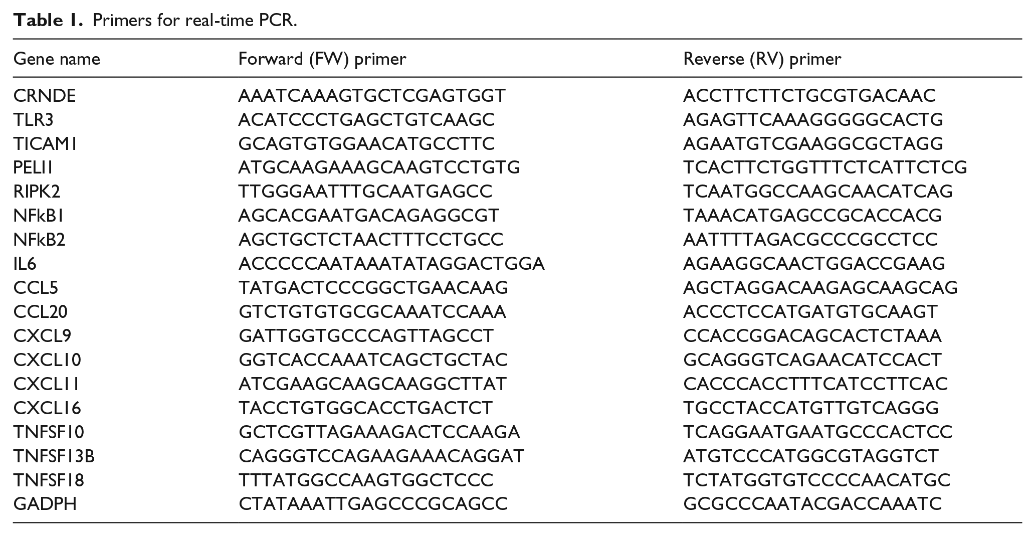
Total RNA was extracted from the cells with Trizol reagent (Invitrogen, Carlsbad, CA, USA) according to the manufacturer’s protocol. For polymerase chain reaction (PCR) Array, 1 µg RNA was reverse transcribed using RT2 First Strand Kit (Qiagen, Hilden, Germany). Quantitative real-time PCR was performed with the TP800 qPCR system (Takara, Osaka, Japan) using RT2 SYBR Green ROX qPCR Mastermix (Qiagen) and RT2 Profiler PCR Array (Human Toll-Like Receptor Signaling Pathway; Qiagen). PCR conditions included an initial step of 95°C for 10 min, followed by 40 cycles of 95°C for 15 s, 55°C for 30 s, and 72°C for 30 s. The quantification of gene expression was performed using the 2−ΔΔCT method. Data analysis was performed using the online software www.SABiosciences.com/pcrarraydataanalysis.php. The real-time PCR assay was repeated three times using different samples. For the other real-time PCR, reverse transcription reaction was performed using PrimeScriptRT reagent Kit with gDNA Eraser (Takara) and real-time PCR was performed using SYBR Premix Ex TaqII (TliRNaseH Plus) (Takara). The primers were synthesized by Invitrogen, and the sequences are shown in Table 1.
Primers for real-time PCR.
Microarray analysis
The RNA integrity number (RIN) number was checked to determine RNA integrity by an Agilent Bioanalyzer 2100 (Agilent technologies, Santa Clara, CA, USA). Total RNA was further purified by RNeasy micro kit (Qiagen) and RNase-Free DNase Set (Qiagen).The expression microarray was performed by Shanghai Biotechnology Corporation (Shanghai, China) using Affymetrix Human Gene 2.0 ST Array (Affymetrix, Santa Clara, CA, USA). The arrays were scanned using the GeneChip Scanner 3000 (Affymetrix). Array data export processing and analysis were performed using Command Console Software (Affymetrix).
Western blot
Proteins were extracted from cells with RIPA lysis buffer (Applygen, Beijing, China) and quantified using a BCA Protein Assay Kit (Applygen). A total of 30 µg micrograms of protein lysates were separated on sodium dodecyl sulfate polyacrylamide gel electrophoresis (SDS-PAGE), and transferred onto a polyvinylidene difluoride (PVDF) membrane (Millipore, Billerica, MA, USA). The membrane was blocked with 5% nonfat milk and incubated with diluted primary antibody (1:1000–2000, Cell Signaling Technology, Danvers, MA, USA) overnight at 4°C, followed by incubation with a horseradish peroxidase (HRP)-conjugated secondary antibody (1:2000, Transgen Biotech, Beijing, China) for 1 h at room temperature. The membranes were developed using chemiluminescent HRP substrate (Millipore). Glyceraldehyde-3-phosphate dehydrogenase (GAPDH) was used as a control (1:2000, Transgen Biotech).
Statistical analysis
Data were presented as the mean ± SD from at least two independent replicates. Statistical analysis was performed using SPSS statistical software. Two-tailed Student’s t-test was used to analyze the differences between the two groups. The differences were considered statistically significant at p < 0.05.
Results
Over-expression of CRNDE resulted in immune response activation and cytokine production
The expression patterns of whole mRNAs were analyzed by microarray to elucidate the changes between the CRNDE over-expression group (transfected with pcDNA3.1(+)-CRNDE) and the control group (transfected with pcDNA3.1(+)). In the CRNDE over-expression group, the expression of 19 genes decreased, while the expression of 70 genes increased as compared to the control group (fold change ⩾ 1.5, p < 0.05) (Figure 1(a)). Cluster analysis of the differentially expressed genes indicated that three copies of the CRNDE over-expression group and three copies of the control group were closely clustered together. However, there was obvious difference between the two groups (Figure 1(b)). Expression of cytokines such as IL6, CCL5, CCL20, CXCL9, CXCL10, CXCL11, CXCL16, TNFSF10, TNFSF13B, and TNFSF18 were significantly induced, which was confirmed by real-time PCR, and the two experiments showed high concordance (Figure 2(a)). We used gene ontology (GO) enrichment and Kyoto Encyclopedia of Genes and Genomes (KEGG) pathway databases to analyze these 89 differentially expressed genes. Biological analysis showed that nearly all functions of CRNDE were related to immune process and cytokine response (Figure 1(c)). According to KEGG analysis, 18 pathways were enriched (p < 0.05), many of which were related to inflammatory diseases and signaling (Table 2, Figure 1(d)).

Differential expression patterns of HA cells with or without CRNDE transfection. (a) Volcano plot shows significant differences between the CRNDE over-expression group and the control group. Red dot, up-regulated >1.5 fold and p < 0.05; green dot, down-regulated >1.5 fold and p < 0.05. (b) Cluster analysis of differentially expressed genes between the CRNDE over-expression group and the control group. (c, d) Top 10 significant GO terms and pathways and their enrichment scores. (c) Biological analysis showed that nearly all functions of CRNDE were related to the immune system and stimulus, and cytokine response. (d) Pathway analysis in KEGG database indicated that many pathways were associated with cytokines, immune response, and inflammation. Among these, TLR signaling was the most enriched pathway. (e) Volcano plot shows the differentially expressed genes in the PCR array. Many of these genes were up-regulated, but only two genes were down-regulated. Cut off value: FC > 1.5, p < 0.05. Red dot, up-regulated >1.5 fold; green dot, down-regulated >1.5 fold.

Expressions of NF-κB and several cytokines are positively associated with CRNDE expression. (a) Real-time PCR of 10 differentially expressed cytokine genes validated the microarray results. These genes were significantly up-regulated in CRNDE over-expressed HA cells. (b) Western blot showed that the expression levels of TLR3, TICAM1, and members of the NF-κB family were increased in CRNDE over-expressed HA cells. (c) Expression of key genes in the TLR pathway and cytokines were decreased in CRNDE knockdown U87MG cells.
KEGG analysis of differentially expressed genes.

CRNDE up-regulated majority of the key factors in the TLR-signaling pathway
The microarray data showed that the TLR-signaling pathway was significantly enriched (Table 2). We used RT2 Profiler PCR Array to confirm the microarray results, which contained 84 genes related to the TLR pathway. A total of 41 genes were up-regulated >1.5 times, of which 13 genes were significantly up-regulated (p < 0.05) (Table 3). Only two genes were down-regulated >1.5 times. The results showed that over-expression of lncRNA CRNDE increased the expression of the majority of the genes in the TLR pathway (Figure 1(e)). Moreover, after over-expression of lncRNA CRNDE, key factors in the MyD88-independent TLR-signaling pathway were up-regulated, such as TLR3, TICAM1, receptor-interacting serine-threonine kinase (RIPK), protein pellino homolog 1 (PELI1), and NF-κB. In order to verify whether CRNDE participated in the same signaling pathway in the glioma cell line, we established CRNDE gene knockdown U87MG cells. After transfecting three CRNDE siRNAs in the U87MG cells, expression of genes related to the TLR-signaling pathway decreased. Expression of four TLR pathway genes, two NF-κB genes, and four cytokine genes decreased 0.5–0.8 fold in the CRNDE knockdown cell line as compared to the NC (Figure 2(c)).
Differentially expressed genes in the TLR pathway after CRNDE transfection using PCR array.

CRNDE induced expression of NF-κB
Both microarray and PCR data showed that CRNDE induced key factors of the TLR-signaling pathway and NF-κB at the transcriptional level. We used Western blot to confirm if CRNDE activated MyD88-independent TLR signaling pathway and NF-κB at the protein level. The result indicated that expression of TLR3 (a receptor of TLR pathway) and TICAM1 (molecular adapter) were up-regulated and five members of the NF-κB family including NF-κB1 (P50), NF-κB2 (P52), Rel (cRel), RelA (P65), and RelB were induced in the CRNDE over-expressed cells (Figure 2(b)). In order to rule out nonspecific stimulation, we used reverse complement sequence of CRNDE as an experimental control. After transfecting reverse complement, expression of the target protein showed no obvious changes (Figure 2(b)).
Discussion
Epidemiological studies showed that 25% of tumors are closely related to inflammation. Inflammation promotes the occurrence and development of tumors through complex physiological and biochemical processes.19,20 In this study, we found that nearly all functions of CRNDE were related to the immune system and cytokine response according to gene ontology enrichment analysis. KEGG analysis revealed that inflammatory diseases and signaling pathways were enriched due to over-expression of CRNDE. We speculated that lncRNA CRNDE was closely related to inflammation.
The TLR signaling pathway plays significant roles in inflammation, immune regulation, and tumor formation and development. The TLR family includes 11 receptor proteins (TLR1−TLR11) in humans. Different TLRs serve as receptors for various ligands, including bacterial cell wall components, viral RNA, fungi, or immune modulatory compounds.13,15 Using KEGG analysis, we found significant enrichment in the TLR signaling pathway. The gene chip and PCR arrays showed that over-expression of CRNDE induced key factors of the TLR signaling pathway, especially the MyD88-independent pathway. Among the 84 genes associated with the TLR signaling pathway, 41 were up-regulated >1.5 times.
TLR3 is the only TLR involved in the MyD88-independent pathway. It activates immune cells in response to double-stranded viral RNA.21,22 The stimulation of TLR3 sequentially triggers TICAM1, TBK1, and IRF3 activation, which leads to IFN-β secretion. TICAM1 also activates Receptor-Interacting Protein-1 (RIP1) and TNF receptor associated factor-6 (TRAF6), which may further activate the NF-κB pathway.16,17 In this study, the key factors in the TLR3-related pathway were up-regulated. According to the PCR assay, TLR3, TICAM1, PELI1, and RIPK2 were increased 2.17, 1.77, 1.93, and 1.34 fold, respectively, in the CRNDE-transfected cell line. Western bolt showed the same trend. These results indicated that CRNDE might participate in the TLR3-mediated MyD88-independent pathway.
NF-κB is involved in multiple biological processes such as defense response, cell differentiation, apoptosis, and tumor progression. There are five members in this family (NF-κB1, NF-κB2, RelA, RelB, and Rel). 23 We found that expressions of NF-κB members were obviously up-regulated both at the mRNA and protein levels in the CRNDE-transfected cell line. The PCR assay showed that the expression of NF-κB1, NF-κB2, RelA, and Rel increased 2.21, 1.76, 1.69, and 1.6 fold, respectively, after CRNDE transfection. Western blot demonstrated that all five members of the NF-κB family were induced in the CRNDE over-expression group as compared to the reverse complement control and NC.
Inflammatory microenvironment includes various cells, cytokines, and chemokines, which exhibit complicated interactions with each other. 24 In this study, after CRNDE transfection, expressions of cytokines such as IL6, CCL5, CCL20, CXCL9, CXCL10, CXCL11, CXCL16, TNFSF10, TNFSF13B, and TNFSF18 were significantly up-regulated according to gene chip results and IL1A, IL1B, IL2, IL6, IL10, CCL2, CXCL10, and TNF were up-regulated according to PCR array results. The changes in these cytokine levels triggered inflammation and could stimulate angiogenesis, tumor growth, invasion, and metastasis.
In CRNDE knockdown U87MG cells, the key genes of the TLR pathway, NF-κB, and cytokines were generally down-regulated. This result showed an opposite trend as compared to the CRNDE over-expressed HA cell line. This demonstrated that CRNDE might also participate in the TLR signaling pathway in glioma cells. Many recent studies have indicated that CRNDE played an oncogenic role and promoted cellular proliferation, migration, and invasion, and inhibited apoptosis in glioma and medulloblastoma cells.5,6,8,9 Our previous research also showed that CRNDE knockdown inhibited cell proliferation and migration in U87MG cells.25,26 We speculated that CRNDE triggered inflammation in tumor cells through the TLR pathway, and released transcription factors and cytokines, which promoted tumorigenesis and tumor development. These results suggest a novel link between lncRNA CRNDE and tumors.
Besides TLR3, TLR2, which is involved in the MyD88-dependent pathway, was also induced after transfection of lncRNA CRNDE. TLR2 led to activation of NF-κB and the MAPKs (JNK, p38 MAPK), and finally promoted inflammatory cytokines. This result indicated that CRNDE might participate in the TLR signaling pathway through multiple receptors and might activate both MyD88-dependent and MyD88-independent signaling. However, we did not observe significant up-regulation of the downstream genes of TLR2.
As a stimulant, exogenous double-stranded viral RNA triggers the TLR pathway through TLR3 and finally induces immune response. In order to identify the activation of TLR signaling pathway related to lncRNA CRNDE but not the structure of nonspecific exogenous RNA, we used reverse complement sequence of CRNDE as an experimental control. Western blot showed that after reverse complement transfection, expression of TLR3, TICAM1, and five members of NF-κB showed no obvious changes. This result indicated that CRNDE gene, but not the reverse complement sequence, activated the TLR signaling pathway through TLR3.
In conclusion, lncRNA CRNDE activated TLR3-mediated MyD88-independent TLR signaling pathway through TICAM1, PELI1, and RIPK2, and further activated NF-κB and several cytokines (Figure 3). We speculated that CRNDE triggered inflammation through the TLR3-NF-κB-cytokine pathway that finally resulted in tumorigenesis and tumor development. This study has revealed a new signaling link between CRNDE gene and tumors.

Model showing CRNDE as a stimulant promoting TLR signaling and triggering inflammation and several biological processes.
Footnotes
Contributorship
Haowen Li designed the experiments and drafted the manuscript. Haowen Li, Qi Li, and Chengya Dong performed cellular and molecular biology experiments. Tao Guo performed the microarray data analysis. Yajie Wang and Wenyan He helped to edit the manuscript. All authors read and approved the final manuscript.
Declaration of conflicting interests
The author(s) declared no potential conflicts of interest with respect to the research, authorship, and/or publication of this article.
Ethical approval
Not Applicable.
Funding
The author(s) disclosed receipt of the following financial support for the research, authorship, and/or publication of this article: This study was supported by the National Natural Science Foundation of China (81572474) and the Science and Technology Development Fund Project of Traditional Chinese Medicine of Beijing (JJ2015-14).