
Abstract
The Hippo pathway regulates intrinsic organ sizes by regulating apoptosis and cell proliferation. YAP1 (yes-associated protein 1) is a transcriptional effector of the Hippo pathway. YAP1 expression is reported to be associated with gastric cancer carcinogenesis and malignancy. In this study, we compared the expression of YAP1 in gastric cancer and normal stomach tissues. Tissue microarray analysis was performed in 156 gastric cancer samples, 8 adjacent normal stomach tissues, and 4 normal stomach tissues. We also analyzed the association between YAP1 protein expression and clinicopathological features, such as age, gender, histological differentiation, and clinical stages. We used the ONCOMINE database and the Kaplan–Meier plotter to analyze YAP1 expression status in different clinicopathological parameters of gastric cancer. We also used the Kaplan–Meier plotter to summarize the survival information of YAP1 from a total of 631 gastric cancer patients. YAP1 expression was found to be elevated in gastric cancer tissues compared to normal stomach tissues. YAP1 messenger RNA was found to be upregulated in gastric intestinal-type adenocarcinoma and gastric mixed adenocarcinoma compared to gastric mucosa. YAP1 high expression was found to be correlated to worse overall survival for all gastric cancer patients followed for 20 years. These results indicate that YAP1 can be used to predict the prognosis of gastric cancer. And YAP1 maybe a potential drug target for gastric cancer patients.
Introduction
Gastric cancer (GC) is the second-most common cause of cancer-associated death worldwide. 1 Despite achievements in diagnosis, surgery, adjuvant/post-operative chemotherapy and radiotherapy, GC remains a high malignant tumor with poor survival outcomes.2,3 The median overall survival (OS) period is 12 months for advanced stages of GC in Western countries.2,3 And the 5-year survival rate is below 30%. 4 Therefore, developing new molecular targets and treatment biomarkers is urgently needed to facilitate the diagnosis and treatment of GC.
Located on chromosome 11q22, YAP1 (yes-associated protein 1) has been identified as a candidate oncogene in several types of tumors.5–7 YAP1 is a transcriptional effector component of the Hippo pathway. 8 The Hippo pathway regulates intrinsic organ sizes by regulating apoptosis and cell proliferation. Some researchers reported knockdown of YAP1 to decrease cell proliferation in GC cell lines, 9 while some reported YAP1 to be a tumor suppressor. 10 The role of YAP1 has been controversial and needs to be clarified.
In this study, we analyzed the expression level of YAP1 in GC through immunohistochemistry (IHC) experiments using tissue microarray consisting of 156 GC samples, 8 adjacent normal stomach tissues, and 4 normal stomach tissues. We also analyzed the association between YAP1 protein expression and clinicopathological features, such as age, gender, histological differentiation, and clinical stages.
ONCOMINE is an online database for researchers to analyze data from genome-wide expression analyses. 11 We used the ONCOMINE database to analyze the role of YAP1 in different tumors. YAP1 messenger RNA (mRNA) expression between GC and normal specimens was compared to generate a p value.
The Kaplan–Meier (KM) plotter includes information from a total of 631 GC patients. KM plotter can be used to analyze genes with OS and relapse-free survival information of patients. 12 To date, the database can be used to illustrate data of GC, lung cancer, ovarian cancer (OC), and breast cancer. In this study, we used KM plotter to analyze the prognostic role of YAP1 effector mRNA expression in patients with GC.
Material and methods
Patients and samples
Tissue microarray was provided by AlenaBio (ST2091a; https://www.alenabio.com) authorized by US Biomax. There were 208 samples included in the microarray. With 40 samples of metastatic lymph nodes excluded, 156 GC samples, 8 adjacent normal stomach tissues, and 4 normal stomach tissues were included in the analysis.
IHC
Positive YAP1 staining was defined as brown granules in cytoplasm or nuclei. Briefly, the proportion score was 0 (0%–4%), 1 (5%–25%), 2 (26%–50%), 3 (51%–75%), and 4 (76%–100%). And the intensity score was graded as 0 (negative), 1 (weak), 2 (moderate), and 3 (strong). YAP1 expression was calculated by multiplying the proportion score and the intensity score. The score ranged from 0 to 12. The staining results were categorized into negative (score 0; −), low (score 1–4; +), moderate (score 5–8; ++), and high (score 9–12; +++). The results were evaluated by two independent pathologists.
ONCOMINE database
We used the GC versus normal control specimens to produce a p value using student’s t-test with the ONCOMINE online database (https://www.oncomine.org). We set up the p value to 0.01, fold change to 2, and gene rank to top 10%.
KM plotter
An online database was utilized to evaluate the correlation of YAP1 effector mRNA expression to OS. The database was created with gene expression data and survival information of 631 patients with GC from Gene Expression Omnibus database. Briefly, YAP1 was entered into the database (http://kmplot.com/analysis/index.php?p=service&cancer=gastric) to obtain KM survival plots. Hazard ratio (HR), 95% confidence interval (CI), and log-rank p were determined and displayed.
Statistical analysis
Statistical analysis was performed using SPSS 17.0 software. Associations between YAP1 expression in GC tissues and clinicopathological features were evaluated using chi-square test. The p value was set to 0.05 to show statistical significance.
Results
YAP1 expression was upregulated in GC
YAP1 protein expression was detected using IHC in GC tissues and normal stomach tissues. We detected YAP1 expression in 156 GC samples (Figure 1). We confirmed that the rate of IHC scores −, +, ++, and +++ of YAP1 staining was found in 6.4% (10/156), 42.3% (66/156), 30.8% (48/156), and 20.5% (32/156) of patients. For statistical analysis, − and + were considered as low expression of YAP1, whereas ++ and +++ were considered as high YAP1 expression. In conclusion, 48.7% (76/156) of GC patients demonstrated low YAP1 expression and 51.3% (80/156) of GC patients demonstrated high YAP1 expression (Table 1).

Immunohistochemical staining of YAP1 protein in GC: (a) negative YAP1 staining, (b) low YAP1 expression, (c) moderate YAP1 expression, and (d) high YAP1 expression.
Relation between YAP1 expression and clinicopathological characteristics in gastric cancer.

Association of YAP1 protein expression with the clinicopathological characteristics of GC
We evaluated the association of YAP1 expression and clinicopathological characteristics of GC using chi-square test. As shown in Table 1, high YAP1 expression in GC was significantly correlated to deep invasion depth (p = 0.048) and worse lymph node metastasis (p = 0.003). However, the results did not show statistical difference between YAP1 expression and other clinicopathological features, for example, age, gender, histological differentiation, and clinical stages.
Transcription levels of YAP1 in gastric cancer
ONCOMINE database demonstrated that YAP1 was significantly upregulated in GC vs. normal samples (Figure 2(a)). In DErrico dataset, the transcription levels of YAP1 in both gastric intestinal type adenocarcinoma (GITA) and gastric mixed adenocarcinoma (GMA) were higher than in gastric mucosa (Fold changes were 2.204 and 2.089, respectively) (Figure 2(b) and 2(c)).

YAP1 mRNA expression in tumors (ONCOMINE database). (a) The graph is a representation of the datasets with statistically significant mRNA overexpression (red) or downexpression (blue) of YAP1 gene (cancer vs normal). The p value threshold was set to 0.01. Numbers in squares represent number of analyses meeting the threshold. The gene rank was analyzed based on all genes measured in the dataset. Colors of squares are shown based on the gene rank percentile. YAP1 mRNA analysis in box plots represents YAP1 mRNA expression data derived from ONCOMINE database of normal and GC samples. The p value was set to 0.01 and fold change to 2. (b) Comparison of YAP1 mRNA expression between GM and GITA tissues. (c) Comparison of YAP1 mRNA expression between GM and GMA tissues.
The prognostic value of YAP1 in GC
KM survival information of YAP1 can be found in https://www.kmplot.com. We examined the prognostic value of YAP1 mRNA expression in the database (Figure 3). The Affymetrix ID is valid: 224895_at (YAP1). Survival curves were plotted for all patients (N = 631; Figure 3), for human epidermal growth factor receptor 2 (HER2) positive patients (n = 202; Figure 4(a)), and for HER2-negative patients (n = 429; Figure 4(b)). YAP1 mRNA high expression was discovered to be linked to worse OS in all GC patients followed for 20 years—HR = 1.48 (1.19–1.84) and p = 0.00033. YAP1 mRNA was found not to be correlated to OS of HER2-positive patients—HR = 1.38 (0.95–2.01) and p = 0.09. But, YAP1 mRNA high expression was found to be correlated to worse OS in HER2-negative patients—HR = 1.57 (1.2–2.04) and p = 0.00081.

The prognostic effect of YAP1 mRNA expression in www.kmplot.com. The Affymetrix ID is valid: 224894_at (YAP1). Survival curves are plotted for all GC patients (N = 631).

Association of YAP1 with HER2 status of GC patients. Survival curves are plotted for (a) HER2-positive patients (n = 202) and (b) HER2-negative patients (n = 429).
We then examined the correlation of YAP1 effector with the histology of Lauren classification of GC patients. Survival curves were plotted for gastric intestinal-type adenocarcinoma (GITA) patients (n = 269; Figure 5(a)), for gastric diffuse adenocarcinoma (GDA) patients (n = 240; Figure 5(b)), and for gastric mixed adenocarcinoma (GMA) patients (n = 29; Figure 5(c)). YAP1 mRNA expression was found not to be linked to OS in GITA patients—HR = 1.42 (0.98–2.05) and p = 0.059. YAP1 mRNA high expression was correlated to worse OS of GDA patients—HR = 1.89 (1.34–2.67) and p = 0.00021. However, YAP1 mRNA expression was found to be correlated to better OS in GMA patients—HR = 0.22 (0.06–0.74) and p = 0.0075.

Association of YAP1 with OS of different Lauren classification of GC patients. Survival curves are plotted for (a) GITA patients (n = 269), (b) GDA patients (n = 240), and (c) GMA patients (n = 29).
The correlation of YAP1 mRNA expression to TNM stages of GC patients was then examined (Table 2). The sample size was too small with T1 stage GC patients (n = 14); therefore, the OS was not analyzed in this group. Upregulated YAP1 was significantly correlated to worse OS in T2 (n = 241), T3 (n = 204), and T4 (n = 38) stage patients, with HR = 1.62 (1.03–2.53) and p = 0.034 in T2-stage patients, HR = 1.51 (1.07–2.14) and p = 0.019 in T3-stage patients, and HR = 2.49 (1.02–6.06) and p = 0.038 in T4-stage patients. High expression of YAP1 also contributed to worse OS in N0-, N1-, and N2-stage patients, with HR = 2.46 (1.04–5.81) and p = 0.034 in N0-stage patients (n = 74), HR = 1.59 (1.05–2.39) and p = 0.026 in N1-stage patients (n = 225), and HR = 2.03 (1.29–3.2) and p = 0.0019 in N2-stage patients (n = 121). However, YAP1 high expression was found not associated to OS of N3-stage patients (n = 76)—HR = 0.71 (0.41–1.22) and p = 0.21. YAP1 high expression led to worse OS in N1 + N2 +N3 stage patients (n = 442), with HR = 1.66 (1.27–2.17) and p = 0.00019. As to metastatic status of GC patients, YAP1 upregulation was associated to worse OS of both M0 (n = 444) and M1 (n = 56) stage GC patients, with HR = 1.88 (1.42–2.49) and p = 6.9e−06 for M0-stage patients and HR = 2.01 (1.07–3.77) and p = 0.027 for M1-stage patients.
Correlation of YAP1 mRNA expression with TNM stages of gastric cancer patients.
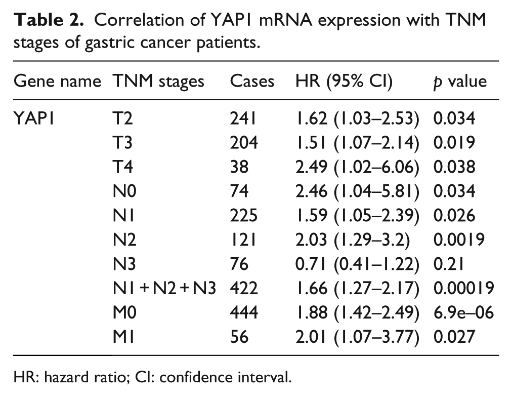
HR: hazard ratio; CI: confidence interval.
To further probe into the correlation of YAP1 transcription factor mRNA expression with other clinicopathological factors, gender (Table 3), differentiation (Table 4), and treatments (Table 5) of GC patients were also investigated. OS of both female and male patients was worse caused by high expression of YAP1 mRNA expression, with HR = 2.11 (1.38–3.24) and p = 0.00046 for female patients (n = 187) and HR = 1.39 (1.02–1.9) and p = 0.036 for male patients (n = 349; Table 3). We then analyzed the OS of differentiation status with GC patients. The sample size was too small in well-differentiated GC patients (n = 5), and we did not analyze the OS in this group. Moderately differentiated GC patients (n = 67) showed no correlation with OS of YAP1 mRNA expression—HR = 1.76 (0.92–3.37) and p = 0.082. But, high YAP1 mRNA expression in poorly differentiated GC patients (n = 121) was associated with poor OS of GC patients—HR = 1.7 (1.05–2.77) and p = 0.03 (Table 3). From Table 4, high YAP1 mRNA expression was correlated to worse OS in surgery-alone GC patients (n = 380)—HR = 1.65 (1.23–2.19) and p = 6e−04. High YAP1 mRNA expression was also correlated to worse OS in 5-fluorouracil (FU)-based adjuvant therapy–treated GC patients (n = 34)—HR = 5.74 (1.83–18.02) and p = 0.00089. YAP1 mRNA expression had no correlation to OS of other adjuvant therapy–treated GC patients (n = 76)—HR = 2.4 (0.99–5.82) and p = 0.045.
Correlation of YAP1 mRNA expression with gender of GC patients.

HR: hazard ratio; CI: confidence interval.
Correlation of YAP1 mRNA expression with differentiation of GC patients.

HR: hazard ratio; CI: confidence interval.
Correlation of YAP1 mRNA expression with different treatments of GC patients.

HR: hazard ratio; CI: confidence interval; FU: fluorouracil.
Discussion
GC causes the second-most high death rate of all cancer-related death worldwide. 1 Despite recent advances in GC treatment, the prognosis remains poor due to chemoradiotherapy resistance and metastasis. 13 To improve the prognosis of GC patients, it is critical to identify novel diagnostic tools and effective approaches.
YAP1 is the core downstream effector of the Hippo pathway. 14 Hippo pathway regulates cell apoptosis, proliferation, and organ sizes.15,16 Therefore, malfunction of the Hippo pathway is a key factor of tumorigenesis.17,18 YAP1 was reported to be elevated in several types of tumors, including OC, prostate cancer, colon cancer, and hepatocellular carcinoma (HCC). In HCC, YAP1 was reported to be activated and hence was used as an independent prognostic marker.19,20 Wang et al. 5 reported that YAP1 expression significantly correlated to pTNM-stage lymph node metastasis in non-small-cell lung cancer (NSCLC). High YAP1 expression led to short OS of NSCLC patients. 5 Xiao et al. 21 described upregulated YAP1 in 74.1% cases of NSCLC tissues. YAP1 was reported to increase OC cell growth and promote tumorigenesis in vitro and in vivo. 6 Therefore, YAP1 was considered a potential target site in OC cells. 6 To date, the role of YAP1 remains to be clarified, and it functions as a tumor marker and a tumor suppressor as well. Yuan et al. 10 reported YAP1 to be a tumor suppressor in breast cancer. They examined YAP1 expression in breast cancer tissues and normal breast tissue using IHC and discovered YAP1 loss in breast cancers. Small hairpin RNA (shRNA)-mediated knockdown of YAP1 in breast cancer cells also demonstrated increased invasiveness and tumor growth in vitro and in vivo, further confirming the role of YAP1 as a tumor suppressor. 10
YAP1 was reported to be elevated in gastric adenocarcinoma. 22 Ectopic expression of YAP1 in MKN45 cells promoted cell proliferation and invasion both in vitro and in vivo. 22 Several studies have demonstrated that high nuclear YAP1 expression is associated with poor prognosis in GC.23,24 Hu et al. 25 reported positive rate of YAP1 in GC was higher than in normal gastric mucosa or atrophic gastritis. Prognosis of GC patients with YAP1 overexpression was poorer than patients with negative YAP1 staining detected using IHC. 25 Therefore, we analyzed the prognostic role of YAP1 effector in GC.
In this study, we discussed YAP1 effector in prognostic value of GC. First, we compared the expression of YAP1 in GC and normal stomach tissues. Tissue microarray analysis was performed in 156 GC samples, 8 adjacent normal stomach tissues, and 4 normal stomach tissues. We found YAP1 high expression in 51.3% (80/156) of GC patients. We also analyzed the association between YAP1 protein expression, invasion depth, and lymph node metastasis. We found that high YAP1 expression in GC was significantly correlated to deep invasion depth and worse lymph node metastasis. We also analyzed the association between YAP1 protein expression and clinicopathological features, such as age, gender, histological differentiation, and clinical stages. We then used ONCOMINE database to clarify YAP1 expression in different types of tumors and different subtypes of GC. Then, KM plotter was applied to analyze the correlation of YAP1 mRNA to OS and other clinicopathological features. We summarized the prognostic value of YAP1 effector in GC patients using KM plotter of 631 GC patients. YAP1 was found to be associated to worse OS for all GC patients followed for 20 years.
Using KM plotter, we found that YAP1 mRNA correlated to worse OS in HER2-negative patients but not in HER2-positive patients. HER2 is a member of tyrosine kinase receptors and was reported to be overexpressed in 30% breast cancers.26,27 And it was reported to be correlated to worse clinical outcomes. 27 HER2 overexpression also occurs in 7%–34% of GC patients and is more common in GITA than in GDA and GMA patients.28–30 However, the association between HER2 positivity and prognosis in advanced GC is controversial. Some work showed poor prognosis associated with HER2 amplification, while others showed no prognostic difference in HER2-positive tumors compared with HER2-negative tumors.31,32 Therefore, further experiments are needed to determine whether YAP1 protein affects the HER2 status or it acts collectively or competitively toward the prognosis of GC. YAP1 mRNA expression was found not to be linked to OS in GITA patients, but correlated to worse OS in GDA patients and better OS in GMA patients detected using KM plotter. It was reported that HER2 overexpression was more common in GITA than in GDA and GMA patients. 28 However, due to limited number of GC specimen, we may also need to expand the GC tissues to further validate the correlation.
As to TNM stages of GC patients, except for T1 and N3 stages, YAP1 mRNA expression correlated to worse OS of T2, T3, T4, N0, N1, N2, N1+N2+N3, M0, and M1 GC patients. By further analyzing the prognostic roles of YAP1 in different clinicopathological features, including gender, differentiation, and treatments, we conclude that YAP1 has critical prognostic values in GC. These results suggest that YAP1 can be used to better predict the prognosis of GC patients and develop new therapies toward treating GC.
Footnotes
Declaration of conflicting interests
The author(s) declared no potential conflicts of interest with respect to the research, authorship, and/or publication of this article.
Funding
This research was supported by funds from the National Basic Research Program of China (973 Program, No. 2013CB911304 to Hui Wang and No. 2015CB553903 to Ding Ma) and Nature and Science Foundation of China (81372805, 81472783, and 81230038).