
Abstract
Crocodile choline, an active compound isolated from Crocodylus siamensis, was found to exert potent anti-cancer activities against human gastric cancer cells in vitro and in vivo. Our study revealed that crocodile choline led to cell cycle arrest at the G2/M phase through attenuating the expressions of cyclins, Cyclin B1, and CDK-1. Furthermore, crocodile choline accelerated apoptosis through the mitochondrial apoptotic pathway with the decrease in mitochondrial membrane potential, the increase in reactive oxygen species production and Bax/Bcl-2 ratio, and the activation of caspase-3 along with the release of cytochrome c. In addition, this study, for the first time, shows that Notch pathway is remarkably deregulated by crocodile choline. The combination of crocodile choline and Notch1 short interfering RNA led to dramatically increased cytotoxicity than observed with either agent alone. Notch1 short interfering RNA sensitized and potentiated the capability of crocodile choline to suppress the cell progression and invasion of gastric cancer. Taken together, these data suggested that crocodile choline was a potent progression inhibitor of gastric cancer cells, which was correlated with mitochondrial apoptotic pathway and Notch pathway. Combining Notch1 inhibitors with crocodile choline might represent a novel approach for gastric cancer.
Introduction
The incidence and mortality rate of human gastric cancer (GC) decreased substantially in many countries during the last few decades.1,2 However, it remains to be the second leading cause of cancer-related deaths worldwide. 3 At present, the main method of treatment for early GC remains to be surgical resection.4,5 Therefore, it is critical to find efficient compounds and therapies to reduce mortality.
The first Notch gene mutation in Drosophila melanogaster has been discovered for a century, 6 and the first characterization of a mammalian Notch gene was described in 1991. 7 The highly conserved pathway of Notch has a small number of signaling members. Four vertebrate Notch genes have been identified: Notch1–Notch4. Furthermore, five ligands, Dll-1, Dll-3, Dll-4, Jagged-1, and Jagged-2, have been discovered. 8 The most extensively characterized Notch pathway is a cell–cell communication system termed as canonical Notch pathway, wherein a membrane-tethered Notch ligand on a signal-sending cell interacts with a transmembrane Notch receptor on a signal-receiving cell. Then, the Notch1 intracellular domain (NICD) is liberated, which subsequently combines with the DNA-binding protein CSL (CBF1/Suppressor of Hairless/LAG-1) and mastermind-like protein (MAML) to induce the transcription of Notch downstream target genes. 9 And non-canonical Notch pathway has been well studied too. 10 Notch pathway plays critical roles in the cellular differentiation and development, including the progression and apoptosis.11,12 Recent studies have suggested that ectopic Notch signaling was implicated in numerous human diseases, including a broad spectrum of cancers. In many cancers, Notch activation can be oncogenic, but there is growing evidence revealing that the same pathway may be a tumor suppressor in hematopoietic cells, skin, and pancreatic epithelium. Notch signaling has been proposed to be a potential target in GC, and the potential therapeutic value of Notch inhibitors such as γ-secretase inhibitor (GSI) is now well appreciated, although its side effects such as thymic atrophy and intestinal goblet cell hyperplasia have been reported. 13 In this study, we explored the role of Notch signaling in the anti-tumor activity of crocodile choline (CCL).
Since the 1980s, bile and bile acids have gained extensive attention in the fields of chemistry and medicine. 14 The medicinal values of animal bile have been well recognized for its enhanced immunity, anti-inflammatory, anti-convulsion, and analgesic effects. 15 Our research concentrated on bile of Crocodylus siamensis, a kind of freshwater crocodile, which is distributed in South East Asia. Previously, we have explored and optimized the technology of bile juice extracting and purifying, and the product was named crocodile choline (CCL). The early study has shown that CCL experted a broad spectrum of anti-cancer efficacy, such as cholangiocarcinoma (CCA) cells and hepatocarcinoma (HCC) cells.2,16 However, its mechanism acting on the GC is still unknown.
In this study, results revealed that CCL suppressed the growth of GC cells and induced apoptosis through the mitochondria-mediated pathway and Notch pathway. These data suggested that CCL might be a potential therapeutic agent optional for human GC.
Materials and methods
Chemicals and reagents
Gallbladders of C. siamensis were supplied by Guangzhou Tuolong bio-technological developmental Co., Ltd . The extracting technology of CCL has been analyzed by our lab already. The powder called CCL was stored at −20°C. As needed, the stock solution of 10 mg/mL was diluted by dimethyl sulfoxide.
Cell culture and viability assay
The human GC cell lines BGC823, MGC803, SGC7901, and MKN28 and normal gastric mucosa epithelial cell line GES1 were maintained in RPMI 1640. The survival rates of cells were assessed by the 3-(4,5-dimethylthiazol-2-yl)-2,5-diphenyltetrazolium bromide (MTT) assay. The absorbance was analyzed at 490 nm using a microplate reader (POLARstar Omega, Germany).
Cell staining, cell colony–formation assay, and invasion ability analysis
Cells were treated for 24 h and harvested, washed with phosphate-buffered saline (PBS) three times, stained, and observed after washing (Nikon TE2000, Japan). In all, 1000, 2000, 4000 cells were seeded in 60 mm plates. The next day, plates were divided into two groups and treated with normal complete medium or medium-added CCL (10 µg/mL). After 24 h, CCL containing medium was dropped, and cells were allowed to form colonies for 2 weeks. Then, the adherence cell colonies were fixed with methanol and stained with Giemsa. Finally, the cell colony numbers were counted.
For the invasion assay, the membranes of the chambers were covered with Matrigel (BD Bioscience, USA) according to the manufacturer’s protocols. Chambers were stained with Giemsa and then photographed and quantified.
Flow cytometry
Cell apoptotic assay
After treatment, the apoptosis of cells was evaluated using Annexin V Apoptosis Detection Kit (Sangon Biotect, China) according to the manufacturer’s instruction. Then, fluorescence of cells was detected by Cytomics FC500 flow cytometry (FCM; Beckman-Coulter, USA).
Cell cycle analysis
After treatment, cells were harvested and washed with PBS. The cells were spinned down and the supernatant was removed. The pellet was fixed in ice-cold 700 mL/L ethanol for 30 min. The cells were washed twice and spinned down again. The pellet was resuspended in binding buffer. The cells were treated with 25 µL propidium iodide (PI; 10 mg/mL) and incubated on ice for 30 min without light.
Measurement of mitochondrial membrane potential (ΔΨm)
Cells were stained with 5,5′,6,6′-tetrachloro-1,1′,3,3′-tetraethylbenzimidazol-carbocyanineiodide (JC-1) at the cell incubator for 20 min, washed twice, and analyzed.
Measurement of reactive oxygen species
The 2′,7′-dichlorofluorescin diacetate (DCFH-DA) is a fluorogenic freely permeable tracer specific for reactive oxygen species (ROS) assay. After treatment, cells were incubated with 10 mmol/L DCFH-DA at 37°C for 30 min in the dark. After incubation, cells were harvested and washed with PBS and detected by FCM.
Cytochrome c release assay and western blot analysis
Mitochondria were isolated from cells using Cell Mitochondria Isolation Kit (Beyotime, China). Proteins from the cytosolic of each sample were further analyzed by western blot using an anti–cytochrome c antibody. Western blot protocols were employed according to Song. 2 Reactive proteins were detected by Bio-Rad Gel Imaging System (USA) with Enhanced Chemiluminescence Western Blot Kit (CWBIO, China). Glyceraldehyde-3-phosphate dehydrogenase (GAPDH) was used as an internal control.
Total RNA extraction, quantitative polymerase chain reaction, and transient protein downregulation by short interfering RNA
Total RNA was extracted from cells using an Isogen RNA extraction reagent RNAiso Plus (Takara, Japan). A volume of 1 µg of RNA was converted into complementary DNA (cDNA) using a PrimeScript™ RT Reagent Kit (Takara, Japan) with (genomic DNA) gDNA Eraser. The cDNA was used as a template for quantitative polymerase chain reaction (qPCR). It was performed by means of the CFX96™ Real-Time PCR Detection System (Bio-Rad, USA) using a SYBR® Premix Ex Taq™ (Takara, Japan). These genes were analyzed and the primers were designed according to the reported cDNA sequences (Table 1). Quantitative values were normalized to the GAPDH expression.
Primer sequences (5′-3′) for qPCR.

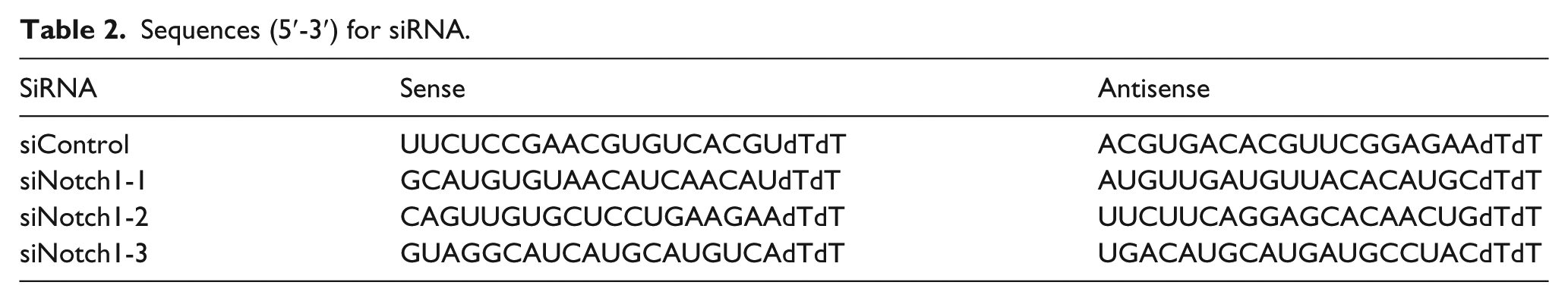
Cells were transfected with negative control short interfering RNA (siRNA) or Notch1 siRNA using lipofectamine 2000 (Invitrogen, USA) according to the manufacturer’s instruction with some adjustments. Sequences for siRNA were purchased from Biomics (Jiangsu, China; Table 2).
Sequences (5′-3′) for siRNA.
The xenograft model, hematoxylin–eosin staining of pathological paraffin sections, and immunohistochemistry
Nude mice (BALB/c) were purchased from SLRC Laboratory Animal Co., Ltd (Shanghai, China). About 1 × 10⁶ BGC823 cells were injected into nude mice. The tumor size was measured every 3 days. Tumor volumes were determined according to the following formula: A × B2/2. When the tumor reached a volume of 100 mm3, the mice were randomized to control and treated groups (n = 6 per group); then, control groups received corn oil (every other 3 days, intragastric (i.g.) administration) and treated groups received 100 mg/kg CCL (every other 3 days, i.g. administration). Mice were sacrificed at day 21. All manipulations were approved by the Animal Care and Use Committee of the Laboratory Animal Center of Xiamen University.
The tumor volume at day n was expressed as relative tumor volume (RTV). 2 After fixed with 10% neutral formalin, paraffin-embedded tissue sections were deparaffinized and rehydrated, stained with hematoxylin and eosin. Immunohistochemistry of tumor tissues collected from control and CCL-treated mice was performed using the DAB Detection Kit (Polymer; Maixin Biotechnologies, China).
Statistical analysis
All experiments were performed in at least triplicate. The results are expressed as mean ± standard deviation (SD). All statistical analyses were performed using the SPSS version 17.0. The p value <0.05 was considered statistically significant.
Results
CCL inhibited GC proliferation, colony formation, and invasion in vitro
The cytotoxicity of CCL was determined by MTT assay in vitro. First, for normal gastric mucosa epithelial cell line GES1, the cell survival rate had no significant change compared with the control group (Figure 1(a)). Then, for GC cell lines, we examined the efficacy of CCL on BGC823, MGC803, SGC7901, and MKN28 cells, with IC50 values of 21.56, 23.9, 28.4, and 33.1 µg/mL, respectively (Figure 1(b)). The most sensitive BGC823 cell line was employed for further study.

Crocodile choline (CCL) inhibited gastic cancer (GC) cell proliferation. (a) The cell survival rate of normal gastric mucosa epithelial cell line GES1 had no significant change compared with control group. (b) After exposure to different concentrations of CCL for different time, the IC50 values of various GC cell lines were measured. (c) Morphological changes of cells stained by Giemsa, Hoechst 33258, and AO/EB after exposure to CCL. Magnification: 200×. (d) Colony assay to detect the ability of colony-forming. (e) CCL inhibited the invasion of BGC823 cells. Magnification: 100×.
For Giemsa staining, the structure of control cells was integral with plenty cytoplasm, and deep-dyed big nuclei were clearly observed. While CCL-treated cells shrunk and turned round, the chromatin was condensed. For nuclear staining with Hoechst 33258, chromatin condensation and the destructive fragmentation of nuclei were detected in many CCL-treated cells, and those cells showed brighter blue light than control cells, indicating the pyknotic and deep-dyed nuclei. For acridine orange/ethidium bromide (AO/EB) staining, the control cells emitted green fluorescence and cell structures were integral, while quite a few treated cells displayed orange and red fluorescence and were smaller in size (Figure 1(c)). Our data indicated that the CCL-treated BGC823 cells displayed typical morphological features of apoptosis: shrunken nuclei, condensed chromatin, and round cells. The effect of the colony-forming inhibition of CCL was then investigated. The BGC823 cells are normal cancer cells that show the phenomenon of adherence. Based on the colony numbers we counted (Figure 1(d)), the colony-forming efficiency of the experimental group has a significant reduction compared with the control group. Transwell assay was used to simulate the matrix environment of extracellular space, which was the classical method of cell invasion analysis. As shown in Figure 1(e), after the treatment with the different concentrations of CCL, the number of cells passing through chambers decreased dramatically.
CCL induced apoptosis and caused cell cycle arrest in BGC823 cells
Treated with increasing CCL concentrations (Figure 2(a) and (b)), the rates of apoptosis and necrosis were raised from 3% to 27% (bp < 0.01). These results further demonstrated that CCL could induce obvious apoptosis in BGC823 cells. The changes in cell cycle distribution after treatment were also examined by FCM. As shown in Figure 2(c), the percentage of cells in G0/G1 phase decreased, while the percentage of cells in G2/M phase increased remarkably. These results suggested that CCL arrested BGC823 cells at G2/M phase and suppressed cell proliferation. Meanwhile, the protein levels of Cyclin B1 and CDK-1 decreased after treatment (Figure 2(d)), which were required for the transition of G2/M phase.

Effects of CCL on cell apoptosis, cell cycle distribution, ΔΨm, and ROS. (a) Assessment of apoptosis using flow cytometry. (b) The quantification of cell apoptotic proportion. (c) Cell cycle analysis. (d) Western blot to analyze the expressions of cell cycle regulating proteins after treated with CCL. (e) Flow cytometry analysis of ΔΨm, the increase rates of green fluorescence indicated reductions in ΔΨm. (f) Effect of CCL on the ROS levels in cells. The arrows indicate the raising of ROS. (g and h) The quantification of the changes of ROS and ΔΨm (bp < 0.01 vs control, n = 3).
Involvement of the mitochondria-mediated intrinsic pathway in CCL-induced apoptosis in BGC823 cells
JC-1 is a fluorescent probe for ΔΨm detection which generates red fluorescence when the level of ΔΨm is high and generates green fluorescence when the level of ΔΨm is reduced. The effect of CCL on ΔΨm in BGC823 cells was also examined by FCM. As shown in Figure 2(e) and (h), with the CCL concentration increased, the percentage of green fluorescence raises from 2% to 17% (bp < 0.01), which means a collapse of ΔΨm in BGC823 cells. Furthermore, to investigate whether CCL-induced apoptosis of BGC823 cells could be tied to the ROS generation, the intracellular ROS level was examined by FCM. After treatment, the mean of ROS increased from 4 to 56 (bp < 0.01), displayed a dose-dependent manner (Figure 2(f) and (g)). This result demonstrated that CCL could enhance the intracellular ROS level in BGC823 cells. Mitochondria are the primary site of ROS, making them uniquely vulnerable to oxidative damage, and oxidative damage can stimulate and lead to mitochondrial dysfunction.
Based on the study of ΔΨm and ROS changes, we further assessed the expression of proteins related to mitochondria-mediated intrinsic pathway after treatment (20 µg/mL CCL) during the different time periods. Western blot results showed that the treatment increased the ratio value of Bax/Bcl-2, caspase-3 was activated, and cytochrome c elevated obviously in cytosol (Figure 3(a)).

Apoptosis induced by CCL through the mitochondria pathway and Notch pathway. (a and b) Western blot for cell lysis collected to analyze the expression of mitochondria pathway–related and Notch pathway–related proteins after treated with CCL. (c and d) Relative Notch1 and Notch2 mRNA expression levels were examined by qPCR, and were normalized to GAPDH. (e) RT-PCR showing the efficiency of transfection. (f)–(k) The joint effect of CCL and siNotch1 on cell viability, invasion, cell cycle, ROS, and relative proteins, and each lane from left to right: control, sicontrol, siNotch1-3, CCL, siNotch1-3+CCL (ap < 0.05 and bp < 0.01, vs control, n = 3).
Involvement of Notch pathway in CCL-induced apoptosis in BGC823 cells and its concurrent treatment with CCL
To characterize the role of Notch pathway in CCL-treated GC cells, we detected some factors of this pathway (Figure 3(b)). The ligand Jagged-1 was detected with decrease, and intracellular domains NICD, intracellular domain of Notch2 (ICN-2), and some downstream targets were downregulated. The qPCR results also assessed that the membrane receptors Notch1 and Notch2 were also attenuated (Figure 3(c) and (d)). So, we came to a conclusion that Notch pathway also might be responsible for the changes of cells after treatment of CCL.
In addition, we explored the combination of Notch1 siRNA (siNotch1) and CCL treatment (20 µg/mL for 24 h). First, synthetic siNotch1-3 showed the potent downregulation effect, and it was used for further study (Figure 3(e)). Then, we observed that the concurrent treatment significantly decreased cell viability (bp < 0.01), and cells became more sensitive to CCL, with an IC50 value decreasing from 21.56 to 11.9 (Figure 3(f) and (g)). Then, this co-action on the invasion ability of cells, cell cycle, ROS, and relative proteins was analyzed. First, the invasion of cells was detected; with the siNotch1-3 transfection and CCL treatment, the number of cells passing through chambers was attenuated (from 76 to 35), while the combination group showed more significant decrease (from 76 to 13; Figure 3(h)). For cell cycle analysis and ROS measurement, promising joint effects were also achieved. Namely, Notch1 deficiency potentiated the ability of CCL on the G2/M cycle arrest and ROS production (Figure 3(i) and (j)). Moreover, western blot shed light on the details of Notch pathway–related and apoptosis-related proteins. Consistent upregulation/downregulation tendency was achieved by Notch1 deletion and CCL treatment, and the combined manipulation was more effective (Figure 3(k)). Taken together, these results verified the single action of Notch1 deletion on the cell progression and further demonstrated that the joint inhibition of BGC823 cells was more potent than observed with either agent alone.
CCL suppressed tumor growth in vivo
We further assessed the GC xenograft tumors. The weight of bodies and internal organs of nude mice displayed no significant changes compared to control after treatment (Figure 4(a) and (b)), while the volume of tumors demonstrated apparent differences between the treated and the control groups (at the 18th day, ap < 0.05 and at the 21st day, bp < 0.01). And the weight of tumors from CCL-treated was smaller than the control group (bp < 0.01; Figure 4(c)–(e)). To confirm the non-toxicity of CCL, the viscus of mice was stained with hematoxylin–eosin, as shown in Figure 4(g); internal organs had no remarkable changes in organizational structure. To the contrary, treated pathological paraffin sections of xenograft tumors displayed typical apoptotic features: condensed chromatin and pyknotic nuclei (Figure 4(f)). Our data by computation demonstrated that CCL effectively suppressed the growth of BGC823 xenograft tumors (T/C% = 59.85%) without toxic to the normal field.

CCL suppressed the growth of tumor in xenograft models without toxic to viscera. (a) The variational curve of nude mice body weight. (b) Relative visceral coefficient after administration (i.g.) of corn oil and 100 mg/kg CCL. (c) Mean tumor volume measured by caliper on the indicated days. (d) Mean tumor weight at the end of 21 days. (e) Solid tumors excised from treated nude mice compared with the control group. (f and g) Pathological paraffin sections of tumors and viscera stained by Hematoxylin–eosin staining. (h) Western blot of tumor lysis to confirm the expression of apoptosis- and Notch pathway–related proteins, as well as VEGF and PCNA. (i) Immunohistochemistry was performed to measure the expression of Bax, Bcl-2, Casp-3, Notch1, and VEGF in tumor tissues isolated from control and CCL-treated mice. (j) The expression of relative mRNA (ap < 0.05 and bp < 0.01, vs control, n = 6).
Furthermore, the expressions of messenger RNA (mRNA) and proteins obtained from tumors were measured by qPCR and western blot. We examined the apoptosis-related (Figure 4(h) and (j)) and Notch pathway–related (Figure 5) members. The changes of mRNA and proteins were consistent with experiments in vitro, as well as the protein abundance detected by the immunohistochemical examination (Figure 4(i)). Last but not least, VEGF (vascular endothelial growth factor) and PCNA (proliferating cell nuclear antigen), playing critical roles in angiogenesis and proliferation in cancer, decreased significantly.

The illustration of Notch pathway according to the Cell Signaling Technology, Inc. The red arrowheads point to the changes of relative proteins.
Discussion
We previously reported that CCL inhibited the progression and led to the apoptosis of various cancers, such as CCA and HCC. However, its effect and mechanism on GC were still unknown. This study proved that CCL does no harm to normal gastric mucosa epithelial cells. Then, poorly differentiated (BGC823 and MGC803), moderately differentiated (SGC7901), and well-differentiated (MKN28) GC cell lines were treated with CCL, and we found that the proliferation of these cell lines were all inhibited obviously. Furthermore, the cells of lower differentiation level seemed to be more sensitive to CCL, and the poorly differentiated BGC823 cells were found to be the most sensitive one, with the lowest IC50 value. However, many researchers17–19 have claimed that patients with the poorly differentiated type had more significant invasion, much more lymph node involvement, and more advanced stage than patients with the well-differentiated type, and had more aggressive features, poorer prognosis, and lower survival rates. Given this, poorly differentiated BGC823 cell line was used for further study.
One of the best-studied mechanisms of apoptosis is the mitochondria-mediated intrinsic pathway. Our findings elucidated in GC cells were in line with previous studies showing that the mitochondria-mediated intrinsic pathway was involved in CCA and HCC. After exposure of CCL, the intracellular ROS level increased dramatically, ΔΨm was collapsed and the cytochrome c got released into the cytoplasm from mitochondria. In addition, western blot results also indicated that the ratio of Bax/Bcl-2 was increased and the activity of caspase-3 was activated, promoting the release of cytochrome c. Mitochondria have the matrix, surrounded by the inner membrane (IM), and the intermembrane space, surrounded by the outer membrane (OM). The IM is almost impermeable in physiological conditions, therefore allowing the respiratory chain to create an electrochemical gradient (Δψm), the mitochondrial membrane potential. Δψm derives from electron-transport-chain-mediated pumping of protons and is vital for ATP production. 20 The changes in Δψm have been positioned as early, obligate events in the apoptotic signaling. 21 Mitochondrial control of apoptosis was thought to be associated with ATP production, Δψm, and mitochondrial membrane permeability for the release of certain apoptotic factors. 20 In the intrinsic pathway, outer stress or signals result in the permeabilization of OM, which leads to loss of Δψm and release of apoptosis-related proteins such as cytochrome c. Then, cytochrome c combines with caspase-9 and Apaf-1 and activates downstream caspases to initiate apoptosis. 22
Ectopic Notch pathway can often lead to the malignancy of cancer. Du et al. 23 revealed that NICD protein was overexpressed in GC tissues and was associated with overall patient survival, tumor metastasis, and an advanced tumor stage. In fact, the expressions of Notch1 and Hes1 were significantly higher in GC tissues than in normal tissues. 23 In addition, a number of studies24,25 have shown that the activation of Notch pathway played a critical role in GC initiation and proliferation. However, the expression of Notch pathway changed by CCL, and its roles remain unknown in GC. This study has, for the first time, showed that typical Notch ligands and Notch receptors were remarkably deregulated by CCL in vitro and in vivo. As we all know, recently, various biopharmaceutical or genetic Notch pathway inhibitors such as GSIs have been suggested to the treatment of cancer. 26 However, accumulation evidence has suggested that potent GSIs were effective to inhibit γ-secretase activity in GC cell lines but were limited in their ability to induce apoptosis. 27 What about CCL? What about the combination of CCL and Notch pathway inhibitors? Our results revealed that this concurrent treatment led to dramatically increased cytotoxicity than observed with either agent alone on GC cells. SiNotch1 sensitized and potentiated the ability of CCL to suppress the cell progression and invasion of GC cell. These studies did not employ a potent Notch inhibitor for combination treatment, but they might also represent a novel approach of combination therapy. Further studies will strengthen studies on a xenograft mouse tumor model, which may serve as an effective anti-tumor treatment against GC in vivo.
Accumulating evidence has revealed the crosstalk between the Notch pathway and other signalings, such as the Wnt, 28 and the mechanisms of interactions were divided into three different categories. Perumalsamy et al. first observed the link between mitochondrial dynamics and Notch signaling. Their study 29 positioned the mitochondrion as an integral part of a Notch-activated signaling cascade that regulated cell progression, and NICD inhibited mitochondrial fragmentation and controlled Bax oligomerization to avoid apoptosis. Moreover, ROS and Ca2+ emerged as the signal to regulate Notch pathway.30,31 However, Notch signaling was found modulating mitochondrial respiration through a complicated way in many cancers such as breast cancer. 32 Thus, the crosstalk is bidirectional, and the mutual effect and actions between mitochondrial pathway and Notch signaling arouse attention increasingly.
In our study, Notch1 knockdown greatly increased ROS generation and caused G2/M cell cycle arrest and consequently enhanced CCL-induced cell apoptosis. As we know, ROS plays a causal role in mitochondria apoptotic pathway, while Tormos et al. 33 suggested that ROS played an early, causal role in cellular differentiation. Increased mitochondrial mass and cellular ROS content have been correlated with cellular differentiation in various systems. Notch inhibits ROS production, and conversely, ROS generation could affect Notch pathway. Kim et al. 34 reported that Notch deletion led to G2/M cell cycle arrest and apoptosis through interaction with the Cyclin B1 and CDK-1 complex. Our data were in concordance with the previous study, and the CCL-affected crosstalk between mitochondria pathway and Notch pathway was detected deeply. Collectively, the enhancement of CCL-induced apoptosis by Notch1 deficiency could be partly attributed to the interaction between the two pathways, and more attention should be given on their coordinated approach to the cellular differentiation, progression, apoptosis, and so on.
The xenograft model was employed to confirm the consistency with the vitro assays and determine the non-toxic and efficient of CCL. Our data demonstrated that 100 mg/kg body weight dose of CCL every 3 days was moderately effective (T/C% = 59.85%) and did not have toxic side effects. CCL also effectively inhibited the expression of PCNA and VEGF, which played critical roles in angiogenesis and proliferation in cancer. The molecular mechanism demonstrated the consistency between vitro and vivo after treated with CCL. Therefore, our data suggested that CCL was a safe, natural, and effective compound for GC.
Collectively, our demonstration in BGC823 cells that CCL changed the levels of ΔΨm and ROS and regulated the expression of relative proteins suggested that mitochondria-mediated intrinsic pathway and Notch pathway were involved in it. However, the type of action and the exact mechanism by which CCL came into play still keep unclear. Regardless, these findings not only highlight the efficient anti-tumor activity of CCL but also suggest a novel approach of combination treatment.
Footnotes
Acknowledgements
X.-M.M. and Q.-R.F. contributed equally to this work.
Declaration of conflicting interests
The author(s) declared no potential conflicts of interest with respect to the research, authorship, and/or publication of this article.
Ethical approval
All manipulations were approved by the Animal Care and Use Committee of the Laboratory Animal Center of Xiamen University.
Funding
This work was supported by the Natural Science Foundation of China (No. 81571418); the National Science Foundation for Fostering Talents in Basic Research of the National Natural Science Foundation of China (No. J1310027); the Natural Sciences Foundation of Fujian Province, China (No. 2016J05105); the Natural Sciences Foundation of Guangdong Province, China (No. 2016A030310374).