
Abstract
Background and Objectives:
Attention-deficit/hyperactivity disorder (ADHD) is coupled with subtle variations in fronto-striato-cerebellar brain circuits. Although advanced neuroimaging supports these findings, conventional MRI—being broadly accessible and cost-effective—has constrained proven utility in ADHD. This study was done with the objective of describing the frequency and nature of structural brain abnormalities detected on conventional MRI among children diagnosed with ADHD.
Materials and Methods:
A retrospective study of 154 children aged 1–12 years diagnosed with ADHD (Diagnostic and Statistical Manual of Mental Disorders-5 criteria) at a tertiary care centre in Chennai between January and December 2024 was conducted. MRI (3T, T1-weighted, T2-weighted, and FLAIR sequences) was assessed. Children with comorbid psychiatric or neurological conditions, TBI, metallic implants, or incomplete imaging were excluded. MRI findings were evaluated qualitatively with a focus on the frontal lobe, basal ganglia, and cerebellum.
Results:
The cohort (mean age 6.29 ± 3 years) was primarily male (83.8%). Comorbidities included autism spectrum disorder (66.2%), speech delay (12.3%), and seizures (3.9%). MRI was reported as normal in 94.8% of cases. Frontal lobe hyperintensities were seen in 3.3%, cerebellar anomalies in 1.9%, and no basal ganglia abnormalities were observed. Only 2.6% of scans showed potentially ADHD-related findings. Incidental observations (e.g., cavum septum pellucidum/vergae) were rare and statistically unrelated to comorbidities.
Conclusion:
Conventional MRI notices structural brain changes in a small proportion of children with ADHD. Routine use in typical ADHD cases may have limited clinical value unless atypical features are present.
Introduction
The global prevalence of Attention-Deficit Hyperactivity Disorder (ADHD) among children and adolescents was 8%.[1] Characterised by symptoms of inattention, hyperactivity, and impulsivity, ADHD can significantly impact academic performance, social interactions, and overall quality of life.[2] While the exact aetiology of ADHD remains unclear, growing evidence suggests that structural and functional abnormalities in specific brain regions, such as the prefrontal cortex, basal ganglia, and cerebellum, may play a critical role in its pathophysiology.[3]
Neuroimaging studies, particularly those using magnetic resonance imaging (MRI), have provided valuable insights into the neural correlates of ADHD, offering a window into the structural and functional alterations associated with the disorder.[4] Conventional MRI, which includes T1-weighted, T2-weighted, and fluid-attenuated inversion recovery (FLAIR) sequences, is widely used in clinical practice to assess brain anatomy and detect structural abnormalities. While advanced imaging techniques like functional MRI (fMRI) and diffusion tensor imaging (DTI) have been extensively utilised in ADHD research, conventional MRI remains an accessible and cost-effective tool for evaluating brain structure in clinical settings.[5]
Despite its widespread use, the role of conventional MRI in identifying structural abnormalities in children with ADHD has not been thoroughly explored. This study aims to address this gap by retrospectively analysing conventional MRI findings in children diagnosed with ADHD.
Background and Justification
The Neurobiological Basis of ADHD
ADHD is increasingly recognised as a disorder with a strong neurobiological basis. Structural neuroimaging studies have consistently reported volumetric differences in key brain regions involved in attention, impulse control, and motor coordination. For instance, reductions in the volume of the prefrontal cortex, a region critical for executive functions, have been frequently observed in children with ADHD.[6] Similarly, abnormalities in the basal ganglia, particularly the caudate nucleus and putamen, have been linked to the motor and inhibitory control deficits characteristic of ADHD.[7]
The cerebellum, traditionally associated with motor coordination, has also been implicated in ADHD, with studies reporting reduced cerebellar volume and altered connectivity among affected individuals.[8]
While these findings highlight the potential of neuroimaging in understanding ADHD, most studies have relied on advanced imaging techniques, which are not always available in routine clinical practice. Conventional MRI, on the other hand, is widely accessible and can provide valuable information about brain structure. However, its role in ADHD research has been underexplored, particularly in the context of identifying structural abnormalities that may serve as diagnostic markers.
The Role of Conventional MRI in ADHD
Conventional MRI offers several advantages in the study of ADHD. It is non-invasive, widely available, and provides high-resolution images of brain anatomy. T1-weighted imaging is particularly useful for assessing grey matter volume and cortical thickness, while T2-weighted and FLAIR sequences are effective in detecting white matter abnormalities and lesions.[9]
Despite these advantages, the use of conventional MRI in ADHD research has been limited, with most studies focusing on advanced techniques like fMRI and DTI. This study aims to bridge this gap by systematically analysing conventional MRI findings in children with ADHD.
Study Objectives
This study has two main objectives:
To identify and describe conventional MRI brain imaging findings in children diagnosed with ADHD. By retrospectively reviewing MRI scans of children with ADHD, this study aimed to catalogue the structural abnormalities observed in this population. This will provide a comprehensive overview of the brain regions most commonly affected in ADHD. To explore the frequency and distribution of structural abnormalities in specific brain regions (e.g., prefrontal cortex, basal ganglia, cerebellum).
The study will focus on key brain regions implicated in ADHD, such as the prefrontal cortex, basal ganglia, and cerebellum, to determine the prevalence and distribution of structural abnormalities in these areas.
By achieving these objectives, this study seeks to contribute to a deeper understanding of the structural brain alterations associated with ADHD and evaluate the clinical utility of conventional MRI in its diagnosis and management.
Methodology
A retrospective descriptive observational study was conducted to evaluate the structural brain abnormalities in children diagnosed with ADHD using conventional MRI imaging. The study was carried out from January 2024 to December 2024 at a tertiary care centre with advanced neuroimaging facilities. Children diagnosed with ADHD who attended the tertiary care centre during the study period (January 2024 to December 2024) were included in the study. The inclusion criteria comprised children aged 1–12 years diagnosed with ADHD according to the Diagnostic and Statistical Manual of Mental Disorders, 5th Edition (DSM-5) criteria, availability of conventional MRI brain imaging reports, and absence of other neurodevelopmental or psychiatric disorders. Exclusion criteria included a history of traumatic brain injury, documented neurological or psychiatric comorbidities, presence of metallic implants or other contraindications for MRI, and incomplete or inadequate MRI reports and MRI images with artefacts. All children who met the inclusion and exclusion criteria were considered for analysis.
Ethical approval for the study was obtained from the Institutional Ethics Committee, Biomedical Research, Apollo Hospitals, AHEL, Chennai. Confidentiality and anonymity of patient data were maintained throughout the study. A retrospective review of clinical MRI reports of children diagnosed with ADHD was conducted by consecutive sampling. Relevant imaging findings such as grey matter volume changes, white matter abnormalities, and cortical thinning were extracted. Image analysis included qualitative assessment of MRI findings (e.g., normal vs. abnormal brain structures) and identification of structural abnormalities in key brain regions associated with ADHD, such as the prefrontal cortex, basal ganglia, and cerebellum.
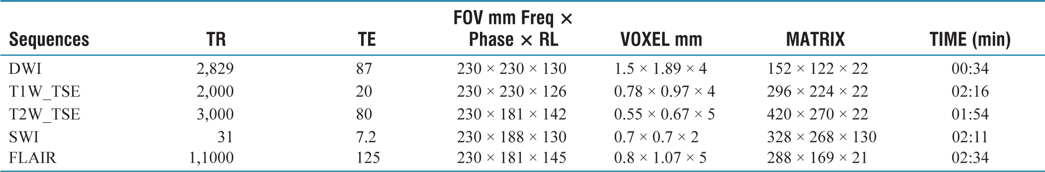
MRI examinations were performed using a three-Tesla Magnet MRI scanner (PHILIPS MR System Ingenia) to ensure high-resolution imaging for accurate assessment of structural brain changes. The sequences and imaging parameters used are tabulated below. The study aimed to provide insights into the neuro-anatomical variations in ADHD children, contributing to a better understanding of the disorder’s structural brain correlates.
Data collected were entered in MS Excel and analysed using SPSS version 16. Descriptive statistics were used to summarise the frequency and distribution of MRI findings.
Results
This study included 154 children aged 1–12 years with ADHD who underwent MRI. The mean age was 6.29 + 3 years. The majority of them were in the age group 5–10 years (51.9%), 33.8% of the participants were under five years of age, and the rest (14.3%) were >10 years of age. 83.8% of the study participants were male children, and the remaining 16.2% were female children.
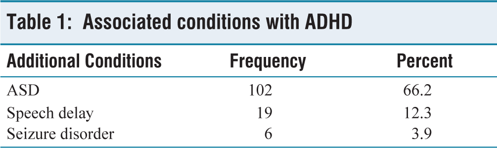
Table 1 shows the associated conditions with ADHD; the majority of them had autism spectrum disorder (ASD) (66.2%), followed by speech delay (12.3%) and seizure disorder (3.9%).
Associated conditions with ADHD
MRI Findings Among Children with ADHD
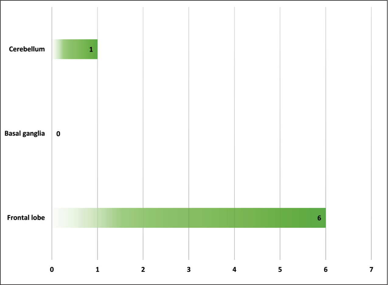
Abnormal MRI findings were observed in the frontal lobe in six children (3.9%), in the cerebellum in one child (0.65%), and no abnormal findings were observed in the basal ganglia [Figure 1]. Non-specific findings were also observed, which were negligible and are tabulated below.
Site of abnormal MRI findings among the ADHD children
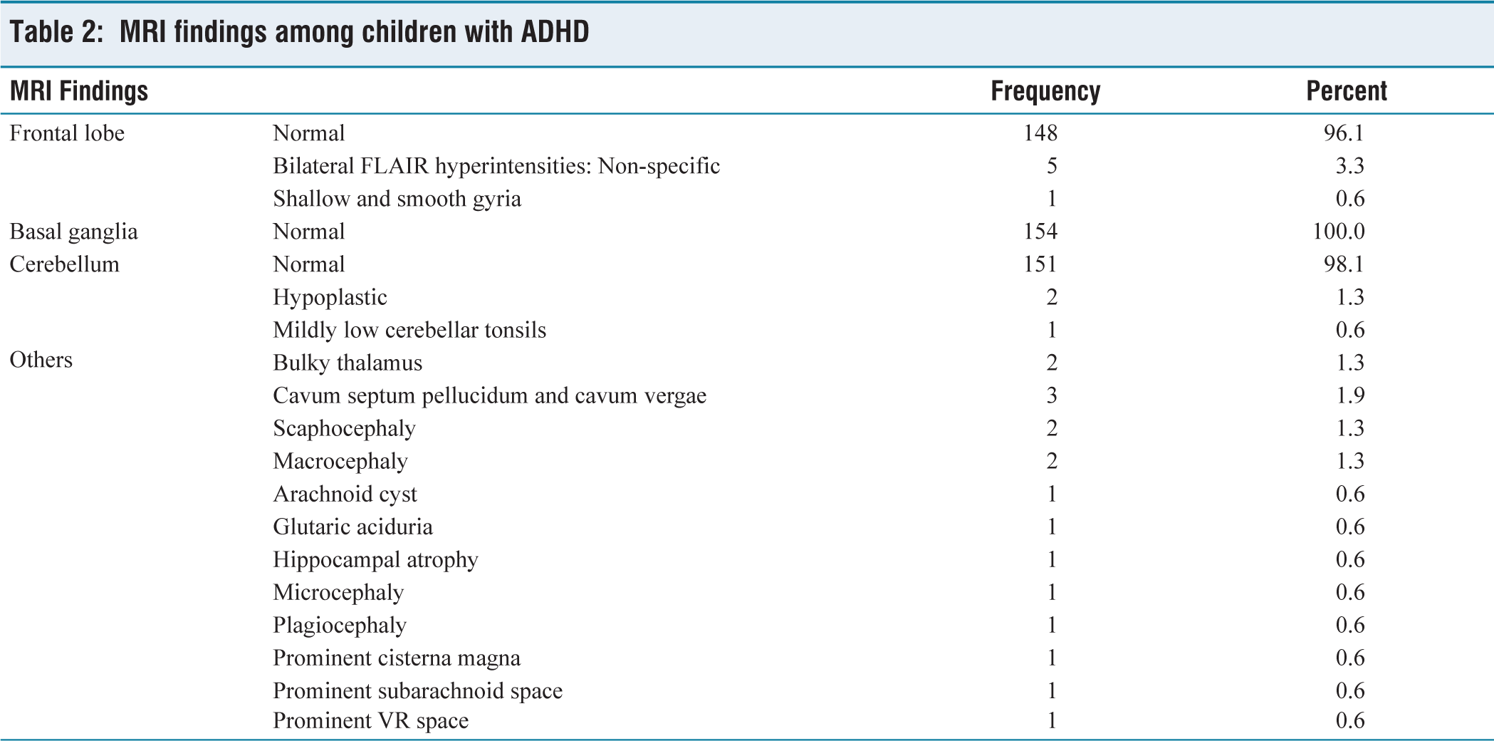
MRI findings in the frontal lobe reveal that 96.1% of the MRIs were normal, 3.3% had non-specific bilateral FLAIR hyperintensities, followed by shallow smooth gyria in 0.6%.
MRI findings in the basal ganglia were normal in all participants (100%). In the cerebellum, 98.1% (n = 151) were normal, while 1.3% (n = 2) had a hypoplastic cerebellum. Other MRI findings observed among the ADHD children were Bulky thalamus (1.3%), Cavum septum pellucidum and cavum vergae (1.9%), Macrocephaly (1.3%) and scaphocephaly (1.3%).
MRI findings observed among children with ADHD were tabulated and listed in Table 2. Of the 154 study participants who were subjected to MRI, only 2.6% had specific features related to ADHD, such as shallow and smooth gyria (n = 1), hypoplastic cerebellum (n = 2) and low-lying cerebellar tonsils (n = 1). The rest of the MRIs were widely normal, and a few had non-specific findings which were neither exclusive nor confirmatory for ADHD. Figures 2-7 shows the key MRI findings among the study participants.
MRI findings among children with ADHD
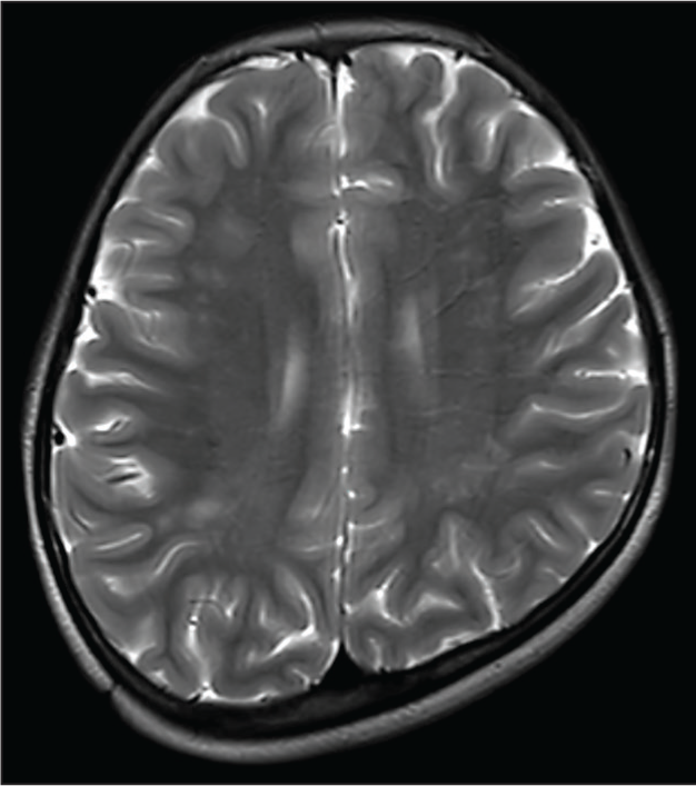
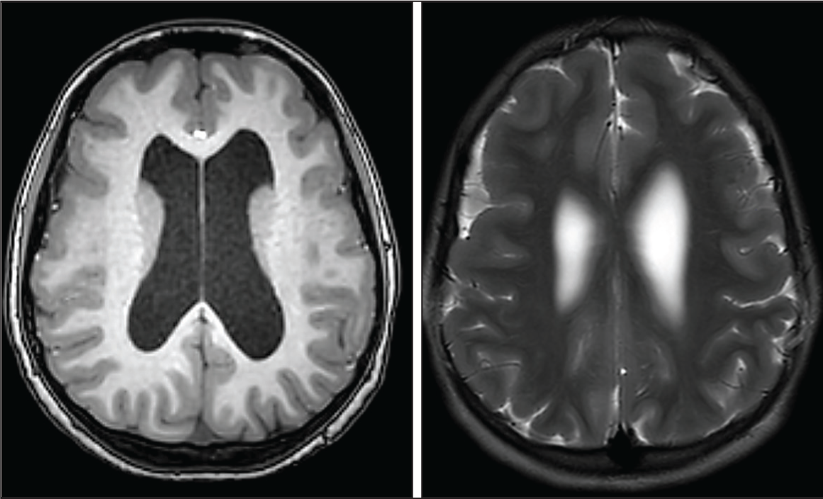
Axial T2 showing plagiocephalic skull with discrete areas of hyperintensity in bilateral frontal and parietal white matter, which are non-specific
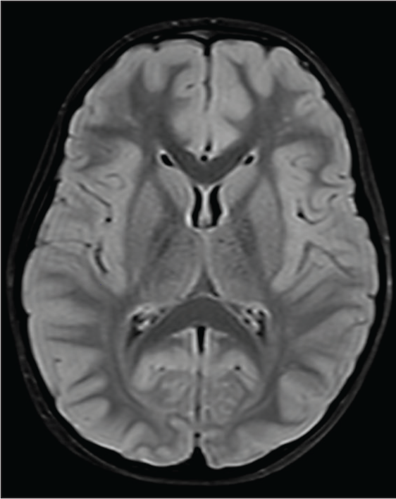
Axial FLAIR image showing bilateral non-specific frontal FLAIR hyperintensities
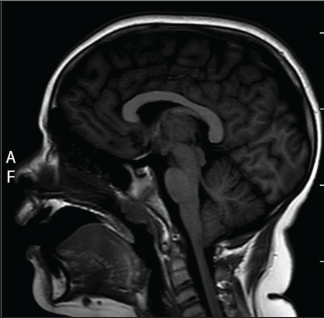
Sagittal T1 image showing low-lying cerebellar tonsils
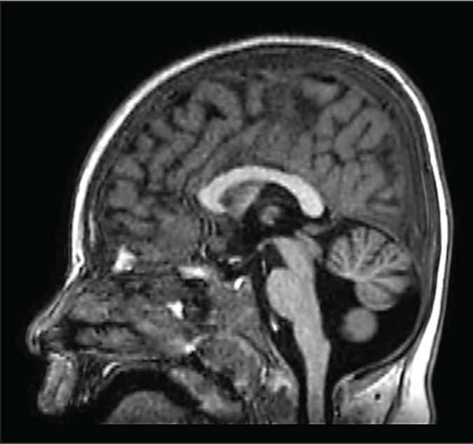
Sagittal T1 image showing Ponto-cerebellar hypoplasia
Axial T1 and T2 weighted images showing shallow and smooth gyri with cortical thickening
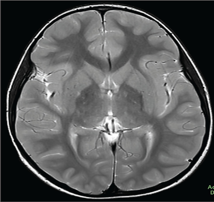
Axial T2-weighted image showing bilateral symmetrical mild Bulky thalamus
Moreover, there was no statistically significant association between associated clinical conditions like ASD, Seizure disorder, speech delay and MRI findings for ADHD at any sites by χ 2 test statistics, with a P value >.05.
Discussion
This retrospective analysis assessed conventional MRI brain findings among 154 children, clinically diagnosed with ADHD, with a special focus on the structural anomalies in areas concerned in the disorder, specifically the frontal lobe, basal ganglia, and cerebellum.
The majority of the study participants in our study were male children (83.8%) and in the age category of 5–10 years, which represents the global epidemiological profile, signifying a greater prevalence of ADHD among boys and in early to middle childhood.[1,10]
Our study population closely lines up with that of earlier neuroimaging literatures, which mostly consider children between 6 and 12 years of age, the phase in which the symptoms of ADHD are more evident and the phase where diagnosis occurs more frequently. However, there is literature to substantiate the work on a varied age group, like Hoogman et al.[11] who studied a wider cross-sectional population involving children to adults, and Shaw et al.[12] who studied 4–18 years involving both children and adolescents to assess the pattern of cortical development. Our study sample had a significant proportion of ASD (66.2%) as an associated condition, when compared to most other earlier studies, which often tend to ignore such comorbidities to assess ADHD-specific neuro-anatomical findings. This inclusion mirrors real-world clinical intricacy; however, it may also add to heterogeneity in radio-imaging findings.
Our study findings reveal that conventional MRI has detected anomalous findings in the frontal lobe among 3.9% of participants and 1.9% of participants in the cerebellum, while all MRIs revealed no abnormalities in the basal ganglia. The most common observation was bilateral FLAIR hyperintensities in the frontal lobe, which were non-specific. Largely, >97% of the MRIs were without any significant findings related to ADHD, emphasising the reduced sensitivity and questionable utility and role of conventional MRI in ADHD.
These findings are, however, in contrast to the earlier literature employing advanced imaging modalities. Shaw et al.[12] described delay in cortical maturation and decreased cortical thickness, especially in the prefrontal cortex, among children with ADHD. Similarly, Nakao et al.[13] highlighted the reduction in grey matter in the prefrontal cortex and basal ganglia, while Hoogman et al.[11] reported reduced volumes in the basal ganglia (Amygdala, Caudate, and Putamen). These studies used high-end advanced volumetric and surface-based morphometric evaluation, which have feasibility constraints with the conventional MRI.
Norman et al.[14] and Valera et al.[7] also described structural abnormalities in the frontal-striatal-cerebellar circuits, with a special focus on decreased cerebellar and caudate volumes. However, our study appreciates no such abnormalities, probably attributed to the diminished sensitivity of traditional qualitative assessment using conventional MRI sequences.
Notably, few incidental and non-specific observations such as macrocephaly, cavum septum pellucidum and prominent subarachnoid spaces were noted, while their clinical significance in ADHD is inconclusive. These findings were supported by the observations of Castellanos et al.,[8] who stated variability in incidental abnormal findings in MRI among children with ADHD.
Our study findings highlight that, while conventional MRI can be a valuable tool for detecting gross structural anomalies, it lacks the degree of sensitivity to spot subtle, yet clinically substantial neuro-anatomical findings correlated with ADHD. This opens the gate for evidence generation on the utility of advanced imaging techniques, such as DTI and fMRI in ADHD.
Furthermore, the lack of a significant association between associated conditions like ASD, speech delay and structural abnormalities in MRI advocates that conventional MRI observations are unlikely to aid as a standalone radio-imaging marker for ADHD or its associated clinical conditions. This is in line with the current accord that diagnosis of ADHD remains largely clinically driven, with imaging modalities providing a supportive role rather than a diagnostic role.
Conclusion
This retrospective analysis aimed to describe the findings of conventional MRI among children with ADHD. While the majority of MRIs were normal, a meagre proportion displayed non-specific observations in the frontal lobe. Remarkably, no structural abnormalities were detected in the basal ganglia, an area usually involved in the pathophysiology of ADHD. These observations convey that conventional MRI may have a restricted role in identifying the subtle changes, such as thinning of the cortex or volumetric reductions in certain regions of the brain, usually described in ADHD.
Limitations
A single-centre approach in a retrospective design carries the limitation of availability and quality of the image data, which could potentially contribute to inter-subject variability and under-reporting. Relatively limited sample size and lack of a control or comparative group limit further exploration. Also, due to its retrospective nature and completeness of available data, ADHD subtype-specific analysis was not feasible within the current dataset.
Recommendations
A larger multicentric study with robust methodology involving standardised image interpretation protocols and application of advanced imaging modalities is expected to throw light on the areas of uncertainty in the arena of ADHD.
Footnotes
Acknowledgements
We thank the institution (Apollo Hospitals, Greams Road, Chennai) for rendering us the required support in shaping this work.
Declaration of conflicting interests
The authors declared no potential conflicts of interest with respect to the research, authorship and/or publication of this article.
Funding
The authors received no financial support for the research, authorship and/or publication of this article.
Institutional ethical committee approval number
This study has obtained ethics committee approval form the Institutional Ethics Committee, Biomedical Research, Apollo Hospitals, AHEL, Chennai, carrying the approval number: AMH-C-S-067/04-25, Dt:29/04/2025.
Informed consent
Not applicable.
Credit author statement
Tamilselvan T: Participated in conceptualisation, Methodology, Data collection, Data analysis, Literature Search, and Manuscript Preparation.
Aishwarya B: Involved in Conceptualisation, Literature Search, Validation, Supervision, and manuscript revision.
All the authors have reviewed and approved the manuscript.
Data availability
The datasets used and/or analysed during the current study are available from the corresponding author upon request.
Use of artificial intelligence
We acknowledge the use of an AI tool (ChatGPT-4.0-OpenAI) solely for language refinement and content editing purposes, and we declare no role of AI in conceptual design, methodology or scientific observations.