
Abstract
Background and Aims:
Birth injuries in newborns can range from mild soft tissue damage to fractures of long bones and even bleeding within the brain. Bilateral auricular haematomas are uncommon clinical entities found in newborn babies.
Methods:
We present a rare case of a 12-days old baby with bilateral auricular haematoma.
Results:
This baby underwent incision and drainage with bolster dressing under general anaesthesia.
Conclusion:
Birth trauma is an extremely rare cause of manifesting bilateral auricular haematoma. In auricular haematoma, blood collecting in the subperichondrial space compromises the vascular supply of the adjacent cartilage and results in cartilage necrosis and tissue destruction. It needs prompt identification and treatment to prevent cartilage necrosis of the pinna and deformity. Treatment of auricular haematoma needs drainage and evacuation of the haematoma.
Introduction
Birth injury sometimes adversely affects the foetus during the delivery process.[1] The traumatic injuries during birth may be divided into hypoxia to the foetus and injuries of the face due to mechanical forces.[1] Auricular haematoma is an uncommon complication of birth trauma. Auricular haematoma is a collection of blood that separates the perichondrium from the cartilage of the pinna.[2] Auricular haematoma most commonly occurs by blunt trauma to the pinna, though it may develop spontaneously or from conditions such as autoimmune disease and infection.[2] The shearing forces between the anterior perichondrium and underlying cartilage are responsible for the development of haematoma.[2] There are several treatment options for preventing the complications of auricular haematoma and returning the auricle to its pre-trauma form. The improper drainage of haematoma or late treatment can result in cauliflower deformity of the pinna.[3]
Case report
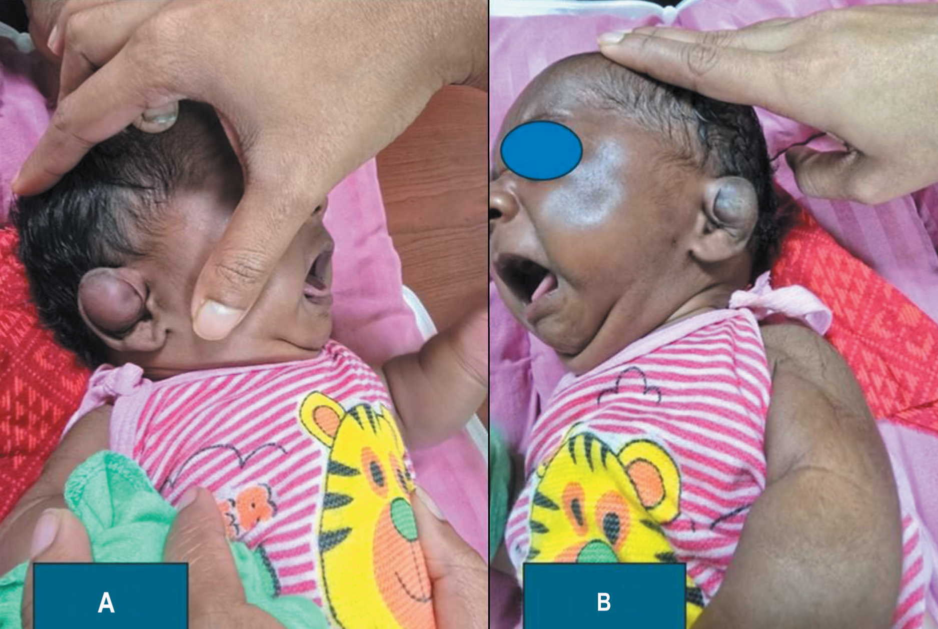
A 12-day-old male infant with an unremarked medical history presented with a bulging auricle/pinna on both sides. There was a history of mild bruising of both auricles just following delivery. The mother first noticed this swelling a day prior and mentioned it during a consultation at the outpatient department. The infant had a normal physical examination by the paediatrician, showing no signs of bruising anywhere on the body except in left pinna, both during the hospital stay and at follow-up visits, which included careful monitoring of weight gain and jaundice. The parents denied any insect bites or environmental allergies. As per birth history, the baby was born at 39 weeks and 3 days via spontaneous vaginal delivery to a 30-year-old, gravida 1 para 1. The baby was delivered small for gestational age, weighing 2,600 grams, with Apgar scores of 8 and 9 at one and five minutes, respectively. The baby received the hepatitis B vaccine, vitamin K after birth. The physical examination of the baby showed a healthy, well-nourished infant who was not in any apparent distress. He had only boggy swellings on the anterior wall of the bilateral pinna with mild bruising on the left side [Figure 1]. No additional bruises, scars, rashes or patterned injuries were observed. The needle aspiration confirmed the haematoma of the pinna. The bleeding parameters of the baby were within normal limits. Parents also denied any anticoagulant treatment. The baby underwent incision and drainage along with bolster dressing, under general anaesthesia. At the outpatient department of otolaryngology follow-up, the baby showed satisfactory healing and no re-accumulation of the haematoma following removal of the bolster dressing after seven days.
(A and B) Newborn baby showing bilateral auricular haematoma
Discussion
Auricular haematoma typically results from injury to the anterior part of pinna.[4] This trauma can occur from a direct impact on the pinna, such as in road traffic accidents, contact sports like boxing or through a traumatic piercing at the upper part of the helix.[4] Birth injury usually occurs during the birth process at the transit through the birth canal.[5] Trauma to the foetus or neonates during the delivery process can be due to several factors such as placenta, foetus, mother and/or instrumentation.[5] Birth-related trauma is commonly caused by challenging vaginal deliveries, especially when the shoulder is in a vertex presentation, or the arms are extended in a breech delivery. Other contributing factors include macrosomia, shoulder dystocia and the use of delivery instruments such as forceps or a vacuum.[6] Birth trauma to the pinna results in extravasation of blood between the cartilage and perichondrium causing a soft, doughy swelling of the pinna. The lack of subcutaneous fat protecting the perichondrium, which covers the cartilage, makes the anterior surface of the pinna particularly vulnerable to trauma. The blood supply to the pinna comes from the posterior auricular and superficial temporal arteries, both of which originate from the external carotid artery.[6] When trauma affects the pinna, it damages the perichondrium and capillaries, causing the perichondrium to separate from the cartilage. This separation creates a space that allows blood to accumulate. The site of the haematoma is classically seen between the perichondrium and cartilage of the pinna; however, in some cases, haematoma can be seen within the cartilage itself. Once blood accumulates in this space, it leads to vascular compromise of the adjacent cartilage and venous congestion resulting in histologic changes and cartilage deformity, called cauliflower ear.[7] Necrosis of the cartilage leads to changes in the normal histologic structure of the cartilage framework of the pinna. Auricular haematoma contains blood and leads to abscess formation following infection. The pinna or external ear is not only involved in hearing but also responsible for providing aesthetics of the face and head. Any change in the shape of the pinna is noticeable and so the patient require repair. In auricular haematoma, the pinna appears reddish blue in colour. When the pinna swelling is complete, it is hard to touch.[8] Auricular haematoma is usually seen in young adult males. In this case, the auricular haematoma was found in a newborn baby due to birth injury. The incidence of birth trauma has been reported reduced over time due to improvements in obstetric care and proper prenatal diagnosis. If treatment of the auricular haematoma is delayed or inadequate, cauliflower ear deformity may arise due to complications. If the auricular haematoma is not treated in time, the blood becomes organised, and the pinna remains permanently thickened due to the development of new fibrocartilage.[9] This deformity of the pinna is also called a Wrestler’s ear or Cauliflower’s ear. One study showed that 12% of patients with auricular haematoma presented with perichondrial reaction, 20% showed thickening of the pinna and 4% presented deformity of the pinna those were managed with window procedure.[10] Sometimes, patients with auricular haematoma are presented with cosmetic deformity of the pinna following attempts of aspiration followed by the development of an abscess of the pinna with cartilage necrosis and discoloration of skin and tenderness. The important differential diagnosis of auricular haematoma includes haemangioma, perichondritis, pseudocyst, seroma, keloid, Winkler disease (relapsing perichondritis), erysipelas and skin cancer.[11,12] These lesions of the pinna often resemble an auricular haematoma that needs proper evaluation and management. The history and clinical presentations are helpful to give the diagnosis of the auricular haematoma. Needle aspiration often confirms the diagnosis of auricular haematoma. Early identification of obstetric factors and stoppage of traumatising manoeuvres can reduce the incidence of birth injuries such as facial trauma or auricular haematoma. Aspiration and drainage of an auricular haematoma is a straightforward procedure that facilitates recovery. The drainage of the auricular haematoma is done via a surgical incision on the most prominent area over the helix followed by debridement. In case of a large auricular haematoma presenting for more than six hours, blood begins to coagulate; it becomes more difficult to aspirate through simple needle aspiration, requiring a surgical incision and drainage for removal. This treatment can include an incision and drainage along the helix and irrigation of subperichondrial space.[7] To avoid the recurrence of auricular haematoma, it is recommended to maintain pressure on the treated site of the pinna for five to seven days. Proper compression with a splint or bolster should be applied after drainage to prevent the formation of dead space.[13] Recently, steroids or OK-432 applications are considered as an important option.[14] A report showed that good outcomes occur with fibrin glue haemostatic material.[15] Sclerotherapy with OK-432 induces strong inflammation at the injection sites in auricular haematoma without any evidence of deformity. Sclerotherapy by OK-432 does not need hospitalisation or leave any scar on skin of pinna.[16] In this case, incision and drainage of auricular haematoma were done in both pinna with bolster dressing under general anaesthesia.
Conclusion
Bilateral auricular haematoma in newborns is a very uncommon complication due to birth injury. Infants with auricular haematoma need prompt consideration for further evaluation of child abuse or any haematological disorders. Prompt drainage of the auricular haematoma prevents re-accumulation of blood. The cauliflower ear is a permanent pinna deformity that occurs when the auricular haematoma is not completely drained, recurs or is left untreated.
Footnotes
Acknowledgements
The author is thankful to all nursing officers of the ENT department of AIIMS, Bhubaneswar, India, for timely help with patient care.
Declaration of conflicting interests
The author declared no potential conflicts of interest with respect to the research, authorship and/or publication of this article.
Funding
The author received no financial support for the research, authorship and/or publication of this article.
Institutional ethical committee approval number
Not applicable as this is a case report.
Informed consent
The parents of the infant were informed that data from the research would be submitted for publication, and they consented.
Credit author statement
SKS was involved in patient management, data collection, writing and drafting.
Data availability
The data are available with the due consent of the patients.
Use of artificial intelligence
Nil.