
Abstract
Sir,
Antibodies directed against nuclear antigens (ANA) give rise to different immunofluorescence patterns. Commonly, homogenous, fine speckled and nucleolar patterns are observed in patient with autoimmune illnesses. Uncommonly, Golgi-like pattern is reported, albeit with different illness including autoimmune- lupus, primary Sjögren syndrome (pSS) and nonautoimmune diseases—viral infections. Rarely, bullous pemphigoid (BP) is reported. 1 We report a case of uncommon association of BP and small vessel vasculitis, which showed Golgi-like immunofluorescence pattern on Hep-2 cell.
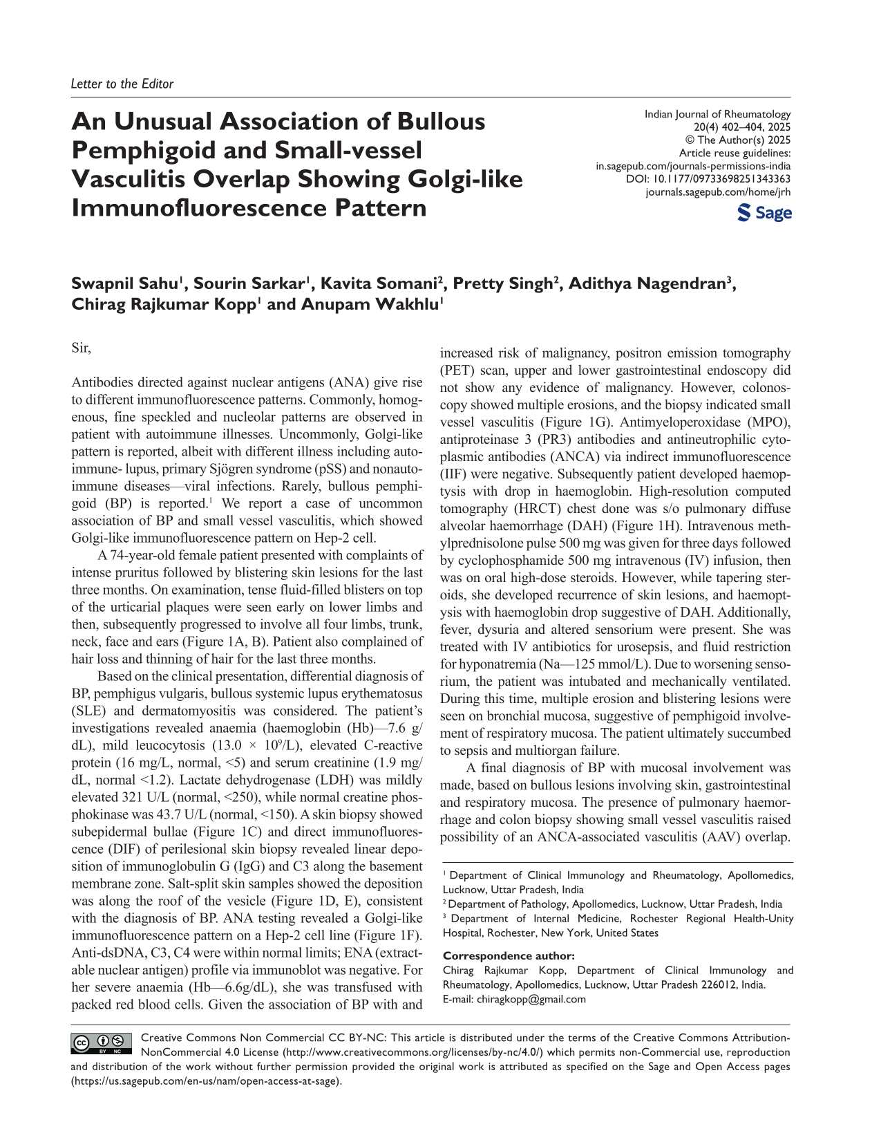
A 74-year-old female patient presented with complaints of intense pruritus followed by blistering skin lesions for the last three months. On examination, tense fluid-filled blisters on top of the urticarial plaques were seen early on lower limbs and then, subsequently progressed to involve all four limbs, trunk, neck, face and ears (Figure 1A, B). Patient also complained of hair loss and thinning of hair for the last three months.
Based on the clinical presentation, differential diagnosis of BP, pemphigus vulgaris, bullous systemic lupus erythematosus (SLE) and dermatomyositis was considered. The patient’s investigations revealed anaemia (haemoglobin (Hb)—7.6 g/dL), mild leucocytosis (13.0 × 10⁹/L), elevated C-reactive protein (16 mg/L, normal, <5) and serum creatinine (1.9 mg/dL, normal <1.2). Lactate dehydrogenase (LDH) was mildly elevated 321 U/L (normal, <250), while normal creatine phosphokinase was 43.7 U/L (normal, <150). A skin biopsy showed subepidermal bullae (Figure 1C) and direct immunofluorescence (DIF) of perilesional skin biopsy revealed linear deposition of immunoglobulin G (IgG) and C3 along the basement membrane zone. Salt-split skin samples showed the deposition was along the roof of the vesicle (Figure 1D, E), consistent with the diagnosis of BP. ANA testing revealed a Golgi-like immunofluorescence pattern on a Hep-2 cell line (Figure 1F). Anti-dsDNA, C3, C4 were within normal limits; ENA (extractable nuclear antigen) profile via immunoblot was negative. For her severe anaemia (Hb—6.6g/dL), she was transfused with packed red blood cells. Given the association of BP with and increased risk of malignancy, positron emission tomography (PET) scan, upper and lower gastrointestinal endoscopy did not show any evidence of malignancy. However, colonoscopy showed multiple erosions, and the biopsy indicated small vessel vasculitis (Figure 1G). Antimyeloperoxidase (MPO), antiproteinase 3 (PR3) antibodies and antineutrophilic cytoplasmic antibodies (ANCA) via indirect immunofluorescence (IIF) were negative. Subsequently patient developed haemoptysis with drop in haemoglobin. High-resolution computed tomography (HRCT) chest done was s/o pulmonary diffuse alveolar haemorrhage (DAH) (Figure 1H). Intravenous methylprednisolone pulse 500 mg was given for three days followed by cyclophosphamide 500 mg intravenous (IV) infusion, then was on oral high-dose steroids. However, while tapering steroids, she developed recurrence of skin lesions, and haemoptysis with haemoglobin drop suggestive of DAH. Additionally, fever, dysuria and altered sensorium were present. She was treated with IV antibiotics for urosepsis, and fluid restriction for hyponatremia (Na—125 mmol/L). Due to worsening sensorium, the patient was intubated and mechanically ventilated. During this time, multiple erosion and blistering lesions were seen on bronchial mucosa, suggestive of pemphigoid involvement of respiratory mucosa. The patient ultimately succumbed to sepsis and multiorgan failure.
(A) Peeled Off Bullous Haemorrhagic Skin Lesions Over Trunk and (B) Left Ear on Erythematous Skin Base. (C) Histopathology (200×) of Skin Biopsy Sample Showed Subepidermal Vesicle, Filled with Fibrin and Inflammatory Cells Infiltrate Chiefly Comprising of Eosinophils (arrow). Superficial Dermis Showed Oedema and Perivascular Aggregates of Lymphocytes, Plasma Cells, and Few Eosinophils. Basal Cell Vacuolisation Is Also Noted. Direct Immunofluorescence (DIF) Shows Linear Positivity (3+) Along the Roof of Bulla for C3 (D) and IgG (E). (F) Antinuclear antibody (ANA) on Hep-2 Cell Shows Golgi-like (Polar) Pattern. (G) Histopathology (200×) Biopsy from Small Intestine Shows Leukocyte Infiltration of Vessel Wall. (H) High-Resolution Computed Tomography Scan (HRCT) Shows Ground Glass Opacities and Consolidation in Lung Parenchyma.

A final diagnosis of BP with mucosal involvement was made, based on bullous lesions involving skin, gastrointestinal and respiratory mucosa. The presence of pulmonary haemorrhage and colon biopsy showing small vessel vasculitis raised possibility of an ANCA-associated vasculitis (AAV) overlap. However, vasculitis on biopsy can also be seen with BP, and haemorrhagic bullae lining the respiratory tract explains the haemoglobin drop, but not the findings seen on CT scan. Hence, a possibility of overlap with small vessel vasculitis cannot be excluded. Haemoglobin decline can occur due to haemoptysis from the blistering mucosal lesions in the lungs, which can mimic pulmonary haemorrhage. Additionally, anti-MPO and anti-PR3 antibodies were negative by enzyme-linked immunosorbent assay (ELISA), and ANCA was negative by IIF. While coexisting AAV and BP have been reported, these typically show positive ANCA serology. 2 Small vessel vasculitis can cause bullous-like lesions, and the colon mucosal biopsy confirmed vasculitis, and moreover, AAV can show Golgi-like ANA pattern.3,4 However, the skin biopsy DIP showed C3 and IgG deposits, which is characteristic for BP, not seen in vasculitis. Nonetheless, we cannot completely rule out possibility of small vessel vasculitis overlap. Golgi-like (or polar) ANA pattern shows a ribbon-like staining with polar distribution of immunofluorescence in cytoplasm and is classified as AC-22 by international consensus on ANA patterns. 5 In the Golgi complex, the autoantigens include golgin-67,97 and p115, golgin-95/gm130, golgin-160/GCP170, golgin-245/p230 and giantin/macrogolgin. 4 The anti-Golgi complex autoantibodies (AGA) were first reported in pSS, then in lupus, rheumatoid arthritis, mixed connective tissue disease, ataxia and human immunodeficiency virus (HIV) infection. More than 50% patients harbour autoantibodies against giantin/macrogolgin, having the highest molecular weight—345kDa—among all the Golgi autoantigens. Our hypothesis is that the AGA might cause aberrant transport of hemidesmosome antigens—BP230 and BP180, which are targets involved in BP, that may lead to exposure of antigenic sites. The AGA may also cross react with these antigenic epitopes. AGA by causing protein trafficking disruption and immune cell activation by binding to Golgi proteins aberrantly expressed on the surface of cells may lead to cellular injury, causing inflammation and tissue damage. In conclusion, the association of BP with extensive mucosal involvement and small vessel vasculitis is rare, but the presence of a Golgi-like ANA pattern is an important diagnostic clue. Our case emphasises the importance of ANA patterns in the evaluation of blistering disorders. The Golgi-like ANA pattern may serve as a useful indicator, especially when biopsy and serological tests are inconclusive in diagnosing BP. Additionally, while extensive blistering lesions can also occur in conditions such as lupus and dermatomyositis, the presence of a Golgi-like ANA pattern may help differentiate these diseases and point more specifically toward BP.
Footnotes
Declaration of Conflicting Interests
The authors declared no potential conflicts of interest with respect to the research, authorship and/or publication of this article
Funding
The authors received no financial support for the research, authorship and/or publication of this article.
Informed Consent
A written informed consent from her husband was taken.