
Abstract
Presentation
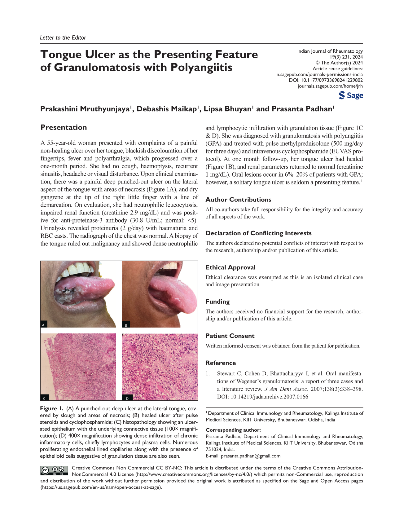
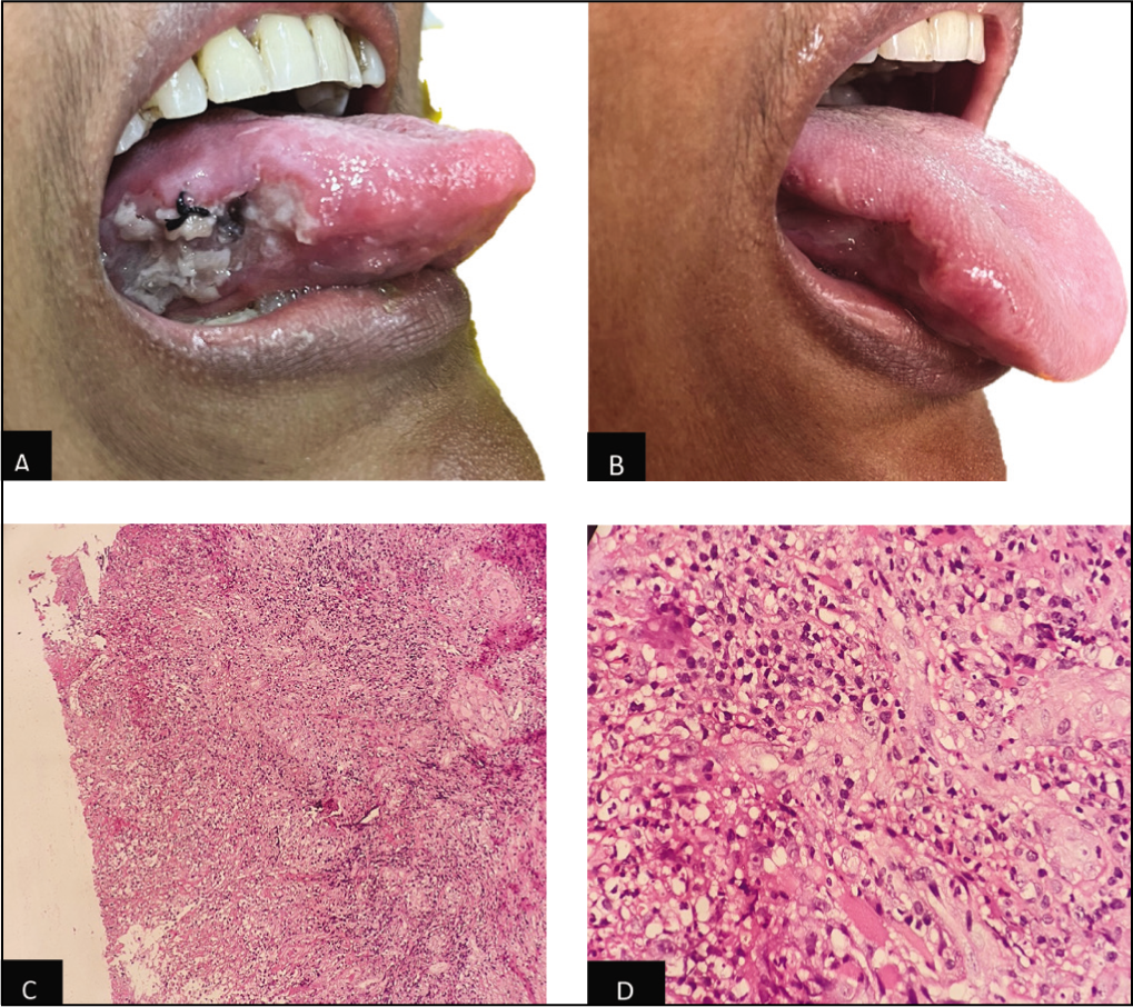
A 55-year-old woman presented with complaints of a painful non-healing ulcer over her tongue, blackish discolouration of her fingertips, fever and polyarthralgia, which progressed over a one-month period. She had no cough, haemoptysis, recurrent sinusitis, headache or visual disturbance. Upon clinical examination, there was a painful deep punched-out ulcer on the lateral aspect of the tongue with areas of necrosis (Figure 1A), and dry gangrene at the tip of the right little finger with a line of demarcation. On evaluation, she had neutrophilic leucocytosis, impaired renal function (creatinine 2.9 mg/dL) and was posit-ive for anti-proteinase-3 antibody (30.8 U/mL; normal: <5). Urinalysis revealed proteinuria (2 g/day) with haematuria and RBC casts. The radiograph of the chest was normal. A biopsy of the tongue ruled out malignancy and showed dense neutrophilic and lymphocytic infiltration with granulation tissue (Figure 1C & D). She was diagnosed with granulomatosis with polyangiitis (GPA) and treated with pulse methylprednisolone (500 mg/day for three days) and intravenous cyclophosphamide (EUVAS protocol). At one month follow-up, her tongue ulcer had healed (Figure 1B), and renal parameters returned to normal (creatinine 1 mg/dL). Oral lesions occur in 6%–20% of patients with GPA; however, a solitary tongue ulcer is seldom a presenting feature. 1
(A) A punched-out deep ulcer at the lateral tongue, covered by slough and areas of necrosis; (B) healed ulcer after pulse steroids and cyclophosphamide; (C) histopathology showing an ulcerated epithelium with the underlying connective tissue (100× magnification); (D) 400× magnification showing dense infiltration of chronic inflammatory cells, chiefly lymphocytes and plasma cells. Numerous proliferating endothelial lined capillaries along with the presence of epithelioid cells suggestive of granulation tissue are also seen.
Author Contributions
All co-authors take full responsibility for the integrity and accuracy of all aspects of the work.
Footnotes
Declaration of Conflicting Interests
The authors declared no potential conflicts of interest with respect to the research, authorship and/or publication of this article.
Ethical Approval
Ethical clearance was exempted as this is an isolated clinical case and image presentation.
Funding
The authors received no financial support for the research, authorship and/or publication of this article.
Patient Consent
Written informed consent was obtained from the patient for publication.