
Abstract

To the Editor,
Immunoglobulin G4-related disease (IgG4-RD) is a multiorgan immune-mediated condition that mimics many malignant, infectious and inflammatory disorders. 1 Erdheim-Chester disease (ECD) is a rare non-Langerhans histiocytic multisystem disorder. ECD most commonly manifests as multifocal sclerotic lesions of the long bones demonstrating sheets of foamy histiocytes on biopsy, with or without histiocytic infiltration of extra-osseous tissues. 2 Clinical symptomatology and histopathology help in the diagnosis and differentiation of both diseases. However, ECD is a close differential of IgG4-related disease in presentation and organ involvement. We report a rare case of IgG4-related disease with a biopsy showing features of IgG4 RD plus ECD and how clinico-radiological features along with therapy helped in the diagnosis. Written consent from the patient was taken for publishing this case including images.
A 64-year-old male patient presented to our OPD with a history of swelling in the right upper eyelid for the past eight months and a history of bilateral submandibular areas for the past two months. He reported a painless nodular swelling on the lateral aspect of the right upper eyelid that progressively increased in size and caused the narrowing of the lateral palpebral fissure. The patient also had two small, painless oval swellings in the submandibular area. There were no other accompanying symptoms like visual disturbances, redness of eyes, sicca symptoms, purpura, epistaxis, nasal polyps, arthritis, photosensitivity oral ulcer, Raynaud’s phenomenon, fever, weight loss, dry cough, shortness of breath. The patient was previously diagnosed with Gilbert syndrome at the age of 15 (asymptomatic since then).
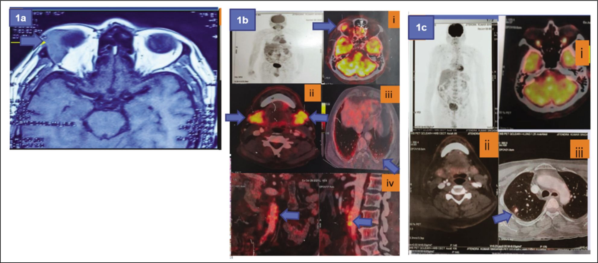
(a) MRI Brain Showing Heterogeneously Enhanced Soft Tissue in Superolateral Quadrant of Right Orbit Without Any Evidence of Bone Erosion (Arrow Pointing). (b) PET CT Before Therapy Showing Metabolic Uptake in Right Orbit (i), Submandibular Gland (ii), Basal Areas of Lungs (iii), Walls of Abdominal Aorta (iv )(Arrows Pointing). (c) PET CT After Six Months Showing Complete Resolution in Right Orbit (i), b/l Submandibular Gland (ii), b/l Basal Lung Fields Except for a Residual Patch in Right Middle.
The 2019 ACR EULAR Classification criteria for IgG4-related diseases strictly mentions the exclusion criteria according to which biopsy showing markers of Macrophage or histiocytic disorder should not classified as IgG4. However, the problem with our index case is that the biopsy showed features of both IgG4RD and Erdheim-Chester.
4
Elevated IgG4 levels and IgG4 staining are non-specific in biopsy as they can be seen in both these diseases. However clinical picture and some features on PET CT (major salivary gland involvement, infrarenal aorta involvement), IgG4/CD 138+ plasma cells >40% on IHC pointed more towards IgG4RD.
5
Overall, the resolution of symptoms with therapy suggested IgG4-related disease and this case highlights the approach and how therapy helped us in solving the case.
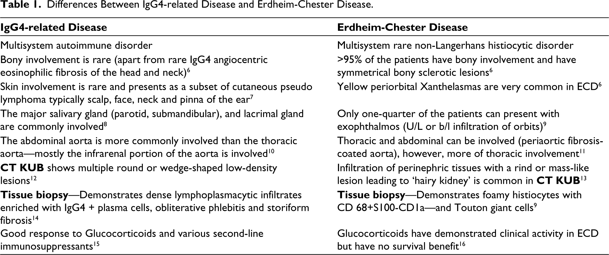
Differences Between IgG4-related Disease and Erdheim-Chester Disease.
Footnotes
Declaration of Conflicting Interests
The authors declared no potential conflicts of interest with respect to the research, authorship and/or publication of this article.
Funding
The authors received no financial support for the research, authorship and/or publication of this article.