
Abstract
Background
Diabetic foot ulcer, a severe complication occurring in diabetic patients, lacks effective remedies at present.
Purpose
The objective of this research is to investigate the impacts of exogenous irisin on diabetic wounds in mice and explore the associated mechanisms.
Materials and Methods
An animal model was constructed by administration of streptozotocin (STZ) (50 mg/kg) in mice to simulate a diabetic condition. A full-thickness excisional wound was produced on the dorsal skin of the mice to create a wound for the observation of the wound healing process. Cutaneous administration of 0.1 mL of exogenous irisin solution (1 µg/mL and 3 µg/mL) was applied to the injured tissue to assess the impact of exogenous irisin on the wound. Hematoxylin and eosin (H&E) staining assay was applied to evaluate histological changes in the skin tissue. The expression levels of endogenous irisin, adenosine 5′-monophosphate-activated protein kinase (AMPK), nuclear factor kappa B (NF-κB), and inflammatory factors in the skin tissue of the mice were measured by enzyme-linked immunosorbent assay (ELISA).
Results
The results showed that irisin levels are positively correlated with the progress of wound healing. The STZ mice exhibited decreased pAMPK, increased NF-κB, and increased inflammatory cytokines. Exogenous irisin treatment restored these abnormal expressions in STZ mice. However, co-treatment with the AMPK inhibitor, Compound C, abolished exogenous irisin’s function of promoting wound healing and relieving inflammation.
Conclusion
Taken together, exogenous irisin possesses a therapeutic effect on skin wounds in STZ-induced diabetic mice, which is related to the regulation of inflammatory factors mediated by the AMPK–NF-κB pathway.
Introduction
Diabetes, a prevalent chronic metabolic disease, has affected an estimated population of 600 million worldwide by 2022, and its incidence is still increasing year by year (Lang et al., 2021). Serious complications may develop in diabetic patients, and diabetic foot ulcer (DFU) represents the most frequent complication. DFU affects at least 15% of people with diabetes, with approximately 20% experiencing non-traumatic amputation or even death (Hart et al., 2017). At present, there are no successful clinical remedies for DFU aside from enhancing diabetes management or resorting to surgical intervention (Bellary et al., 2021). Therefore, there is an urgent requirement for studies investigating the underlying mechanisms of DFU and developing effective treatments.
Because of the insufficient ability in glucose metabolism, diabetic patients exhibit chronic hyperglycemia. Hyperglycemia impairs all stages of the wound-healing cascade, including hemostasis, inflammation, proliferation, and remodeling (Patel et al., 2019). Overexpression of inflammatory mediators in diabetic wounds, such as interleukin-1β (IL-1β), interleukin-6 (IL-6), and tumor necrosis factor-alpha (TNF-α), affects the replication of fibroblasts and epithelial cells (Dardmah & Farahpour, 2021). Thus, wound healing often stalls at the inflammation phase, resulting in delayed wound healing (Geerlings & Hoepelman, 1999). Therefore, promoting wound cells by shortening the inflammation phase may improve DFU.
Irisin, a muscle-derived protein, has been shown to increase with exercise in humans and animals. In response to exercise, this myokine is released into the bloodstream subsequent to proteolytic processing of its cellular precursor (Boström et al., 2012). It is well known that muscle and blood irisin can improve the body’s energy expenditure and can alleviate metabolic diseases such as diabetes and obesity. In recent years, irisin has also been found to have anti-inflammatory effects (Belviranli et al., 2016; Deng et al., 2020; Jiang et al., 2021; Trettel et al., 2023). A prior study illustrated the potential of irisin to mitigate neuroinflammation in cerebral hemorrhage mice. The positive effects of irisin were achieved by increasing the expressions of integrin αVβ5, pAMPK, and Bcl-2, and decreasing the expressions of IL-1β, TNF-α, MPO, and Bax following cerebral hemorrhage via integrin αVβ5/AMPK signaling pathway (Wang et al., 2022). In Jiang et al.’s study (2020), irisin was shown to protect rats from spinal cord injury, which was associated with adenosine 5′-monophosphate-activated protein kinase (AMPK)/nuclear factor kappa B (NF-κB)-mediated suppression of IL-1β, IL-6, TNF-α, inducible nitric oxide synthase (iNOS), and cyclooxygenase-2 (COX-2). In another study performed by Jiang et al. (2021), irisin protects against endometritis in female mice. The protective effect was also associated with the AMPK/NF-κB pathway. According to the aforementioned studies, irisin has therapeutic effects on multiple diseases in animal models, the mechanism of which is associated with the modulation of AMPK and inflammatory factors. However, whether irisin can alleviate diabetic wounds is unknown.
This study sought to evaluate the therapeutic potential of exogenous irisin in promoting wound healing in streptozotocin (STZ)-induced diabetic mice model. The expression of AMPK and its downstream factors, including NF-κB, IL-6, TNF-α, COX-2, and iNOS, was detected to explore the possible mechanisms.
Materials and Methods
Animals and Treatment
Male ICR mice (20–25 g, 10 weeks old), obtained from the Experimental Animal Center of Wenzhou Medical University, were kept in a room under standard conditions (temperature: 25°C ± 1°C; humidity: 40%–60%). The experimental procedures were approved by the Laboratory Animal Ethics Committee of Zhejiang Pharmaceutical University (No. ZYLL 202308011) and performed pursuant to the National Institutes of Health Guide for Care and Use of Laboratory Animals.
Experimental Design
Part 1
This part is to establish an animal model of diabetes with wound and explore the possible correlation of endogenous irisin with wound healing. For the animal grouping, a total of 24 mice were randomly assigned into the sham and STZ-treated groups. Each group was further divided into three subgroups based on different time points (3-day, 7-day, and 14-day post-wound injury, n = 4 per subgroup). Diabetes was triggered by injecting STZ (50 mg/kg) for 5 days in mice. Fresh STZ (sigma) solution (5 mg/mL, i.p.) was prepared in a sodium citrate buffer (pH = 4.5) (Zhao et al., 2018). Mice with blood glucose levels exceeding 16.7 mmol/L for 1 month were deemed as having diabetes (Ding et al., 2022). After the diabetic condition was established, a full-thickness excisional wound measuring 1 cm in diameter was made on the dorsal side of the mice. The observation period of the wound-healing process was 14 days. Images of the wound areas were taken and analyzed using ImageJ software (National Institutes of Health). Skin tissue samples surrounding the wounds were collected for subsequent analysis of irisin levels.
Part 2
This part is to evaluate the effects of exogenous irisin (sigma) on wound healing. Thirty mice were randomly assigned into five groups (n = 6 for each group): sham group, STZ group, 1 µg/mL irisin-treated STZ group, 3 µg/mL irisin-treated STZ group, and 3 µg/mL irisin + AMPK inhibitor (Compound C, 10 mg/kg, i.p.)-treated group. The experimental protocol was the same as that in Part 1. For the AMPK inhibitor-treated group, Compound C was administered 30 min before the application of exogenous irisin. The dosage of Compound C was based on a previous study (Jiang et al., 2019). Exogenous irisin was administered using a cotton swab to apply 0.1 mL of the irisin solution onto the wound. Skin tissue samples surrounding the wounds were collected on the 14th day post-injury.
Histology Assay
Histological changes were evaluated through hematoxylin and eosin (H&E) staining. Specifically, sections of dorsal skin tissue from mice were collected and immediately fixed using 5% paraformaldehyde. Then, the paraffin-embedded tissue sections were dewaxed by two 5-min immersions in xylene. After thorough water rinsing, nuclei were stained with hematoxylin for 5 min, then rinsed. Sections were differentiated in acid alcohol for 2 s and blued in tap water. The cytoplasm was counterstained with eosin for 1 min. Post-eosin staining, sections were dehydrated through 70%–100% ethanol, cleared in xylene for two 2-min periods, and a coverslip was applied with a resinous medium for microscopic examination.
Enzyme-linked Immunosorbent Assay (ELISA)
Levels of NF-κB p65, COX-2, iNOS, pAMPK, AMPK, IL-6, and TNF-α in the skin tissue were assessed by the corresponding ELISA kits (Abcam, USA). The irisin level was assessed using an ELISA kit obtained from Phoenix Pharmaceuticals, Burlingame, CA, USA. The tissue was weighed and sheared, then RIPA lysate was added (450 µL of lysis solution per 50 mg of tissue) and placed on ice for grinding. Following centrifugation at 12,000 rpm for 5 min, the supernatant was obtained. For ELISA analysis, diluted protein standards and samples were added to a 96-well ELISA plate, then incubated with biotin-conjugated antibodies. After washing with wash buffer, wells were supplemented with premixed avidin-horseradish peroxidase complex. The reaction was finally stopped using a stopping solution. Optical density (OD) was measured at 450 nm for irisin, pAMPK, AMPK, IL‑6, TNF‑α, COX‑2, and iNOS, while absorbance for NF‑κB p65 was detected at 405 nm.
Statistical Analysis
Statistical analysis was performed by one-way ANOVA, followed by Tukey’s test for multiple group comparisons. The data were expressed as mean ± SD. p < .05 was considered a significant difference.
Results
The Correlation Between the Expression of Endogenous Irisin and Wound Healing in STZ-induced Diabetic Mice
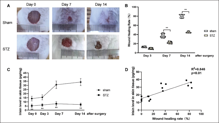
As shown in Figure 1, STZ-treated mice exhibited significantly delayed wound closure (Figure 1A), and the wound healing rates were significantly lower in the STZ-treated mice (Figure 1B) relative to sham-operated mice (p < .01 at both day 7 and day 14). Interestingly, the endogenous irisin level in the epidermal tissue of the sham mice significantly increased from day 7 after injury (p < .01, Figure 1C), whereas the irisin level in the STZ-mice was persistently low for 2 weeks after injury and were significantly lower compared with the sham mice (p < .01, Figure 1C). Correlation analysis found that endogenous irisin level had a positive correlation with the wound healing rate (R2 = 0.846, p < .01, Figure 1D). These findings suggest that irisin may be involved in the wound healing process, and the phenomenon that diabetic wounds are difficult to heal may be associated with the low level of endogenous irisin expression in diabetic mice.
Exogenous Irisin Treatment Accelerates Wound Healing Process in STZ-induced Diabetic Mice
To further confirm the effects of irisin on diabetic wounds, exogenous irisin was given in the STZ mice. As observed on day 14, the exogenous irisin treatment (3 µg/mL) improved the wound healing rate (p < .01, Figure 2A and 2B). The efficacy of exogenous irisin on wound healing became evident from day 7 post-injury and remained consistent throughout the healing process. In the H&E staining assay, thinner epidermis and less collagen were observed 14 days after injury in STZ mice, while administration of exogenous irisin significantly promoted the formation of new epidermal tissue and collagen deposition (Figure 2C). Moreover, exogenous irisin treatment rescued the decreased irisin expression in the epidermal tissue of STZ mice (p < .01, Figure 2D).
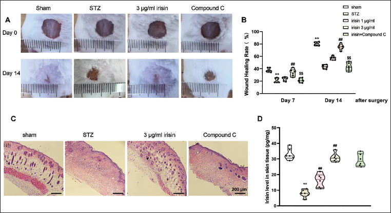
Interestingly, the effect of improving wound healing of exogenous irisin was reversed by co-treatment with Compound C (Figure 2A). The results of H&E staining assay also revealed that pretreatment with Compound C restored the therapeutic function of exogenous irisin (Figure 2C). These findings suggest that irisin promotes wound healing, and this effect is related to its regulation of AMPK expression.
Exogenous Irisin Treatment Increases pAMPK and Inhibits NF-κB p65 Expression in Diabetic Mice
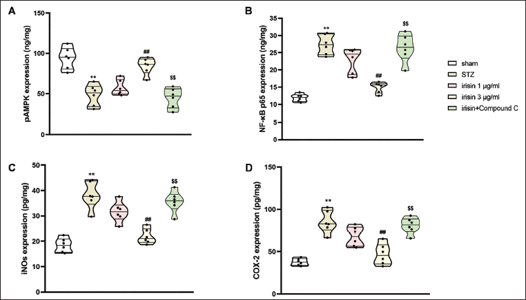
AMPK serves as a key regulator in both energy metabolism and inflammatory progression, exerting its effects via modulating NF-κB activity. STZ-treated mice exhibited significantly decreased pAMPK expression in the epidermal tissue, an effect that was markedly reversed by exogenous irisin (p < .01, Figure 3A). Meanwhile, NF-κB p65 was overexpressed in the epidermis of these diabetic animals (p < .01, Figure 3B). Exogenous irisin treatment markedly attenuated the abnormal expression of NF-κB p65 (p < .01). Both the upregulation of pAMPK and inhibition of NF-κB p65 by exogenous irisin were restored by Compound C. These findings suggest that irisin’s function of promoting wound healing is associated with the regulation of NF-κB-mediated by AMPK.
The Effects of Exogenous Irisin on the Expressions of Phosphorylated Adenosine 5′-Monophosphate-activated Protein Kinase (pAMPK) (A), Nuclear Factor Kappa B (NF-κB) (B), Inducible Nitric Oxide Synthase (iNOS) (C), and Cyclooxygenase (COX)-2 (D) in the Skin Wounds of Diabetic Mice. Values are Expressed as the Mean ± SEM with Six Mice in Each Group. ** p < .01 Versus the Sham Group; ##p < .01 Versus the Streptozotocin (STZ) Group; $$p < .01 Versus the Irisin-treated Group.
The results in Supplementary Figure 1 show that STZ, exogenous irisin, and Compound C treatment did not influence the total AMPK levels, and the changes of pAMPK/AMPK ratio were consistent with those of the pAMPK levels, as shown in Figure 3A. This demonstrated that exogenous irisin’s effect on pAMPK is through the phosphorylation of AMPK, not directly by influencing total AMPK levels.
Exogenous Irisin Treatment Inhibits Overexpressed Inflammatory Mediators in Diabetic Mice
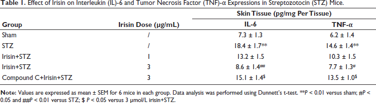
To investigate the mechanism of irisin’s positive effect on STZ mice, the levels of inflammatory factors, including IL-6, TNF-α, iNOS, and COX-2, in the epidermal tissue of mice were detected. As shown in Figure 3C, iNOS expression was markedly elevated in the epidermal tissue of STZ mice (p < .01). In addition, there was a notable elevation of COX-2 level in the STZ mice (p < .01, Figure 3D). Treatment with exogenous irisin effectively attenuated these aberrant expressions. Similarly, significant increases in the levels of IL-6 and TNF-α were observed in the epidermal tissue of STZ mice (p < .01 for TNF-α and IL-6, Table 1). However, the overexpression of these inflammatory mediators was overturned by the pretreatment with exogenous irisin (3 µg/mL). However, the inhibition of irisin on inflammatory mediators was restored by Compound C. Collectively, exogenous irisin’s function of promoting wound healing is associated with the modulation of inflammatory factors regulated through AMPK–NF-κB pathway.
Effect of Irisin on Interleukin (IL)-6 and Tumor Necrosis Factor (TNF)-α Expressions in Streptozotocin (STZ) Mice.
Discussion
This study first investigated exogenous irisin’s function on diabetic wounds. The results indicate that irisin serves as a pivotal regulator in the wound healing process. Local administration of exogenous irisin effectively accelerated cutaneous wound repair in STZ‑induced diabetic mouse model. The positive function of exogenous irisin in diabetic wounds is achieved by expediting the deposition of collagen. The results of our biochemical experiments indicate that exogenous irisin’s therapeutic function for diabetic wounds is associated with the regulation of inflammatory mediators, including IL-6, TNF-α, iNOS, and COX-2 via the AMPK–NF-κB pathway.
Irisin is a myokine first found in skeletal muscles and can be released into the blood circulation following exercise. This myokine is known to modulate key metabolic pathways in vivo, such as lipid and glucose metabolism (Zhang et al., 2014). Increasing evidence has shown that irisin can also be synthesized in other tissues such as bone, nerve, thyroid glands, and skin (Colaianni et al., 2019; Gençer Tarakçı et al., 2016; Momenzadeh et al., 2021). However, the physiological role of irisin in non-muscle tissues is not fully understood. In Tarakci’s study, irisin immunoreactivity was detected in both the dermal and epidermal layers of skin from crested porcupines, though the precise effect of irisin on the skin was not determined in this study. In our study, we explored the function of irisin in skin wound repair. The irisin level in the epidermal tissue was positively correlated with the wound healing rate, indicating that irisin may be a critical factor contributing to wound healing. The local administration of exogenous irisin in the STZ mice promoted the formation of new epidermal tissue and collagen deposition and accelerated the healing process of diabetic wounds, which confirmed the positive effect of irisin on skin wounds. Our result is in line with Tarakci’s result that irisin synthesized in skin possesses immunoreactivity. Several studies have demonstrated that irisin exerts its positive effects by modulating AMPK (Li et al., 2019; Sánchez et al., 2022). Our data also revealed that exogenous irisin intervention promoted the level of AMPK. Along with the promotion of the AMPK level, the wound healing rate was improved after exogenous irisin treatment, while co-treatment of AMPK inhibitor reversed exogenous irisin’s positive effects. This result suggests that the effects of exogenous irisin in promoting diabetic wound healing are achieved by the activation of AMPK. AMPK is an important protein kinase involved in both energy metabolism and inflammatory regulation (Jiang et al., 2020). For example, Kim et al. (2021) found that Valdecoxib alleviates insulin resistance in skeletal muscle by inhibiting inflammation through the AMPK pathway. Jung et al. (2021) revealed that Meteorin-like proteins ameliorate LPS-induced inflammatory responses in human umbilical vein endothelial cells through an AMPK-dependent pathway. On the other hand, inflammatory factors, including IL-1β, IL-6, TNF-α, COX-2, and iNOS, are downstream factors regulated by NF-κB, a nuclear transcription factor (Mahdiani et al., 2022). Based on the interactions between AMPK, NF-κB, and inflammatory factors, we detected the levels of NF-κB, IL-6, TNF-α, COX-2, and iNOS in the skin tissue of the diabetic mice to further investigate the mechanism of exogenous irisin’s effects. Results showed that exogenous irisin treatment decreased the expression of NF-κB, IL-6, TNF-α, COX-2, and iNOS in epidermal tissues of the diabetic mice, while these changes were abolished by the inhibition of AMPK, suggesting that exogenous irisin’s function on wound healing was associated with the suppression of IL-6, TNF-α, COX-2, and iNOS regulated by AMPK–NF-κB pathway. These findings were like that of Xiang et al.’s study, describing that activation of AMPK to inhibit NF-κB nuclear translocation ameliorated Fuchsin adjuvant-induced skin inflammation in mice (Xiang et al., 2019).
Notwithstanding these notable findings, the current study has certain limitations. Although our findings indicate that the beneficial effect of exogenous irisin on diabetic wounds may be mediated through the AMPK–NF-κB, whether the therapeutic effects also depend on other mechanisms remains unknown. The beneficial effects of exogenous irisin may also be achieved by anti-apoptosis, promoting tissue regeneration, or inducing blood glucose and lipid metabolism. It is our aspiration that the impact of exogenous irisin on the healing of diabetic wounds be comprehensively elucidated in the future, because studies specifically investigating irisin’s function in wound healing are limited.
Conclusion
The current study indicates that exogenous irisin represents a promising agent for improving STZ-mediated diabetic wound healing. The beneficial effect of exogenous irisin appears to be related to the suppression of inflammatory mediators such as COX-2, iNOS, IL-6, and TNF-α through the regulation of the AMPK–NF-κB pathway. Our findings indicate that exogenous irisin represents a promising therapeutic agent for diabetic wound management. However, more studies are required to validate its efficacy.
Footnotes
Abbreviations
AMPK: Adenosine 5′-monophosphate-activated protein kinase; ANOVA: Analysis of variance; Bax: Bcl-2-associated X protein; Bcl-2: B-cell lymphoma 2; COX-2: Cyclooxygenase-2; DFU: Diabetic foot ulcer; ELISA: Enzyme-linked immunosorbent assay; H&E: Hematoxylin and eosin; i.p.: intraperitoneal; ICR: Institute of Cancer Research; IL-1β: Interleukin-1β; IL-6: Interleukin-6; iNOS: Inducible nitric oxide synthase; MPO: Myeloperoxidase; NF-κB: Nuclear factor kappa B; NF-κB p65: Nuclear factor kappa-B p65; OD: Optical density; pAMPK: Phosphorylated adenosine 5′-monophosphate-activated protein kinase; RIPA: Radioimmunoprecipitation assay; SD: Standard deviation; STZ: Streptozotocin; TNF-α: Tumor necrosis factor-alpha.
Acknowledgments
None.
Data Availability
The data of this study can be obtained from the corresponding author upon reasonable request.
Declaration of Conflicting Interests
The authors declared no potential conflicts of interest with respect to the research, authorship, and/or publication of this article.
Ethical Approval
All the experimental procedures were approved by the Laboratory Animal Management and Ethics Committee of Ningbo Drug Control Institute and performed pursuant to the National Institutes of Health Guide for Care and Use of Laboratory Animals.
Funding
The authors disclosed receipt of the following financial support for the research, authorship, and/or publication of this article: This work was financially aided by Zhejiang Provincial Natural Science Foundation of China under Grant No. LTGY24H150003 for Xuefeng Yu, the Science and Technology Project of Zhejiang Medical and Health Department (2020KY289), the Science and Technology Project of Ningbo Yinzhou District (2022AS036) for Xi Jiang, and Key Discipline Projects in Yinzhou District of Ningbo for Yubo Yang and Xi Jiang.
Informed Consent
Not applicable.
Supplementary Material
References
Supplementary Material
Please find the following supplemental material available below.
For Open Access articles published under a Creative Commons License, all supplemental material carries the same license as the article it is associated with.
For non-Open Access articles published, all supplemental material carries a non-exclusive license, and permission requests for re-use of supplemental material or any part of supplemental material shall be sent directly to the copyright owner as specified in the copyright notice associated with the article.