
Abstract
Background
Psoralen is a biomaterial that promotes bone growth and repair and has been widely used in clinical orthopedic surgeries.
Purpose
This study aims to explore the role and mechanism of polydopamine nanoparticle-encapsulated psoralen (PSO/PDA NPs) in the recovery of motor function after spinal cord compression.
Materials and Methods
A Compressive Spinal Cord Injury (CSCI) rat model was constructed to explore the effects of PDA NPs and PSO/PDA NPs on spinal cord compression, and how PSO/PDA NPs regulate miR-140-5p, improve motor function of spinal cord compression, and 740 Y-P and LY294002 were used to explore the mechanism of PI3K/Akt signaling in PSO/PDA NPs to promote the recovery of motor function of spinal cord compression. Further experiments were conducted on CSCI rats to verify its mechanism through the combined intervention of miR-140-5p inhibitor and 740 Y-P.
Results
PSO/PDA NPs have a good effect in the rehabilitation after spinal cord compression, and can inhibit miR-140-5p expression, upregulate PI3K/Akt signal, inhibit the apoptosis rate of neurons in CSCI rats, and inhibit the expression of platelet activating factor (PAF), promote Bcl-2 expression, thereby reducing Bax expression.
Conclusion
PSO/PDA NPs downregulate miR-140-5p and promote PI3K/Akt pathway, thereby accelerating the recovery of related functions after spinal cord compression, laying the foundation for the development of drugs targeting CSCI.
Introduction
Spinal cord compression refers to a disease that causes spinal cord compression and then corresponding neurological dysfunction (Patnaik et al., 2020). Early treatment requires taking large doses of drugs, which makes people suffer from the toxicity and side effects of the drugs, and current surgical treatment is ineffective (Sabroe et al., 2021).
miR-140-5p is a kind of microRNA (Ding et al., 2021). Some studies have shown that miR-140-5p can play a protective and repair role in neural stem cells by regulating the ERK/MAPK (Xu et al., 2022) signaling pathway. miR-140-5p binds to the 3’UTR of ERK1/2 and inhibits translation, thereby inhibiting the ERK/MAPK pathway. And this inhibitory effect mainly occurs in the cytoplasm. When the expression level of miR-140-5p increases, it will inhibit ERK1/2 phosphorylation, thereby inhibiting the ERK/MAPK pathway (Luo et al., 2020). In this process, the biological functions of cells are affected. In terms of nerve cell survival, miR-140-5p is involved in the regeneration and repair process after spinal cord injury by regulating PTEN and Sp3 (Fu et al., 2023). By targeting and inhibiting Sp3, miR-140-5p relieves Sp3’s transcriptional repression of the BDNF gene, thereby promoting the expression and release of BDNF, thereby promoting the repair and regeneration of the damaged spinal cord. During the growth of neuronal axons, the key role of the PI3K/Akt pathway is to maintain microtubule transport and axonal growth (Wang et al., 2022). Akt can phosphorylate GABA receptors and increase their phosphorylation levels, thereby promoting the release of GABA from axon terminals (Chen, Huang, et al., 2023). In addition, Akt can also phosphorylate dentate gyrus granule cells and increase their survival, thus ensuring axonal plasticity and synapse formation. In cases of spinal cord compression, axons may be damaged, and PI3K/Akt can be involved in regulating the regeneration and growth of nerve axons (Sun et al., 2020). After spinal cord injury, the phosphorylation level of Akt increases rapidly, which indicates that PI3K/Akt signaling is involved in responding to spinal cord injury. This phosphorylation can promote the phosphorylation of mammalian target of rapamycin (mTOR), thereby inhibiting its autophagy (Jadaun et al., 2022). After spinal cord injury, this inhibition can promote neuronal survival and synaptic repair, contributing to the recovery of neurological function. In addition, studies have shown that in mechanically injured mouse spinal cord neurons, the phosphorylation level of AKT is increased, which inhibits the phosphorylation of mTOR and thereby increases the level of autophagy. This increased autophagy can help clear damaged organelles and pathological protein aggregates, reduce apoptosis, and promote the repair and regeneration of spinal cord neurons (Fu et al., 2020). Although it plays a great role in spinal cord compression, its exact mechanism of action is still not clear.
Psoralen is a type of fatty tissue found in the bone marrow. Some studies have explored injecting psoralen into spinal cord injury sites to promote the survival and regeneration of damaged nerve cells (Jamalis et al., 2020). Psoralen contains a variety of cytokines and growth factors, which can affect cell survival, proliferation, and differentiation, and help promote the regeneration and repair of damaged nerve cells. In addition, psoralen contains some mesenchymal stem cells, which have multi-directional differentiation potential and can differentiate into various cell types, such as nerve cells and supporting cells, which can help fill damaged areas and promote the regeneration of nerve tissue. Other studies have shown that the components in psoralen have anti-inflammatory and anti-oxidant effects (Yin et al., 2022), providing a more favorable microenvironment for the spinal cord after injury and promoting repair. Therefore, it is critical to deeply explore the protective mechanism of psoralen on spinal cord compression injury and to prevent spinal cord injury.
Studies have found that the bioavailability of minerals in psoralen varies due to factors such as chemical form and proportion, making it less stable in the body and prone to dissolution, thereby limiting its effect (Buhimschi et al., 2020) and reducing the therapeutic effect. Therefore, it is very important to find ways to improve the shortcomings of psoralen. PDA nanoparticles have good stability and can stably encapsulate bioactive substances to prevent their premature release or degradation, thereby increasing the action time of psoralen in the body. Meanwhile, it can achieve targeted delivery through surface chemical modification, precisely guiding the encapsulated psoralen to the bone tissue sites in need of repair, thereby enhancing the therapeutic effect. In addition, PDA nanoparticles can regulate the release rate of psoralen during the encapsulation process of psoralen, which is conducive to providing an appropriate concentration of growth factors and signaling molecules, and promoting the proliferation and differentiation of bone cells. Therefore, in this study, polydopamine nanoparticles (PDA NPs) were used as the carrier material to delay the metabolism of Psoralea corylifolia and enhance its repair effect.
Materials and Methods
Experimental Materials
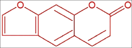
Psoralen (specification: 5 mg, molecular formula: C21H18O5, the molecular structure is shown in Figure 1), LY294002 (PI3K inhibitor) and 740 Y-P (PI3K activator) were purchased from Shanghai Yuanye; secondary antibodies (Shanghai Youningwei Biotech); Transmission Electron Microscope (TEM) was purchased from Shanghai Carl Zeiss, and inverted microscope (Shanghai Leica Microsystems).
Psoralen Molecular Structure.
Preparation and Characterization of PSO/PDA NPs
Preparation of PSO/PDA NPs
Add an appropriate amount of PSO to distilled water and stir to completely dissolve it to obtain a uniform PSO solution; then pour the polystyrene solution into the ultrasonic cleaner to remove bubbles and insoluble impurities to form a uniform polystyrene precursor. Solution, transfer the cleaned solution to a water bath, keep the temperature at 70°C–80°C, and continue stirring to further diffuse the polystyrene molecular chains and form nanoparticles. Add the PSO extract to the prepared polystyrene solution and mix evenly. Ultracentrifugation was used to remove unreacted substances, and finally, the nanoparticle sample was dried to obtain a composite sample of PSO/PDA NPs.
Nanoparticle Characterization
Dip a small amount of the nanoparticle dispersion liquid and evenly drop it on the conductive adhesive. After natural drying, spray gold for 1 min. Then use TEM to observe the shape and structure of the nanoparticles and analyze their potential with dynamic laser scattering.
Construction and Grouping of the Compressive Spinal Cord Injury (CSCI) Model Rats
After anesthesia with 2% sodium pentobarbital, rats were fixed on a rat board in a prone position with their heads lowered. The occipital neck was disinfected, and the skin was prepared. A longitudinal incision was made from the external occipital protuberance to C3 to reveal spinal cord, cut off the intervertebral ligaments, use copper wire and debending needle guidance, insert the air bag into the atlanto-occipital space, located outside the C2 and C3 lamina, rinse with normal saline and suture, and then place the air bag on the head and back of the rat. Secure properly. Twenty-four hours after the operation, connect the end of the balloon catheter to a hand pressure pump (with a three-way tube) and pressurize it with iohexol injection. The pressure is about 600 kPa. After 12 h, the balloon catheter is suctioned to negative pressure and is taken out, housed in a separate cage, and the bladder of the rat is deflated according to specific times to help it urinate.
The rats that were successfully modeled were included in the model group, and after intervention with PSO, they were set as the PSO group. After intervention with the same dose of PDA NPs and PSO/PDA NPs, they were set as the PDA NPs group and PSO/PDA NPs group, respectively. In addition, LY294002, 740 Y-P, PSO/PDA NPs+LY294002, PSO/PDA NPs+740 Y-P, miR-140-5p mimic, miR-140-5p inhibitor, PSO/PDA NPs+miR-140-5p inhibitor and PSO/PDA NPs+miR-140-5p mimic, miR-140-5p inhibitor+740 Y-P, miR-140-5p inhibitor+740 Y-P+LY294002, PSO/PDA NPs+miR-140-5p inhibitor+740 Y-P group.
SCI Behavioral (Basso–Beattie–Bresnahan (BBB)) Score
One day before administration, 7 days, and 14 days after administration, the rat’s limb motor function was evaluated using the BBB score. The cumulative maximum score was 22 points, which was proportional to the rat’s lower limb motor function.
Hematoxylin–Eosin (HE) Staining
Tissues were fixed in 10% formalin, embedded in embedding medium, and cut into 10 µm cross sections at 20°C. Place the slide on the tissue surface, dry overnight at 4°C, and then freeze at −20°C. The prepared cross sections were stained with HE, and photos were collected using an optical microscope (Nikon ECLIPSE Ti-S, Ruike Zhongyi Co., Ltd.).
Apoptosis Index
Fourteen days after administration, the rats were killed, and spinal cord samples from the compressed area were collected. Under sterile conditions, they were crushed mechanically and made into cell suspensions. They were centrifuged, and the cell concentration was adjusted to 2 × 10⁵/mL, 5 µL of Annexin-V and 5 µL of LPI to 400 µL of cells for 0.17 h, and used flow cytometry to detect the number of apoptotic cells.
Enzyme-linked Immunosorbent Assay (ELISA) Test
Detect the content of platelet activating factor (PAF) in plasma and damaged tissue, and measure PAF in different types of spinal cord tissue according to the instructions in the ELISA kit.
Transmission Electron Microscopy (TEM) to Observe the Ultrastructure of Myelinated Nerve Fibers
Spinal cord tissue samples were extracted from the injury site, fixed in glutaraldehyde, and sectioned into sections with a thickness of approximately 50–100 nm. Lead citrate and uranyl acetate staining were used. After the staining was completed, transmission electron microscopy was used to observe the ultrastructure of myeloid nerve fibers.
Polymerase Chain Reaction (PCR) Detection
Gastric cancer tissue was ground and homogenized using liquid nitrogen, total messenger ribonucleic acid (mRNA) was extracted, and transcribed into complementary deoxyribonucleic acid (cDNA). The analysis was performed using real-time PCR system analysis (Shanghai Unico) using primers in Table 1, and glyceraldehyde-3-phosphate dehydrogenase (GAPDH) was a reference. Relative levels were estimated using the 2−∇∇Ct method.
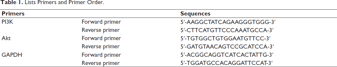
Lists Primers and Primer Order.
Western Blotting Analysis of Protein Expression
Use TRIzol reagent to extract total protein from spinal cord tissue and detect its concentration. Heat the electrophoresed total protein to 100°C and incubate it for 5 min, then use SDS-polyacrylamide gel for electrophoresis (120 V, 100 min), transfer to a membrane, and block. The primary antibodies of rabbit origin anti-Bcl-2 (1:1,000, Abcam #ab32124) and mouse origin anti-Bax (1:1,000, CST #5023) were used and incubated overnight at 4°C; HRP-labeled secondary antibody (1:5,000, at room temperature for 1 h), after ECL development, quantitative analysis was conducted through ImageJ, and this was repeated three times.
Statistical Analysis
The data obtained in each of the above experiments were analyzed using Statistical Package for the Social Sciences (SPSS) 21.0 and GraphPad Prism software. If there are no special requirements, p < .05 is used as the test standard.
Results
Successfully Constructed CSCI Rat Model and PSO/PDA NPs Nanoparticles
It can be observed that the rats in the experimental group showed spastic tail swinging, unilateral limb and body retreat, thrashing, unilateral or bilateral limb paralysis, etc., indicating that CSCI modeling was successful. The lesions of the injured spinal cord measured by magnetic resonance imaging (MRI) are shown in Figure 2A. During the process of model establishment, no rats died, and the success rate of model establishment reached 100%.
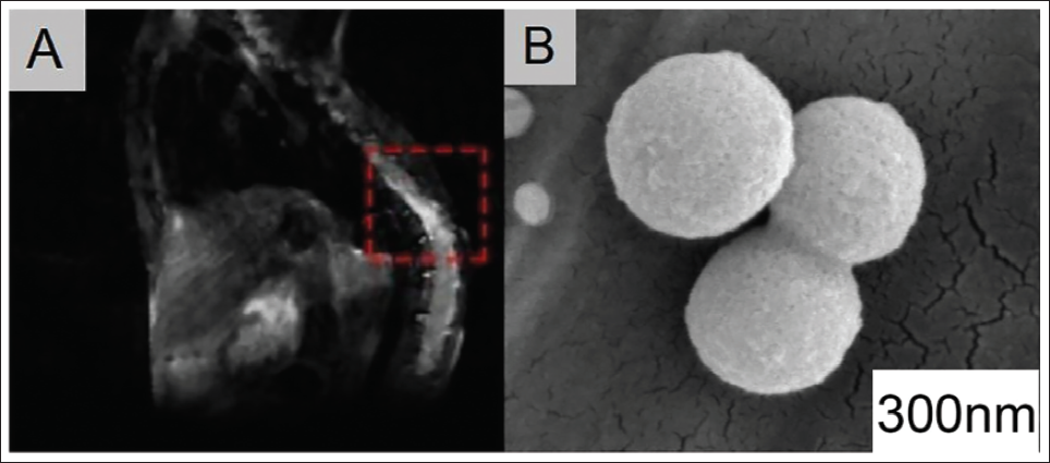
The TEM image shows that the particles of the prepared PSO/PDA NPs composite material are round spheres with uniform size and normal shape (Figure 2B).
PSO/PDA NPs Can Promote Motor Function Recovery After Spinal Cord Compression via the PI3K/Akt Pathway
In order to explore the effect of PSO/PDA NPs on the progression of spinal cord compression, we conducted a group experiment on CSCI rats and found that under the intervention of PSO, PDA NPs, and PSO/PDA NPs, the BBB score increased significantly (Figure 3A), and the nerve apoptosis index and PAF level of neuronal cells decreased significantly (Figure 3B,C); further observation of the HE staining results of rats in each group showed that drug intervention significantly reduced the phenomenon of neuronal nuclear condensation (Figure 3D); myelin sheath ultrasonic microstructural findings showed that in the model group, axonal swelling, myelin structure thickening, disorder, and delamination were seen in the model group; after PSO, PDA NPs, and PSO/PDA NPs intervention, although the myelin structure still showed pathological changes such as disorder and delamination, However, it has been significantly reduced (vs. Model group, Figure 3E). Among the above indicators, the PSO/PDA NPs group had the best therapeutic effect.
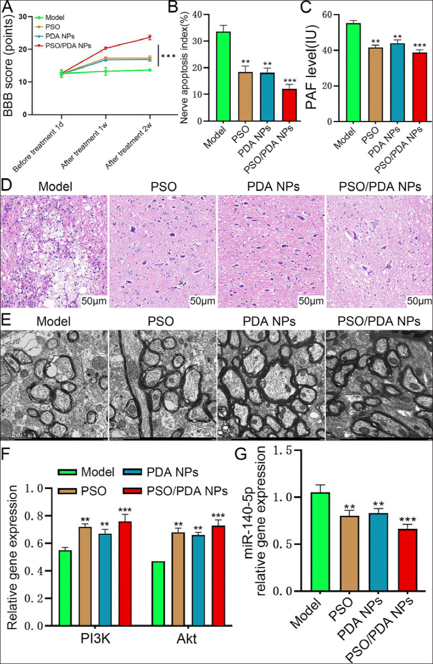
In addition, PCR test results showed that under the intervention of PSO, PDA NPs, and PSO/PDA NPs, PI3K and Akt in the PI3K/Akt pathway was upregulated, and miR-140-5p was downregulated (Figure 3F,G), and with PSO/PDA. The group under the NPs intervention was the most obvious. This indicates that the process of inhibiting the progression of spinal cord compression and improving motor function under the intervention of PSO/PDA NPs may be related to the expression of miR-140-5p and the PI3K/Akt pathway.
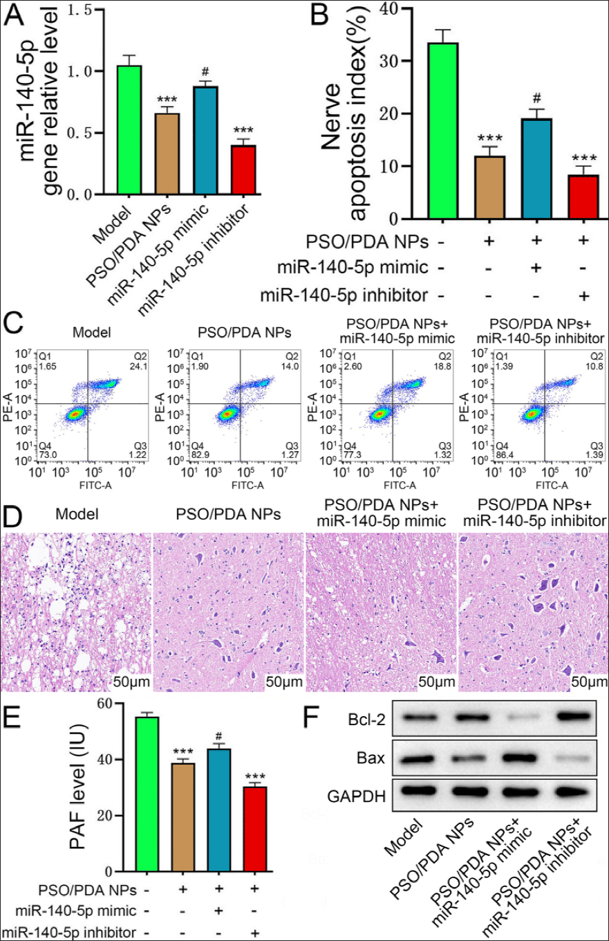
miR-140-5p is Involved in the Process of PSO/PDA NPs Promoting the Recovery of Motor Function from Spinal Cord Compression
On the basis of the above studies, we further studied the effect of PSO/PDA NPs on miR-140-5p and found downregulated miR-140-5p, which was consistent with the trend of the miR-140-5p inhibitor group, suggesting that PSO/PDA NPs can mediate miR-140-5p downregulation (Figure 4A). After the intervention of PSO/PDA NPs+miR-140-5p inhibitor, rat neuronal cell apoptosis and PAF levels were inhibited, and the levels were the lowest (vs. the other three groups, Figure 4B,C,E), and the pathological damage was also improved most significantly (vs. the other three groups, Figure 4D). At the same time, this group had the highest Bcl-2 protein expression and the lowest Bax (Figure 4F), indicating that neuronal apoptosis was significantly inhibited. When PSO/PDA NPs were combined with miR-140-5p mimic, the above trend was reversed.
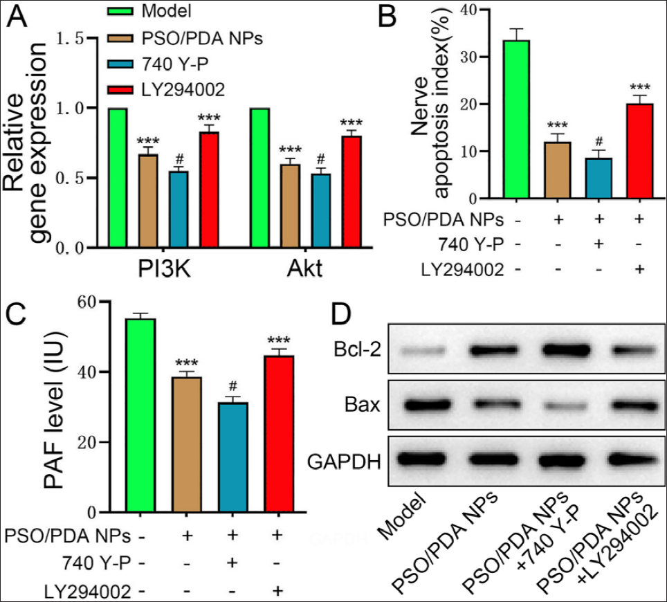
The PI3K/Akt Pathway is Involved in PSO/PDA NPs’ Role in Motor Function
In order to explore the effect of PSO/PDA NPs on the PI3K/Akt signaling pathway, it was found upregulated PI3K and Akt in PSO/PDA NPs group, which was consistent with the trend of the 740 Y-P group, suggesting that PSO/PDA NPs can mediate PI3K/Akt pathway activity, which was reduced (Figure 5A). After the intervention of PSO/PDA NPs+740 Y-P, the apoptosis index and PAF level of rat neurons were inhibited, and the levels were the lowest (vs. the other three groups, Figure 5B,C). At the same time, the Bcl-2 level in this group was the highest, and Bax was the lowest (Figure 5D), indicating that neuronal apoptosis was significantly inhibited. When PSO/PDA NPs were combined with LY294002, the above trend was reversed.
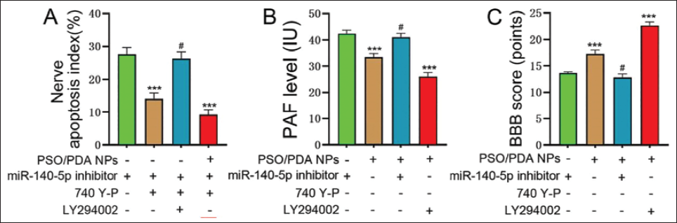
PSO/PDA NPs Regulate PI3K/Akt Signaling Through miR-140-5p to Promote Motor Function Recovery from Spinal Cord Compression
Another experiment was conducted on CSCI rats, and it was found that under the intervention of miR-140-5p inhibitor+740 Y-P, the neuronal apoptosis index and PAF level showed a downward trend (vs. miR-140-5p inhibitor group, p < .05, Figure 6A,B), the BBB score showed an upward trend (Figure 6C), and after adding LY294002 intervention on this basis, the above level was reversed, indicating that miR-140-5p can activate PI3K/Akt pathway. Moreover, the neuronal apoptosis index and PAF level of the PSO/PDA NPs+miR-140-5p inhibitor+740 Y-P group were the lowest, and the BBB score was the highest, suggesting that PSO/PDA NPs have an inhibitory effect on miR-140-5p, thereby upregulating PI3K/Akt signaling activity, which improves motor function in CSCI mice.
Discussion
Studies have shown that psoralen has poor stability in the body and is easily dissolved and absorbed, resulting in limited use (Ma et al., 2021). Therefore, this study uses polydopamine nanoparticles as carrier materials to delay the dissolution and metabolism of the drug by wrapping psoralen, thereby prolonging its action time in the body and enhancing its repair effect. Psoralen is a furanocoumarin compound with anti-inflammatory and anti-oxidant activities. In addition, polydopamine nanoparticles have good adhesion and bioactivity, can tightly combine with bone tissue, and provide a microenvironment conducive to bone cell adhesion and growth. Wrapping psoralen can promote the directional growth of bone cells on the surface of the material, accelerate the formation of new bone, and enhance the bone repair effect. Based on this advantage, we successfully prepared PSO/PDA NPs.
In order to prove that PSO/PDA NPs can promote motor function recovery after spinal cord compression, we found through animal experiments that under the intervention of PSO/PDA NPs, the neuronal apoptosis index and PAF level of rats were lower than those treated with PDA NPs or PSO alone. And it has the most significant effect on improving the myelin structure. Further PCR test results found that under drug intervention, PI3K and Akt were upregulated, and miR-140-5p was downregulated. And the PSO/PDA NPs group is the most obvious. This is because psoralen can activate PI3K/Akt signaling (Salem et al., 2021) and promote phosphorylation and activation of Akt, thereby inhibiting the activity of caspase and preventing apoptosis. Psoralen can reduce the phosphorylation levels of downstream factors, MAPKAPK-2 (Chen, Guo, et al., 2023) and MAPKAPK-3 (Sun et al., 2022), thereby activating the p38 MAPK pathway, reducing Bax, and increasing Bcl-2, further inhibiting the cell apoptosis process, and PSO/PDA NPs strengthened this result. It also further suggests that the process of inhibiting the progression of spinal cord compression and improving motor function under the intervention of PSO/PDA NPs may be related to the PI3K/Akt pathway. In addition, the reduction of miR-140-5p leads to increased c-Met, thereby enhancing HGF/c-Met signaling and promoting the proliferation and neurogenesis of NSCs and improving spinal cord diseases.
Transforming growth factor (TGF)-β signaling can reduce miR-140-5p expression by increasing the modification of H3K27me3 (Zhang et al., 2022) and inhibiting the transcription factor of miR-140-5p, thereby increasing BDNF (Mahajan & Sitasawad, 2021). Downregulation of miR-140-5p can relieve the inhibition of HIF-1α (Liu et al., 2022) and upregulate its expression. The activation of HIF-1α, in turn, promotes the expression of VEGF, thereby increasing angiogenesis and neurogenesis, and improving spinal cord diseases (Liu et al., 2021). Under the intervention of PSO/PDA NPs, we found that miR-140-5p expression was downregulated. After adding miR-140-5p inhibitor on this basis, rat neuronal cell apoptosis, PAF level dropped to the lowest, Bcl-2 protein expression was the highest, Bax was the lowest, and pathological damage was improved most obviously. When PSO/PDA NPs were combined with miR-140-5p mimic, the above phenomenon trend was reversed.
Upregulation of PI3K/Akt pathway can lead to Akt activation (Turke et al., 2012), which can then enhance the stability of Nrf2, increase its transcriptional activity, upregulate the expression levels of SOD and GPx, scavenge free radicals in the body, neutralize harmful substances, and prevent oxidative stress reactions such as membrane lipid peroxidation, thereby preventing nerve cell damage and pain. In addition, VEGF can combine with VEGFR to increase signal transduction (Lu et al., 2021), causing PI3K to be activated, which in turn activates its downstream Akt protein kinase, thus inhibiting the transcription and translation process of the TRPV1 gene and reducing the production and function of the TRPV1 channel protein, thereby reducing neuropathic pain. Under the intervention of PSO/PDA NPs, PI3K and Akt were significantly reduced. On this basis, adding 740 Y-P, the apoptosis index and PAF level of rat neuron cells were significantly reduced, and the expression of Bcl-2 protein was the highest, and Bax was the lowest. When PSO/PDA NPs are combined with LY294002, the above phenomenon can be significantly reversed. It shows that PSO/PDA NPs can upregulate the PI3K/Akt pathway, thereby promoting motor function recovery after spinal cord compression. In order to confirm and elucidate this special mechanism of action, we found through animal in vivo experiments that under the combined intervention of miR-140-5p inhibitor and 740 Y-P, the neuronal apoptosis index and PAF level were higher than those using only miR-140-5p. The inhibitor was low, and after adding LY294002 on this basis, the above phenomenon was reversed, indicating that miR-140-5p can activate PI3K/Akt signaling. Moreover, the PSO/PDA NPs+miR-140-5p inhibitor+740 Y-P group had the lowest neuronal apoptosis index and PAF level, and the highest BBB score, indicating that PSO/PDA NPs improve the motor function of CSCI mice by inhibiting miR-140-5p expression is achieved by upregulating PI3K/Akt signaling activity. However, further clinical investigation of the safety of PSO/PDA NPs is needed to ensure their safety.
Conclusion
In summary, PSO/PDA NPs promote the expression of PI3K/Akt and accelerate the recovery of motor function from spinal cord compression by inhibiting the expression of miR-140-5p. However, the limitation of this study lies in the fact that the long-term safety of PSO/PDA NPs was not deeply evaluated. Further preclinical and clinical studies are needed in the future to verify its safety. Furthermore, the experiment was only conducted in a rat model, and whether the results are applicable to humans still needs further verification.
Footnotes
Acknowledgments
The authors gratefully acknowledge the People’s Hospital of Yingshan County Laboratory for providing the necessary equipment for this study.
Declaration of Conflicting Interests
The authors declared no potential conflicts of interest with respect to the research, authorship, and/or publication of this article.
Ethical Approval and Informed Consent
This study was approved by the ethics committee of People’s Hospital of Yingshan County.
Funding
The authors received no financial support for the research, authorship, and/or publication of this article.