
Abstract
Background
Cell division control protein 42 homolog (Cdc42)/neuronal Wiskott–Aldrich Syndrome Protein (N-WASP) is involved in a variety of tumors; however, its regulatory mechanism in breast cancer is unclear.
Objectives
We aimed to explore the expression and role of Phellinus linteus extract in breast cancer and its possible molecular mechanism.
Materials and Methods
Cdc42/N-WASP level was measured in breast cancer tissues and adjacent tissues to explore the relationship between Cdc42/N-WASP expression and clinical relevance. P. linteus extract’s role in downstream signals in breast cancer was studied through immunofluorescence assays. At the same time, 3-(4,5-dimethylthiazol-2-yl)-2,5-diphenyltetrazolium bromide (MTT) and Transwell assays were used to detect the biological behaviors of cells after transfection.
Results
P. linteus extract can significantly reduce the biological behaviors of breast cancer cells, mainly by inhibiting cell proliferation and migration. P. linteus extract further inhibited breast cancer cell biological behaviors by inhibiting downstream Cdc42/N-WASP signaling. Moreover, Cdc42/N-WASP negatively regulated cell invasion and inhibited the degradation of extracellular matrix in breast cancer cells by regulating cortactin (CTTN).
Conclusion
Our results showed that P. linteus extract targets Cdc42/N-WASP and inhibits invasion and formation in breast cancer by regulating CTTN.
Introduction
Breast cancer is a serious problem in China (Khan et al., 2021), and metastases are the major reason for death (Chang et al., 2016). However, the mechanism of metastasis is still unclear (Lee & Dominguez, 2010; Tufail et al., 2021). Extracellular matrix (ECM) degradation involves tumor invasion and metastasis (Etienne-Manneville & Hall, 2001). Generally, infiltrating components are divided into two types of molecules: (a) involved in protein reconstruction; and (b) proteases that degrade cell membranes. ECM remodeling is a major event promoting cancer invasion and metastasis. The important ECM proteins like integrins (b1-, b5-, and b6-integrins), ECM1 protein, and Hic-5 protein are also actively involved in breast cancer development (Jena & Janjanam, 2018). The pathogenesis of breast cancer is very complicated involving abnormal expression of Bcl-2 (Janaghard et al., 2023). Recently, some studies showed that noncoding RNA expression is involved in the development of breast cancer, and its level is associated with tumor size in breast cancer patients (Tan, Cui et al., 2023; Xu et al., 2024). For the treatment, except abemaciclib or doxorubicin (Eralp et al., 2024), some extracts have shown promising therapeutic effects on breast cancer, such as Carthamus tinctorius L. (safflower) extracts (Kaçaroglu et al., 2023), Solanum nigrum L. water extract (Khan et al., 2023).
New evidence reveals a critical role for cortactin (CTTN) in cancer invasion (Baschieri et al., 2014; Nalbant et al., 2004; Yin et al., 2013). CTTN regulates actin assembly and may be a key player in infiltration-associated ECM degradation. Therefore, CTTN is often used as a biomarker of invasion. In addition, CTTN level is related to patient survival (Park et al., 2015). CTTN promotes the development of multiple cancers. Wiskott–Aldrich syndrome protein (WASP) and neuronal Wiskott–Aldrich syndrome protein (N-WASP) regulate actin assembly (Kodani et al., 2009). N-WASP is involved in the link of cell division control protein 42 homolog (Cdc42) to the actin-related protein 2/3 (Arp2/3) complex to promote actin polymerization (Burbelo et al., 1995; Tomasevic et al., 2007).
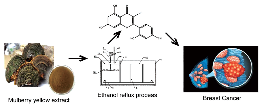
Phellinus linteum extract is a commonly used natural herbal plant and a medicinal plant with multiple biological activities, such as anticancer and anti-inflammatory effects (Abdul-Manan et al., 1999; Sammons et al., 2023). Therefore, this study selected Phellinus linteus extract to evaluate its role in breast cancer. In this study, we found that P. linteus extract targets Cdc42/N-WASP to inhibit the invasion of breast cancer cells by regulating CTTN, thereby inhibiting breast cancer. P. linteum extract is shown in Figure 1.
P. linteum Extraction Process and Research Ideas Diagram.
Materials and Methods
Tissue Sample
The study participants were all from 49 breast cancer patients diagnosed in our hospital from 2018 to 2019. These patients underwent surgical resection of breast cancer during treatment. Staging assessment was performed based on the patients’ clinical examination and imaging data. All patients were in tumor, node, metastasis (TNM) IV stage, and previous treatment history included chemotherapy, targeted therapy, and immunotherapy.
Cell Culture
The breast cancer cell line MCF-7 cell was cultured in 10% fetal bovine serum (FBS) in RPMI 1640 medium.
Scratch Test
A scratch assay was used to measure the migration ability of cells. Briefly, cells were cultured at a density of 1 × 10⁵ mL/cm3 at 37℃, supplemented with 10% FBS (Hyclone, Logan, UT), 100 U/mL penicillin, and 100 U/mL streptomycin. All cells were incubated in a serum-free medium for 12 hours before stimulation. After reaching a confluence of more than 80%, an artificial wound was made, followed by resuspending cells, rinsing with phosphate-buffered saline (PBS), and observing the degree of cell fusion under a microscope. Experiments were performed in triplicates.
Transwell Assay
Cell migration and invasion were measured by the Transwell assay. This was evaluated in modified Boyden chambers with two chambers separated by a membrane. Efficacy was evaluated after crystal violet staining.
Western Blot
Protein was separated by SDS-PAGE for Western blot using NLRP3 antibody (Proteintech, Chicago, USA, 1:1,000, Cat). Glyceraldehyde 3-phosphate dehydrogenase (GADPH) was utilized as a loading control.
miRNA and mRNA Real-time Quantitative Polymerase Chain Reaction (PCR)
RNA was extracted for real-time quantitative PCR (qPCR) with U6 as a control. The –∇∇CT comparison method was used to analyze the results.
Immunofluorescence Assay
After harvesting, fix the tissue in neutral formalin overnight. Dehydrate it in a dehydrator the next day. Take out the tissue, embed it, cool it at 4°C, and trim the slices to the largest surface of the tissue, with a smooth surface. Bake at 65°C. Bake for 1 hour, dewax according to the set procedure, and then wash with PBS buffer. Wash continuously 3 times, with 5 minutes for each time. After washing, prepare sodium citrate buffer for experiments and adjust the citric acid pH concentration to pH 6.0. Boil the prepared solution under high pressure for 5 minutes and soak the tissue with hydrogen peroxide (configured to a final concentration of 0.3%) at room temperature for 30–60 minutes. Absorb the hydrogen peroxide and wash with PBS for 3 × 5 minutes. The hydrogen peroxide should be freshly prepared. 30% hydrogen peroxide storage solution was stored in the dark at 4°C. 10% FBS was used for blocking; half an hour later, the primary antibody against Ki67 (purchased from CST) was applied and incubated overnight, and the next day, secondary antibody was added. The slides were mounted and photographed.
Statistics
GraphPad Prism 6 software was used to process the data, which were expressed as mean ± standard deviation (SD) and assessed by Student’s t-test for two-group comparison and one-way analysis of variance (ANOVA) with Newman–Keuls multiple comparison post hoc analysis for multiple groups comparisons.
Results
P. linteum Extract Inhibits Breast Cancer Cell Proliferation and Invasion
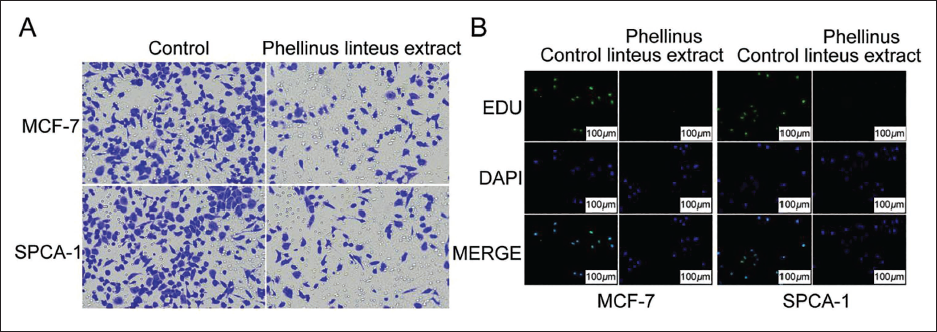
Compared with the control group, P. linteus extract inhibited cell growth. Cells were then cultured for 14 days, and colony formation experiments were performed. The results showed that P. linteus extract can reduce cell invasion. Compared with the control group, P. linteus extract inhibited cell migration and slowed down cell motility (Figure 2A). Further immunofluorescence results showed that the proliferation coefficient was significantly reduced after treatment (Figure 2B).
Phellinus linteus Extract Inhibits Breast Cancer Cell Proliferation and Invasion. (A) The Detection of Cell Invasion Ability, and (B) the Immunofluorescence Detection of Cell Proliferation Ability.
CTTN is Upregulated in Breast Cancer Tissues
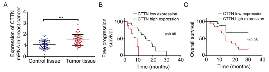
CTTN is known to be overexpressed in a variety of solid tumors and higher in breast cancer tissues, as shown in Figure 3A. Survival analysis showed that in breast cancer patients, a high level of CTTN could predict shorter progression-free survival (p < 0.05) and overall survival (p < 0.05), as shown in Figure 3B and C.
Cortactin is Upregulated in Breast Cancer Tissues. (A) Reverse Transcription-Polymerase Chain Reaction (RT-PCR) Detection of Cortactin (CTTN) Expression in Different Tissues, (B) Survival Time of Different CTTN Expression in Phosphate-buffered Saline (PFS), (C) Survival Time of Different CTTN Expression in Overall Survival (OS).
P. linteus Extract Inhibits Cancer Cell Invasion Through the Cdc42/N-WASP Pathway
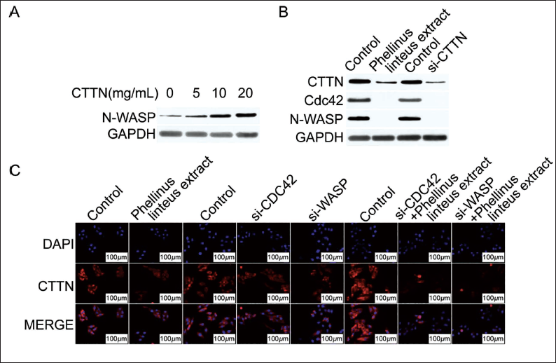
CTTN treatment induced an increase in N-WASP expression. However, it did not upregulate the expression of N-WASP in breast cancer cells mediated by CTTN knockout or P. linteus extract (Figure 4A). Cdc42 protein expression was decreased in MCF-7 cells treated with P. linteus extract or si-CTTN (Figure 4B). Knockdown of CDC42 or N-WASP reduced the percentage of CTTN-induced invasive cells. Obviously, CTTN silencing reduced Cdc42 expression in MCF-7 cells, as shown in Figure 4C. In addition, P. linteus extract mediated the inhibition of the Cdc42/N-WASP pathway and inhibited Arp2/3 activation by inhibiting CTTN, resulting in the inhibited cancer cell invasion.
Phellinus linteus Extract Inhibits the Invasion and Formation of Breast Cancer Cells through the Cell Division Control Protein 42 Homolog (Cdc42)/Neuronal Wiskott–Aldrich Syndrome Protein (N-WASP) Pathway. (A) Western Blot Detects that Cortactin (CTTN) Treatment Induces an Increase in N-WASP Expression, (B) Western Blot Detects the Expression of Phellinus linteus Extract and si-CTTN, (C) Immunofluorescence Detects Cdc42/N-WASP Signal Axis Expression (Magnification is 1:400).

Cdc42/N-WASP Inhibits Breast Cancer Cell Invasion and Metastasis by Regulating CTTN
To explore the role of Cdc42/N-WASP in breast cancer, we transfected breast cancer cells with Cdc42 mimics and found that Cdc42 significantly inhibits cell migration and invasion (Figure 5A). Meanwhile, Cdc42 transfection reduces cell abilities induced by CTTN (Figure 5B). In order to further study whether Cdc42 can regulate CTTN-induced cell invasion, the immunofluorescence method was used to detect the changes in invasiveness after CTTN treatment. As shown in Figure 5C, the cell nuclei are evenly distributed in the cell membrane, but after interference, cell membrane expression was reduced.
Cell Division Control Protein 42 Homolog (Cdc42)/Neuronal Wiskott–Aldrich Syndrome Protein (N-WASP) Inhibits Breast Cancer Cell Invasion and Metastasis by Regulating Cortactin (CTTN). (A) The Effect of Cdc42 Mimics on the Migration and Invasion Ability of Breast Cancer Cells, (B) The Effect of Cdc42 Transfection on the Migration and Invasion Ability Induced by CTTN, (C) Immunofluorescence Method Detects the Changes in Invasiveness after CTTN Treatment (Magnification, A and B are 1:100, C is 1:400).
Discussion
P. linteum extract has been used as a natural plant in Chinese herbal medicine for thousands of years. It is rich in nutrients and health ingredients. P. linteus extract is a plant native to China, Cambodia, and Japan. P. linteum extract has been reported to possess anticancer and antimetastatic activities, as well as stimulate B-cell maturation. Recently, the value of P. linteus extract, which has previously been used as an immunostimulant with anticancer and antimetastatic activities, has emerged as a food ingredient. A mouse model of breast cancer was constructed through an animal model. After treating tumor-bearing mice with P. linteus extract, tumor tissue was collected to determine the role of P. linteus extract in breast cancer development. Some studies showed the highest total polysaccharide content in crude extract, and the extract entities are mainly heteropolysaccharides (Tan, Liu et al., 2023). P. linteus extract polysaccharides mainly contain glucose, with a small proportion of monosaccharides. P. linteus extract has significant DPPH and ABTS free radical scavenging activity and also has a regulatory role in immune cell function in the tumor microenvironment (Zha et al., 2014). Our results show that P. linteus extract can inhibit tumor development and breast cancer cell metastasis by targeting Cdc42/N-WASP.
CTTN is an important invasion molecule that is often used as an invasion marker and plays an important role in cell invasion. First, CTTN may directly enhance actin activity. Second, CTTN can regulate various signaling pathways in the body to regulate cell growth (Kärkkäinen et al., 2010). In addition, CTTN also plays important physiological roles (Artym et al., 2006). Our results demonstrate the upregulation of cortical protein expression and invasion formation in breast cancer cells, indicating a role of CTTN in invadopodia formation.
miR-542-3p regulates CTTN to regulate the growth and invasion of colorectal cancer cells (Bowden et al., 2006), while some studies have reported that miR-134 inhibits the growth and invasion of lung cancer cells by acting on CTTN (Xia et al., 2007). In addition, VEGF-C increases CTTN expression and alleviates the impact of Cdc42/N-WASP on CTTN expression by downregulating dicer-mediated Cdc42/N-WASP maturation (Hsu et al., 2009). All these reports indicate that the effects of CTTN on tumors are regulated by Cdc42/N-WASP, and our findings reveal that Cdc42/N-WASP acts as an inhibitor of tumor metastasis and invasion in breast cancer by directly targeting CTTN. By targeting CTTN, Cdc42/N-WASP regulates the CTTN pathway and inhibits the formation of invasomes (Huang et al., 2012). Cdc42 and Rac undergo prenylation in vivo (Jing et al., 2016), and Cdc42 prenylation is required for activity (Gibcus et al., 2008). It has also been shown that non-prenylated Cdc42 is able to fully activate WASP (Svoronos et al., 2016). Our results are basically consistent with these reports. Most N-WASP activators show dose dependence and become very weak activators at high concentrations (µM).
Cell invasion and migration are key factors in metastasis, and CTTN has been reported to play multiple roles in transporting MMP2 and MMP9 proteases for degradation. In vitro results show that a low expression of CTTN blocks the invasive ability of tumor cells, while overexpression of CTTN promotes the invasion of colorectal cancer. Studies have suggested that the invasion of CTTN in tumor cells depends on the activation of DOCK1-Rac1, which eliminates the impact of CTTN on migration and invasion by inhibiting DOCK1 (Kent & Mendell, 2006; Lim et al., 2003). Our study suggests that Cdc42/N-WASP inhibits breast cancer cell activities by regulating CTTN to modulate cell invasion and migration. Similarly, our study also has certain limitations. In our study, only a single cell line of MCF-7 cells was used; more breast cancer cell lines should be used for the confirmation of the conclusions. In addition, no in vivo validation was performed in the present study, and this is required to be conducted in the future using animal models.
Conclusion
In conclusion, our study reveals the mechanism of targeting CTTN on invasion formation, and miR-134 may become a target for treating glioma.
Footnotes
Abbreviations
Arp2/3: Actin-related protein 2/3; Cdc42: Cell division control protein 42 homolog; CTTN: Cortactin; ECM: Extracellular matrix; FBS: Fetal bovine serum; GADPH: Glyceraldehyde 3-phosphate dehydrogenase; MTT: 3-(4,5-Dimethylthiazol-2-yl)-2,5-diphenyltetrazolium bromide; N-WASP: Neuronal Wiskott–Aldrich syndrome protein; PBS: Phosphate-buffered saline; TNM: Tumor, node, metastasis.
Acknowledgments
This work was supported by the Science and Technology Project of Huainan City (2020067).
Declaration of Conflict of Interests
The authors declared no potential conflicts of interest with respect to the research, authorship, and/or publication of this article.
Ethical Approval
This study was approved by the Ethics Committee of Huainan Chaoyang Hospital.
Funding
The authors received no financial support for the research, authorship, and/or publication of this article.