
Abstract
Background
Pregnancy complicated by diabetes leads to congenital disorders, spontaneous abortion, and neonatal morbidity and mortality, which are termed diabetic embryopathy or fetopathy.
Objectives
In this work, we planned to investigate the therapeutic potentials of nerolidol against streptozotocin (STZ)-induced diabetic embryopathy in rats through attenuating maternal and fetal oxidative stress.
Materials and Methods
The male and female Wistar rats were allowed to mate for a night at a 1:1 ratio. After the confirmation of pregnancy, the rats were administered 40 mg/kg of STZ (i.p.) to initiate diabetes and treated with 20 and 40 mg/kg of nerolidol. The changes in food intake, body weight, blood glucose, and urine output were regularly monitored. The levels of lipid profiles were estimated using assay kits. The oxidative stress and antioxidant biomarkers in the liver tissues and embryonic fractions were measured using assay kits.
Results
The nerolidol treatment considerably depleted the blood glucose and urine output and augmented the body weight and diet intake in diabetic rats. The nerolidol suppressed the cholesterol (Ch), triglycerides (TG), and very low density lipoprotein (VLDL) and elevated the high density lipoprotein (HDL) level in diabetic pregnant rats. The maternal and embryonic oxidative stress levels are effectively reduced by the nerolidol treatment by decreasing the malondialdehyde (MDA) and reactive oxygen species (ROS) levels. The levels of antioxidants were also increased by the nerolidol on both liver and embryonic fractions of diabetic pregnant rats.
Conclusion
Our results exhibited that the nerolidol treatment considerably reduced embryonic and maternal oxidative stress and embryonic deaths. Hence, it can be a hopeful therapeutic candidate to treat diabetic embryopathy.
Introduction
Diabetes mellitus is a heterogeneous metabolic disease characterized by the increased blood glucose in response to decreased insulin production and/or resistance to insulin activity. 1 During the pregnancy, the hyperglycemic condition leads to reproductive aberrations, which elevate the chances of congenital disorders, spontaneous abortion, and neonatal morbidity as well as mortality, which is termed diabetic embryopathy or fetopathy. 2 Diabetes affects more than 5% of all pregnancies and can lead to intrauterine growth retardation. The incidences of spontaneous abortion are more common in diabetic pregnancies than in normal pregnancies. Hyperglycemic condition linked to both fetal and maternal complications during pregnancies complicated by diabetes. 3 Even with many developments in diabetic management and obstetric care, the increased morbidity and mortality risks are augmented during diabetic pregnancies for both the mother and the developing fetus. 4
Several cellular and molecular pathways are dysregulated during embryonic development in diabetic pregnancies. These changes are connected with hyperglycemia, which results in the initiation of a prooxidant environment. 5 The imbalance between the oxidant-antioxidant systems causes oxidative stress in response to higher free radical accumulations that decrease the antioxidant protective mechanism. 6 Hyperglycemia-mediated oxidative stress is a key player in diabetic embryopathy and further facilitates congenital disorders. 7 The developing embryo and fetus are extremely sensitive to oxidative stress because of their premature antioxidant mechanisms. The increased oxidative stress overwhelms the embryonic and fetal defense and leads to embryopathy. 8
The maintenance of euglycemia and supplementation of adequate antioxidants seem to be a hopeful strategy to counteract the oxidative damage to the developing fetus. 9 Dietary supplements containing antioxidants have already been reported to decrease the incidence of diabetic embryopathy in animal models.10, 11 As a result, therapeutic interventions that reduce oxidative stress could be an effective strategy to treat and prevent diabetes-associated complications. 12 In recent times, many drugs have been prescribed to control hyperglycemia; however, the maintenance of euglycemia is poorly achieved. 13 Several plants and their products are being used to treat diabetes and its associated complications since ancient times. 14
Nerolidol is a natural sesquiterpene alcohol that is widely present in the essential oils of many flowers and plants. Nerolidol has been reported to have antiulcer, 15 antioxidant, 16 and antiarthritic, 17 properties. Trindade et al. 18 have reported that the nano-encapsulated nerolidol demonstrated a potential anti-inflammatory activity in zymogen-induced arthritis in mice. Parvez et al. 19 reported that the nerolidol-encapsulated nanoparticles showed anticancer activity against human colorectal cancer cells. Nerolidol effectively ameliorated oxidative injury to the renal and heart tissues. 20 However, the therapeutic properties of nerolidol on diabetic embryopathy have not been studied yet. Hence, in this work, we planned to investigate the therapeutic potentials of nerolidol against streptozotocin (STZ)-induced diabetic embryopathy in rats by attenuating maternal and fetal oxidative stress.
Materials and Methods
Chemicals
The STZ, nerolidol, and other chemicals were obtained from Sigma-Aldrich, USA. To determine the biochemical parameters, the ELISA assay kits were obtained from Thermofisher and R&D Systems, USA, respectively. All the reagents were of an analytical grade.
Experimental Rats
The adult Wistar rats of both sexes were purchased from the institutional animal house and maintained in proper laboratory facilities. Rats were housed in clean polypropylene cabins with 24°C ± 6°C of temperature, 50%–60% of air humidity, and 12/12 h of dark-light sequences. The rats were fed a standard pellet diet throughout the experiments. The male and female rats were allowed to mate for a night at a 1:1 ratio. After that, the vaginal smears were prepared and examined under a microscope to detect the presence of sperms for the establishment of pregnancy. A gestation day (GD) of 0 was labeled on the day of the occurrence of sperms on the vaginal smear. The rats that failed to mate for more than a week were excluded from the experiments.
Treatment Procedures and Interpretations
The pregnant rats were distributed into six groups with six rats in each (n = 6). Group I are control rats and administered citrate buffer. Group II is injected with 40 mg/kg of STZ (i.p.) on the GD 4 to induce diabetes. Groups III and IV are diabetes-induced as specified in Group II and treated with 20 and 40 mg/kg of nerolidol by oral route from GD 5 to 12. The 5% of glucose water was administered to the rats for 48 h. Throughout the study, the food consumption and body weight gain of rats were continuously monitored. The urine output of the rats was measured on the last three days before the scarification. Under standard anesthesia, the three rats were sacrificed on the GD 20, and then blood samples were collected. The collected blood samples were utilized to determine the glucose level and serum preparation. The liver and uterine tissue samples were excised and immersed in a saline solution. By using standard surgical processes, the placenta and embryos were excised and weighed accurately. The placenta, liver, and embryo tissues were stored at –80°C for additional assays.
Preparation of Tissue Homogenates
The collected liver and embryo tissues were washed with saline and then homogenized (10% w/v) using a chilled buffered saline solution. Then homogenized tissues were centrifuged at 6000 rpm for 5 min, and the supernatant was gathered in a clean tube and used for further biochemical assays. By using the Trounce et al. 21 technique, the mitochondrial fractions were prepared and used for the determination of oxidant and antioxidant biomarkers.
Determination of Lipid Profiles and Total Protein Level
The levels of total protein, cholesterol (Ch), triglycerides (TG), high density lipoprotein (HDL), and very low density lipoprotein (VLDL) in the serum of experimental rats were determined using ELISA kits. The assay was performed in triplicates using the protocols recommended by the manufacturer (Thermofisher Scientific, USA), and the outcomes were depicted as mg/dL.
Quantification of Oxidative Stress Markers
The prepared embryo and liver tissue embryo homogenates of control and experimental rats were used to determine the status of malondialdehyde (MDA), reactive oxygen species (ROS), protein carbonyls (PCs), glutathione (GSH), and total thiols (TSH). These markers were quantified with the help of commercial assay kits. All tests were conducted in triplicates using the recommended protocols of the manufacturer (R&D Systems, USA).
Determination of Antioxidant Enzyme Activities
By using the commercially procured assay kits, the activities of antioxidant enzymes such as catalase (CAT), glutathione reductase (GR), superoxide dismutase (SOD), glutathione peroxidase (GPx), and glutathione-S-transferase (GST) in the liver tissue homogenates of experimental rats were measured. All the tests were conducted in triplicates by using the protocols described by the manufacturer (Thermofisher Scientific, USA).
Statistical Analysis
The obtained data from each experiment were analyzed using GraphPad Prism software. The one-way ANOVA and Tukey’s post hoc assay were performed to determine the values, and the final outcomes were illustrated as the mean ± SD of triplicate measurements. The data were set as significant if the p-value was less than 0.05.
Results
Effect of Nerolidol on Body Weight, Urine Output, Diet Intake, and Serum Glucose Level in the Experimental Rats
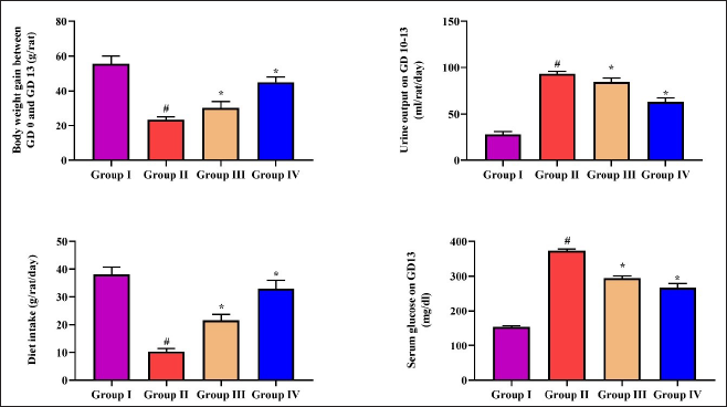
Figure 1 reveals the effect of nerolidol treatment on body weight, urine output, diet intake, and serum glucose level in the experimental rats. The diabetic pregnant rats demonstrated diminished body weight and diet intake, while increased urine output and serum glucose levels were noted. Interestingly, 20 and 40 mg/kg of nerolidol treatment considerably increased the body weight and dietary intake while decreasing urine output and serum glucose in the diabetic pregnant rats (Figure 1). The 40 mg/kg of nerolidol effectively ameliorated the STZ-induced changes in the rats more than the 20 mg/kg treatment.
Effect of Nerolidol on the Embryo and Placental Weight and Implantation in the Experimental Rats
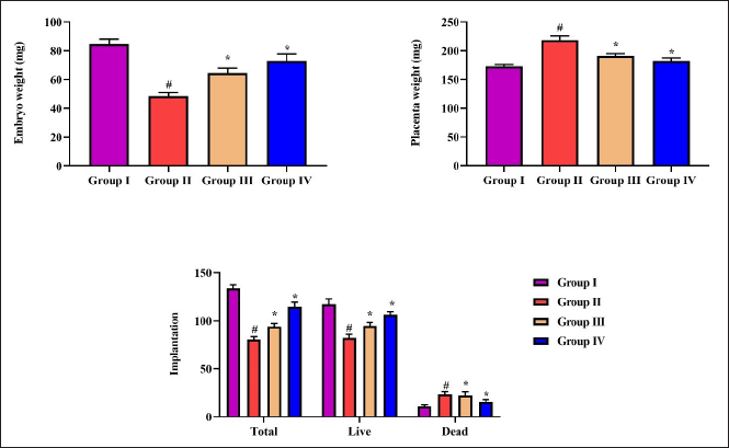
The changes in the weights of placenta and embryo and the live, dead, and total implantation numbers in the control and experimental rats were analyzed, and the outcomes are revealed in Figure 2. The decreased embryo weight and increased placental weight were noted in diabetic pregnant rats. The diabetic pregnant rats also revealed live and total implantation numbers, and an increase in the dead implantation numbers was observed. Meanwhile, 20 and 40 mg/kg of nerolidol considerably improved the embryo weight and reduced the placental weight in diabetic pregnant rats (Figure 2). The nerolidol also augmented the total and live implantation numbers and reduced the dead implantation numbers in diabetic pregnant rats.
Effect of Nerolidol on the Lipid Profiles and Total Protein in the Experimental Rats
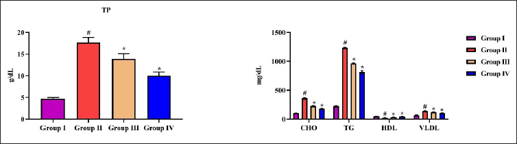
The levels of lipid profiles such as Ch, TG, VLDL, HDL, and total protein were estimated in the serum of experimental rats, and the findings are exhibited in Figure 3. The STZ-induced diabetic pregnant rats revealed increased levels of Ch, TG, VLDL, and total protein and a reduction in HDL. Interestingly, 20 and 40 mg/kg of nerolidol treatment effectively decreased the Ch, TG, VLDL, and total protein in diabetic pregnant rats. The nerolidol also improved the HDL level in diabetic rats (Figure 3). These outcomes proved that the nerolidol ameliorated STZ-induced dyslipidemia in diabetic pregnant rats.
Effect of Nerolidol on the Oxidative Stress Marker Levels in the Liver Tissue Homogenates of the Experimental Rats
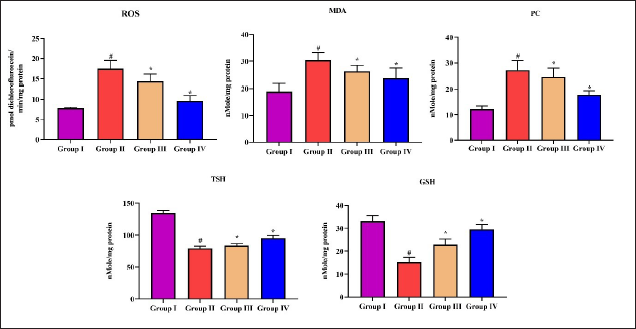
The contents of oxidative stress biomarkers such as ROS, MDA, PC, TSH, and GSH were assessed in the experimental rats, and data were shown in Figure 4. The ROS, MDA, and PC were considerably elevated, and GSH and TSH status were depleted in the liver tissue of diabetic pregnant rats. Meanwhile, the 20 and 40 mg/kg of nerolidol treatment appreciably diminished the ROS, MDA, and PC contents in diabetic pregnant rats. The GSH and TSH levels were also elevated by the nerolidol treatment in the liver tissue of diabetic pregnant rats (Figure 4). These outcomes prove that nerolidol decreased oxidative stress in diabetic pregnant rats.
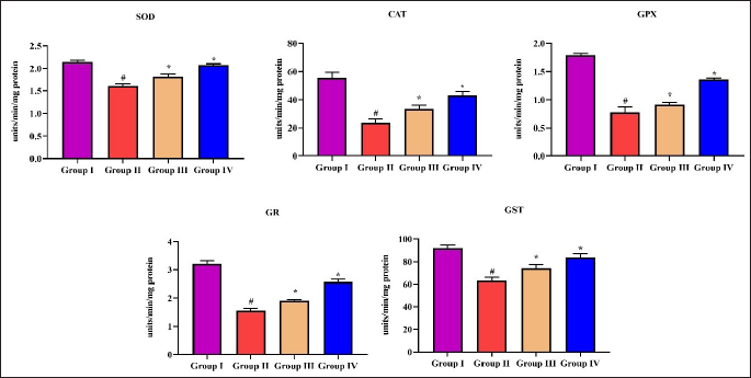
Effect of Nerolidol on the Antioxidant Enzymes in the Liver Tissues of Experimental Rats
The antioxidant enzyme activities such as CAT, SOD, GPx, GR, and GST were determined in the liver tissues of the experimental rats, and the data are illustrated in Figure 5. A remarkable decrease in the CAT, SOD, GPx, GR, and GST levels was noted in the STZ-induced diabetic pregnant rats. Nonetheless, 20 and 40 mg/kg of nerolidol treatment considerably augmented the CAT, SOD, GPx, GR, and GST activities in the liver tissues of the diabetic pregnant rats (Figure 5). These outcomes proved the antioxidant potential of nerolidol against diabetic pregnancy.
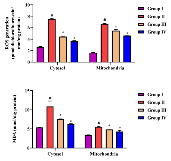
Effect of Nerolidol on the Oxidative Marker Levels in the Embryos of the Experimental Rats
Figure 6 shows the levels of oxidative markers such as ROS and MDA in the embryonic cytosol and mitochondrial fractions of the control and experimental rats. The diabetic pregnant rats revealed an augmented ROS and MDA status in the embryonic cytosol and mitochondrial fractions. Meanwhile, 20 and 40 mg/kg of nerolidol treatments appreciably decreased the ROS and MDA contents in the embryonic cytosol and mitochondrial fractions in the STZ-induced diabetic pregnant rats (Figure 6). These findings proved that nerolidol decreased embryonic oxidative stress in diabetic pregnant rats.
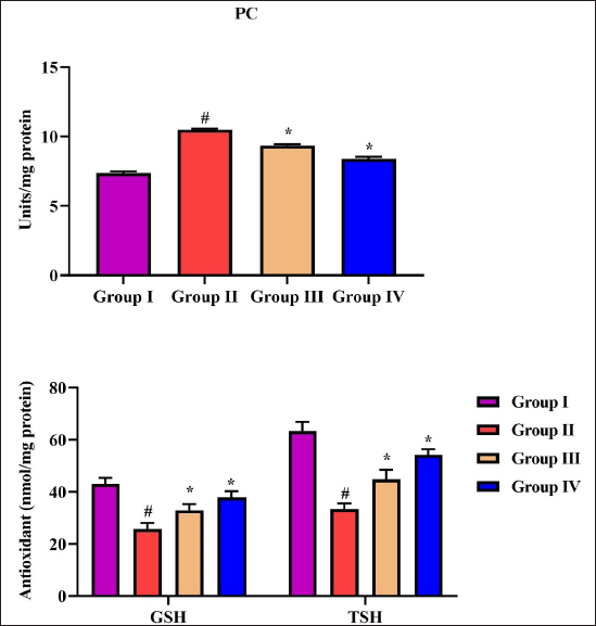
Effect of Nerolidol on the Levels of GSH, TSH, and PC in the Embryos of Experimental Rats
The levels of GSH, TSH, and PC were examined in the cytosol and mitochondrial fractions of embryos in the experimental rats, and the results are revealed in Figure 7. The STZ-induced diabetic pregnant rats demonstrated decreased GSH and TSH, and elevated PC level was observed in the cytosol and mitochondrial fractions of embryos when compared with the control. Interestingly, 20 and 40 mg/kg of nerolidol appreciably augmented the GSH and TSH and reduced the PC level in the cytosol and mitochondrial fractions of embryos in diabetic pregnant rats (Figure 7).
Discussion
Diabetes is considered an important global public health issue, particularly in pregnancy. Pregnancy complicated by diabetes can cause spontaneous abortion, early pregnancy dysfunction, and teratogenesis. 22 The hyperglycemic condition during embryogenesis has been related to several birth defects. These birth defects are mostly found in the cardiovascular system, central nervous system, renal system, and axial skeleton. 23 The higher prooxidant and proinflammatory markers are believed to be major underlying causes of embryopathy in diabetic pregnancy. 24 Oxidative stress is a crucial contributor to diabetes development. It causes insulin deficiency and inhibits insulin signaling via adipocytokine. 25
The impact of oxidative stress in pregnancy and its associated difficulties for mothers and growing fetuses are well-reported. 26 ROS are generated under normal conditions of cellular metabolism and play a major role in cell homeostasis. 27 Moreover, higher ROS accumulation has been reported in diabetics because of prolonged hyperglycemic exposure. 28 The growing embryos are extremely vulnerable to the high ROS level, particularly during the early stages of organogenesis. 29 MDA is a byproduct of lipid peroxidation and a well-known indicator of oxidative stress. 30 MDA can quickly combine with the biomolecules and induce cellular disorders, like pancreatic β-cells, thereby disrupting the glucose metabolism. 31 The elevated level of PC is believed to be a significant indicator of oxidative lipid peroxidation. 32 In this study, our findings revealed that the MDA, ROS, and PC contents were increased in both liver tissue homogenate and embryonic cytosol and mitochondrial fractions of diabetic pregnant rats. Interestingly, the treatment with the nerolidol considerably decreased the MDA, ROS, and PC contents in the liver tissue homogenate and embryonic cytosol and mitochondrial fractions of diabetic pregnant rats. These findings proved that the nerolidol treatment effectively ameliorated the oxidative stress response in both the liver and embryos of diabetic pregnant rats.
Diabetes is tightly connected with increased oxidative stress, thereby decreasing the endogenous antioxidant mechanisms. 33 The antioxidants such as GSH, SOD, CAT, GR, GST, and GPx are vital players in free radical scavenging. It was already reported that these antioxidants were found to decrease in the diabetic condition, which gives protection against oxidative stress by scavenging excessive amounts of ROS. 34 The augmented lipid peroxidation and diminished antioxidant mechanisms have been previously reported in the diabetic pregnant animal model. 35 Similarly, our outcomes exhibited that the antioxidants such as GSH, SOD, CAT, GR, GPx, and GST were found to be depleted in the liver tissue homogenate of the diabetic pregnant rats. The embryonic fractions of diabetic pregnant rats also demonstrated decreased levels of GSH and TSH. Interestingly, the nerolidol effectively augmented the antioxidant markers, that is, GSH, SOD, CAT, GR, GPx, and GST, in the liver tissues of diabetic pregnant rats. The nerolidol also increased the GSH and TSH levels in the embryonic cytosol and mitochondrial fractions of diabetic pregnant rats. These findings confirmed the antioxidant potentials of nerolidol in diabetic pregnancy.
During embryo implantation, ROS plays a major role in uterine homeostasis regulation. The decrease in the implantation rate may exhibit reduced fetus numbers. 36 Similarly, our findings also demonstrate more total and dead implantation numbers in the diabetic pregnant rats than in the control. These findings confirm that hyperglycemic exposure is associated with the loss of implantations. 37 Furthermore, the failures in the embryonic implantation may cause embryonic morphological alterations that inhibit the implantation and cause embryo lethality. 38 Here, we found that the diabetic pregnant rats demonstrated increased dead implantation numbers. This result proved that the hyperglycemic condition in the first trimester of pregnancy has a negative effect on the blastocyte and embryonic development. An earlier study has found that the experimental diabetic model is connected with the considerable increase in post-implantation embryonic deaths. 39 Interestingly, the treatment with nerolidol appreciably increased the live and total implantation numbers in the diabetic pregnant rats. The nerolidol treatment also depleted the dead implantation numbers in the diabetic pregnant rats. These outcomes confirm that nerolidol has significant positive effects during implantation.
Dyslipidemia is an another complication of diabetes, in which increased maternal serum levels of Ch, TG, and VLDL and decreased HDL levels were observed. 40 Dyslipidemia is common in diabetic patients, which causes irregular lipids. 41 The reduction in VLDL, TG, and Ch and the elevation in the HDL level can inhibit or delay the progression of diabetes-associated difficulties. 42 Here, we found that the diabetic pregnant rats demonstrated increased contents of Ch, TG, VLDL, and reduced HDL levels. Interestingly, the treatment with the nerolidol appreciably reduced the Ch, TG, and VLDL levels while increasing HDL levels in the diabetic pregnant rats.
Conclusion
Our findings exhibited that the nerolidol treatment considerably reduced embryonic deaths, decreased serum glucose, and improved body weight. The nerolidol also regulated dyslipidemia and reduced maternal and embryonic oxidative stress by decreasing MDA and ROS levels and increasing antioxidant status. Overall, these outcomes suggested that nerolidol may have therapeutic effects on diabetic embryopathy in STZ-induced rats. Furthermore, we also recommend additional studies in the future to make a clear conclusion about the therapeutic roles of nerolidol against diabetic embryopathy.
Footnotes
Abbreviations
Declaration of Conflicting Interests
The authors declared no potential conflicts of interest with respect to the research, authorship, and/or publication of this article.
Funding
This work was supported by the Department of Obstetrics and Gynecology, The 985th Hospital of the PLA Joint Logistics Support Force, Taiyuan 030001, China.
Statement of Informed Consent and Ethical Approval
We have completed the animal experimental based on the animal ethical guidelines and same experimental procedures were approved by the committee (PLA985-TYH-20210714).
Summary
The levels of antioxidants were also increased by the nerolidol on both liver and embryonic fractions of diabetic pregnant rats.
The nerolidol treatment considerably depleted the blood glucose and urine output and augmented the body weight and diet intake in diabetic rats.
The nerolidol suppressed the Ch, TG, and VLDL and elevated the HDL level in diabetic pregnant rats.
Nerolidol treatment considerably reduced embryonic and maternal oxidative stress and embryonic deaths.