
Abstract
Background
Cardiovascular disease (CVD) is a group of heart disorders, which is a major cause of non-communicable disease-related mortalities worldwide. Myocardial infarction (MI) is an acute disorder due to the poor supply of oxygen and blood to the myocardium. MI is the foremost form of CVD, which is the primary cause of mortality worldwide.
Objectives
Here, we intended to discover the ameliorative properties of the ponicidin against the isoproterenol (ISO)-stimulated MI in rats.
Materials and Methods
About 85 mg/kg of ISO was administered to the rats to trigger the MI and then treated with 25 and 50 mg/kg of ponicidin. The body weight and heart weight of all rats were determined. The total protein, c-reactive protein (CRP), and uric acid levels were examined. The activities of cardiac function markers such as creatine kinase (CK), ALT, AST, and gamma-glutamyl transferase (GGT) were examined. The antioxidants such as glutathione (GSH), GST, and GPx were examined by the previous methods. The status of Na+/K+, Mg2+, and Ca2+ ATPase activities was assessed using kits. The status of Na+, K+, and Ca2+ ions and inflammatory makers such as TNF-α and IL-6 were investigated using respective kits. The histopathological analysis was performed on the heart tissues to detect the histological changes.
Results
The results revealed that ponicidin increased body weight and decreased heart weight in MI rats. The status of CRP and uric acid was decreased and total protein was augmented in the ponicidin-treated MI rats. The AST, ALT, CK, and GGT activities were appreciably decreased in serum and elevated in the cardiac tissues of the ponicidin-administered MI rats. Furthermore, the ponicidin improved the antioxidant levels, decreased the TNF-α and IL-6, and regulated the Na+, K+, and Ca2+ ion transports in the MI rats. The activities of Na+/K+, Mg2+, and Ca2+ ATPase enzymes were remarkably increased in the heart tissues by the ponicidin-treated MI rats. Ponicidin treatment also ameliorated the ISO-stimulated histological alterations in the heart tissue of the MI rats.
Conclusion
Ponicidin treatment appreciably improved the antioxidants, Na+/K+, Mg2+, and Ca2+ ATPase enzyme activities, decreased the inflammatory markers, and regulated the cardiac marker enzyme activities in the MI rats. Hence, it can be a talented therapeutic candidate in the future to treat MI.
Introduction
Cardiovascular disease (CVD) is a group of heart disorders, which are the foremost reason for non-communicable disease-related mortalities across the world. CVD has been reported to cause nearly 18 million mortalities each year globally, which is responsible for 31% of all mortalities around the world. Myocardial infarction (MI) is an acute disorder, which is developed due to the imbalance between the demand and supply of oxygen to the myocardium. 1 MI is the foremost form of CVD, which is the primary cause of mortality globally. 2 The blockage of the coronary artery results in poor blood supply to the heart, which eventually triggers heart muscle infarction and ischemic tissue necrosis. The pathogenesis of MI comprises oxidative stress, inflammation, hyperlipidemia, and depletion of plasma membrane integrity. 3
The inflammatory condition during the development of MI comprises higher pro-inflammatory cytokine expressions. TNF-α is a foremost inflammatory regulator, which stimulates the expression of other inflammatory cytokines and chemokines. It also triggers the up-regulation of interleukin-1β (IL-1β), a gatekeeper of the inflammatory mechanisms coupled with IL-6, which reduces the myocardial basal contractility.4, 5 A increased production of free radicals reacts with the membrane-bound molecules, which leads to the oxidative stress condition.6–8 The cardiomyocytes are damaged when the oxidative stress level was exceeded and the internal antioxidant defensive mechanisms such as catalase (CAT) and glutathione (GSH) were decreased. 9 A previous study highlighted that the increased oxidative stress due to the over-ROS production plays a critical role in the MI progression, witnessed by the elevated cardiac troponins, and augmented pro-inflammatory regulators such as IL-1β, IL-6, and TNF-α. 10 Additionally, MI exhibits impaired activities of potassium-sodium ATPase and alterations in the electrocardiogram with histological changes. 11
The therapeutic agents with antioxidant properties were of great interest due to the devastating evidence that reported the destructive roles of oxidative stress during the progression of MI. 12 Furthermore, the synthetic antioxidants were often reported with several limitations due to their higher toxicity and low aqueous solubility, which eventually shifted the research interests to natural sources. The exploration of natural herbal-derived bioactive compounds was increased in recent times because of their high safety margin, acceptability, and assured effectiveness towards several diseases. 13
Isoproterenol (ISO) is a β-adrenergic agonist that causes a serious strain and necrosis in the myocardial cells. The morphological and pathophysiological abnormalities in the heart tissues of the ISO-induced MI animal models resemble the human MI. 14 ISO stimulates myocardial necrosis via various mechanisms including hypertension, calcium (Ca2+) overload, higher/poor oxygen utilization, dysregulated myocardial cell metabolism, deranged electrolyte milieu, disturbed membrane permeability, hypoxic condition, energy depletion, and increased oxidative stress, which are the primary consequences of ISO exposure. The ISO-induced MI model provides more supported techniques to examine the influence of several possible cardioprotective bioactive agents. 15
Ponicidin is a di-terpenoid compound isolated from Rabdosia rubescens. The earlier reports have highlighted that the ponicidin could trigger apoptosis in several cancer cells, for example, liver, 16 breast, 17 leukemia, 18 colorectal, 19 and gastric, 20 and cancers. Ponicidin also exhibited antibacterial properties, anti-inflammatory as well as anti-viral properties. 21 Cui et al. 22 found that the ponicidin suppressed the growth and induced ferroptosis in pancreatic cancer. However, the ameliorative roles of ponicidin against MI are not been scientifically discussed yet. Therefore, this work was dedicated to discover the ameliorative properties of the ponicidin against the ISO-stimulated MI in rats via alleviating inflammation and ion transport.
Materials and Methods
Chemicals
Ponicidin was bought from Fisher Scientific, Hampton, USA. ISO, sodium chloride (NaCl), and other chemicals were procured from Sigma-Aldrich (USA). The ELISA assay kits for biochemical measurements were procured from Santa Cruz Biotechnology, R&D Systems, USA, respectively.
Experimental Rats
Healthy Wistar rats were employed in this current investigation. This research was approved by Shaanxi Province Geriatric Hospital animal ethical committee Approved No. SXSRM210507. Before conducting the studies, rats were adjusted to the laboratory facility for a week and caged in pathogen-free polypropylene confines. The laboratory conditions were sustained with 25 ± 5°C of temperature, 50±5% of air humidity, and a 12/12 dark/light cycle. All rats were given regular pellet food with pure drinking water throughout the experiments.
Experimental Design
The 1-week acclimatized rats were distributed randomly into five groups (n = 6). Group I was control rats and given only 0.1% NaCl as the vehicle. Group II was administered with ISO (85mg/kg) for two consecutive days to initiate the myocardial damage. Groups III and IV were orally administered with 25 and 50 mg/kg of ponicidin for 1 month. Group V rats were administered with 50 mg/kg of ponicidin alone for a month to assess its effects on normal conditions in rats.
After the completion of studies, rats were anesthetized using ether and sacrificed then blood was drawn via retro-orbital plexus using the capillary tube for the serum preparation. The whole heart was excised surgically and cleansed with 0.9% cold saline and its homogenates were utilized for biochemical analysis. The excised heart tissues were weighed using digital balance to determine the alterations in heart weight.
Measurement of Total Protein, c-reactive Protein, and Uric Acid
The technique by Lowry et al. 23 was utilized to assess the total proteins present in the serum of control and treated rats. The status of c-reactive protein (CRP) and uric acid was determined using the kits by the instructions provided by the manufacturer (ThermoFisher, USA).
Determination of Myocardial Function Markers
The creatine kinase (CK), alanine aminotransferase (ALT), aspartate aminotransferase (AST), and gamma-glutamyl transferase (GGT) activities were assessed with the help of commercially procured ELISA assay kits (R&D Systems, USA). The assays were executed based on the protocol descriptions of the manufacturer.
Quantification of Antioxidant Biomarker Levels
The level of GSH level and glutathione S-transferase (GST) activity in the cardiac tissues of both control and treated rats were investigated by the Moron et al. 24 technique. The Glutathione peroxidase (GPx) activity was examined by the technique of Ellman and Fisches. 25
Determination of Na+/K+, Mg2+, and Ca2+ ATPase Activity
The Na+/K+, Mg2+, and Ca2+ ATPase activities in the myocardial tissues of both control and treated rats were examined by the earlier procedures by Bonting, 26 Ohnishi et al., 27 and Hjertén and Pan, 28 respectively.
Determination of Na+, K+, and Ca2+ Ion Levels
The levels of Na+, K+, and Ca2+ ions present in the myocardial tissues of both control and treated rats were assessed using commercial diagnostic kits (ThermoFisher Scientific, USA).
Quantification of Inflammatory Markers
The marker-specific ELISA assay kits were utilized to examine the status of inflammatory biomarkers like TNF-α and IL-6 in both serum and heart tissues of control and treated rats using the protocol descriptions of the manufacturer (R&D Systems, USA).
Histopathological Analysis
The heart tissues from the control and treated rats were excised and processed with 10% of formalin and then dehydrated by the addition of graded ethanol. After that, the heart tissues were paraffinized and cut into 5 µm sizes using a microtome. The sliced heart tissues were stained with hematoxylin and eosin (H&E) and then mounted on slides, which is examined by using a light microscope at the 40× objective lens.
Statistical Analysis
The values are examined statistically using SPSS software (ver 17.0). The data are assessed by one-way ANOVA and subsequently DMRT. The final values were represented as mean ± SD of triplicates and p ≤ 0.05 were fixed as significant.
Results
Effect of Ponicidin on the Body Weight and Heart Weight of the Experimental Rats
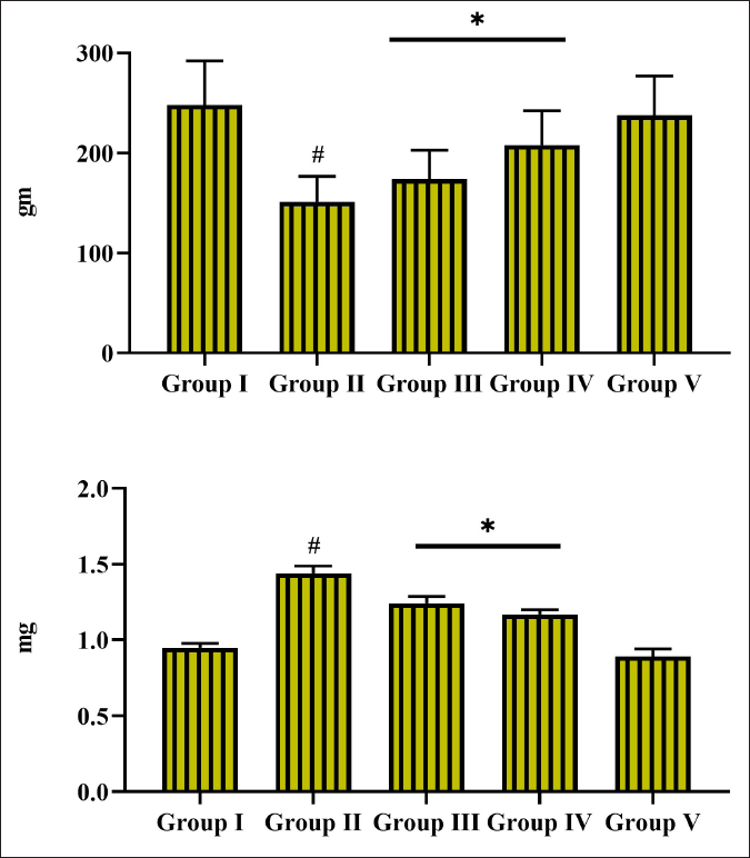
The influence of ponicidin on the alterations in body weight and heart weight was examined and the values are illustrated in Figure 1. The MI rats demonstrated a remarkable decrease in body weight and an improvement in heart weight. However, 25 and 50 mg/kg of ponicidin treatment revealed a considerable improvement in the body weight and a reduction in the heart weight of MI rats (Figure 1). Only mild changes were noted in both body weight and heart weight of 50 mg/kg of ponicidin alone administered rats.
Effect of Ponicidin on the Total Protein, CRP, and Uric Acid in the Experimental Rats
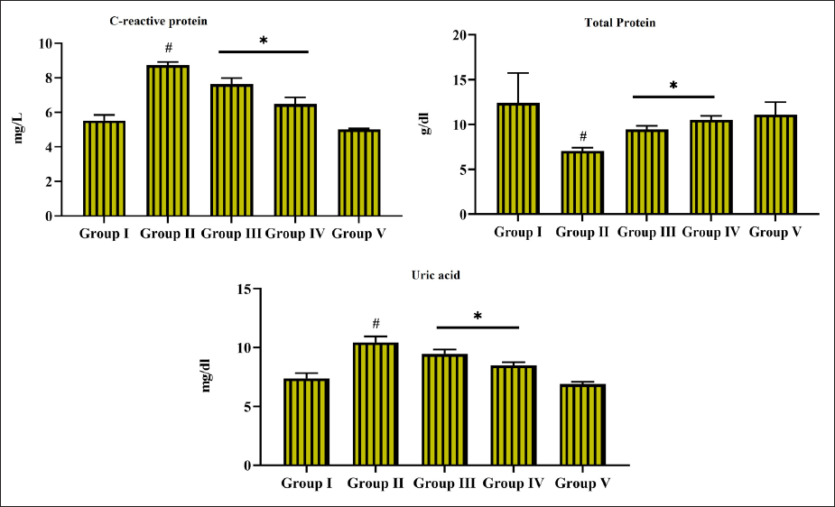
Figure 2 comprises the influence of ponicidin treatment on the status of total protein, CRP, and uric acid. The marked elevation in the CRP and uric acid was noted in the serum of MI rats. The ISO challenge also revealed a notable decrease in the total protein in the MI rats. Meanwhile, the treatment with 25 and 50 mg/kg of ponicidin was remarkably decreased both CRP and uric acid and improved the total protein status in the MI rats. Additionally, the CRP, total protein, and uric acid were remained unchanged or slight changes were noted on the serum of 50 mg/kg of ponicidin alone administered rats (Figure 2).
Effect of Ponicidin on the Activity of Cardiac Function Marker Enzyme Activities in the Experimental Rats
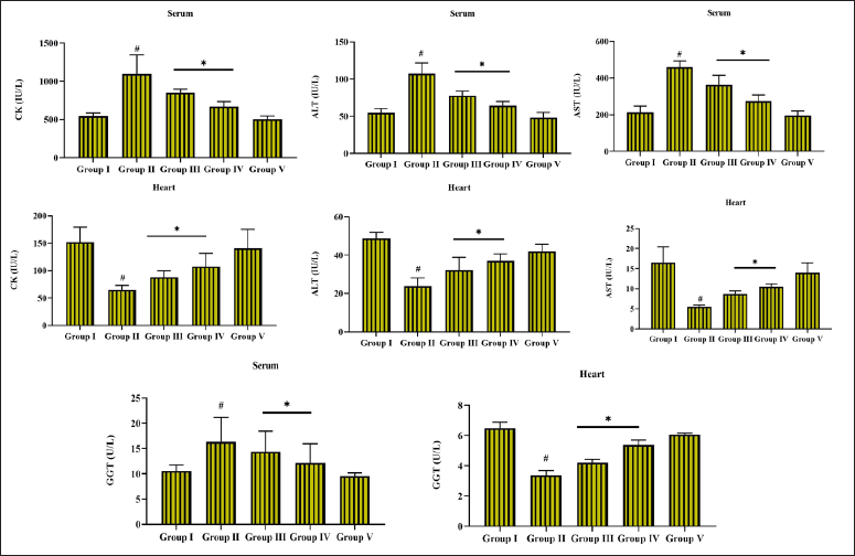
Figure 3 reveals the changes in the cardiac function markers such as GGT, CK, AST, and ALT. The MI rats exhibited a substantial elevation in the CK, ALT, AST, and GGT activities; however, these enzyme activities were found to decrease in the heart tissues. While the treatment with 25 and 50 mg/kg of ponicidin appreciably depleted the CK, ALT, AST, and GGT enzyme activities in the MI rats and at the same time ponicidin augmented these enzyme activities in the heart tissues (Figure 3). About 50 mg/kg of ponicidin alone treated rats revealed only slight alterations in these enzyme activities on both serum and heart tissues.
Effect of Ponicidin on the Antioxidant Biomarker Levels in the Heart Tissues of Experimental Rats
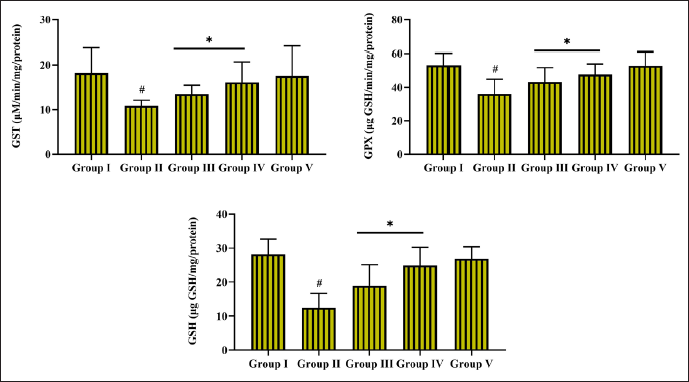
The influence of ponicidin treatment on the status of antioxidant levels, that is, GSH, GST, and GPx is depicted in Figure 4. A remarkable decrease in the GST and GPx and GSH levels was noted in the heart tissues of MI rats. Interestingly, 25 and 50 mg/kg of ponicidin treatment considerably improved the GST and GPx activities and GSH levels in the cardiac tissues of the MI rats. The ponicidin (50mg/kg) alone treatment exhibited mild alterations in the GST and GPx activities and GSH levels in the normal rats (Figure 4).
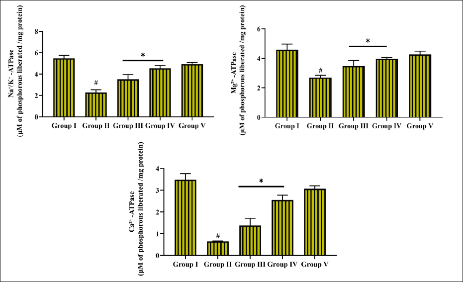
Effect of Ponicidin on the Potassium Na+/K+, Mg2+, and Ca2+ ATPase Activity in the Heart Tissues of the Experimental Rats
Figure 5 displays the influence of ponicidin treatment on the Na+/K+, Mg2+, and Ca2+ ATPase enzyme activities in the heart tissues of control and treated rats. The MI rats revealed a substantial reduction in the Na+/K+, Mg2+, and Ca2+ ATPase enzyme activities in the heart tissues when compared with the control. However, these ATPase enzyme activities were appreciably improved by the treatment with 25 and 50 mg/kg of ponicidin on the MI rats. About 50 mg/kg of ponicidin alone treatment revealed slight variations in the Na+/K+, Mg2+, and Ca2+ ATPase enzymes in the heart tissues of rats (Figure 5).
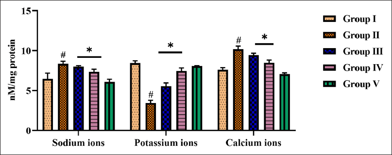
Effect of Ponicidin on the Levels of Na+, K+, and Ca2+ Ions in the Heart Tissues of the Experimental Rats
The status of Na+, K+, and Ca2+ ions from the cardiac tissues of experimental rats is displayed in Figure 6. The Na+ and Ca2+ ions were found elevated while the K+ ion level was decreased in the MI rats. Meanwhile, the treatment with 25 and 50mg/kg of ponicidin appreciably decreased the Na+ and Ca2+ ion status and improved the K+ ion level in the MI rats. The contents of Na+, K+, and Ca2+ ions were remain unchanged or only mild changes were observed in the heart tissues of 50 mg/kg of ponicidin alone treated rats (Figure 6).
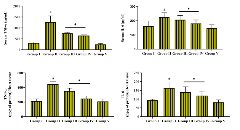
Effect of Ponicidin on the Levels of Pro-inflammatory Markers in the Heart Tissues and Serum of the Experimental Rats
Figure 7 depicts the level of IL-6 and TNF-α in the control and treated rats. The MI rats demonstrated a considerable increment in the IL-6 and TNF-α status in the serum and heart tissues. However, the treatment with 25 and 50 mg/kg of ponicidin treatment substantially decreased the status of IL-6 and TNF-α in the MI rats. About 50 mg/kg of ponicidin alone slightly altered the IL-6 and TNF-α in both serum and heart tissues (Figure 7).
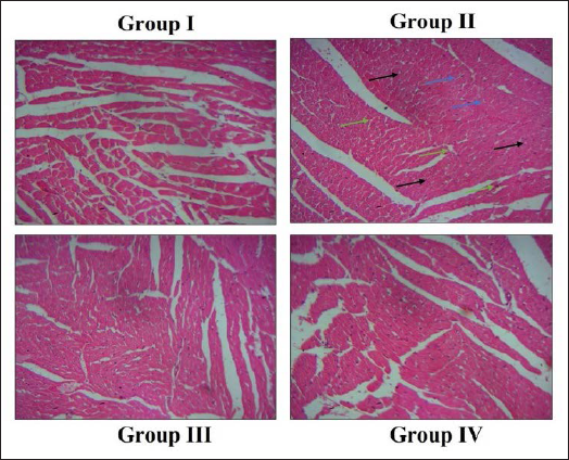
Effect of Ponicidin on Heart Histopathology in the Experimental Rats
As depicted in Figure 8, the heart tissues of the control rats revealed normal histological arrangements. However, the heart tissues of the MI rats demonstrated the histological damage, a high level of inflammatory cell infiltration, and cardiomyocyte necrosis. Interestingly, the treatment with 25 and 50 mg/kg of ponicidin appreciably decreased the ISO-stimulated histological alterations in the heart tissues of MI rats (Figure 8). The ponicidin alone treatment also did not exhibit any major changes in the heart tissues.
Discussion
A heart attack or MI is an imperative cause of mortalities in both developing and developed countries. If the heart does not receive proper blood/oxygen supply due to blockage of the arteries, it will lead to prolonged inadequate blood and/or oxygen supply to the cardiomyocytes and ultimately result in cardiomyocyte necrosis, a condition known as acute MI. 29 The elevation in the heart weight may because of the higher water content with intramuscular spaces (edema) in the cardiac tissues and elevated protein level. It was already been reported that the function of the myocardium may be decreased by roughly 10% because of the increment in the myocardial water content. 30 Similarly, the MI rats demonstrated a remarkable increase in heart weight. However, the ponicidin-treated rats revealed a reduced heart weight (Figure 1).
The myocardium comprises higher levels of diagnostic markers of MI. If the myocardium gets injured, it releases marker enzymes into the extracellular fluids. 31 The cardiac membrane becomes permeable or gets ruptured in response to the poor oxygen/glucose supply, which further leads to the release of marker enzymes. 32 The activity of the CK enzyme is a helpful diagnostic marker in the early detection of MI injury. The release of CK, AST, GGT, and ALT into the blood flow from cardiac tissues may occur if the cardiac cell membrane becomes permeable or damaged. The integrity of the myocardial membrane and its permeability can be changed by ISO exposure. The increased contents of these enzymes in the serum and the decrease in the cardiac tissues directly reflect the number of necrotic cells.33, 34 In agreement with these statements, here our findings demonstrated that the CK, AST, ALT, and GGT activities were found augmented in the serum and the same enzyme activities were elevated in the cardiac tissues of MI rats. Meanwhile, these changes were substantially ameliorated by the ponicidin in the MI rats, which evidences that the ponicidin improved cardiac function (Figure 3).
The serum levels of CRP are usually found to increase in response to inflammatory condition and acute infections. The CRP is a useful marker to detect the risks of cardiac diseases, which is a reliable inflammatory biomarker. 35 Garg and Khanna, 36 reported that the increment in the CRP levels was found in the ISO-exposed animal models, which may be due to the increased inflammatory markers. The current findings revealed that the ISO-exposed rats demonstrated an increased CRP level than the control. Interestingly, the administration of ponicidin appreciably decreased the CRP level in MI rats (Figure 2).
The cardiac tissues are extremely prone to oxidative stress than the other tissues because of their decreased antioxidant protection. Oxidative stress has straight connections to CVDs, which is often connected with the disproportion between oxidant and antioxidant mechanisms. 37 The ISO exposure triggers Ca2+ overload, which eventually disturbs the mitochondrial membrane integrity and produces excess reactive oxygen species (ROS) and free radicals, which facilitates its cardiotoxicity. ROS in myocardial tissues can stimulate several signaling cascades that can result in cardiac hypertrophy. 38 The endogenous antioxidant mechanisms are highly essential to defend the cardiac tissues against oxidative stress. 39 The GST and GPx are some of the most essential antioxidant enzymes, which are participated in the primary defense against oxidative stress and neutralizing free radicals. 40 The tissue-bound antioxidants were decreased in response to the elevation of free radicals. 41 Previous studies also highlighted that the ISO-exposed rats revealed a remarkable reduction in the antioxidant mechanisms.42, 43 The dysregulation of antioxidant mechanisms was observed in MI patients.44, 45 In agreement with this statement, our current results also exhibited a marked reduction in the GSH, GST, and GPx in the cardiac tissues of MI rats. However, the ponicidin treatment exhibited an appreciable increment in the GSH, GST, and GPx activities in the cardiac tissues of MI rats (Figure 4).
During the development of ischemic heart diseases, the inflammatory responses were initiated and regulated by several inflammatory regulators such as IL-1β, IL-6, and TNF-α. 46 TNF-α and IL-6 are the critical regulators, which actively participate in the progression of MI.47, 48 The increment in these pro-inflammatory regulators could speed up the progression of MI. In cardiomyocytes, the intracellular ROS enhances the accumulation of TNF-α and IL-6. The earlier studies are highlighted that the expression of inflammatory mediators is found augmented in both the animal and human models of ISO-stimulated MI. 49 In agreement with these lines, here our current findings witnessed that the TNF-α and IL-6 contents were augmented in both cardiac tissues and serum of MI rats. Interestingly, the administration of ponicidin was appreciably decreased IL-6 and TNF-α in both heart tissues and serum of MI rats (Figure 7).
The stable maintenance of electrolytes in the body is highly necessary for the regular functions of cells/organs, including the heart. The normal regulation of myocardial electrical activity depends on the concentrations of Ca2+, K+, and Na2+; however, ideal contraction of the myocardium needs Ca2+, Mg2+, and phosphorus. 50 The myocardial cytoplasm consists of higher levels of K+ ions that were regulated by the Na+/K+ ATPase. 51 Because of the reduced activity of Na+/K+ ATPase, the concentrations of K+ ions were depleted and Na+ ions were elevated. Ca2+ is the critical player in regulating the myocardial diastolic and systolic functions, and regular myocardial excitability and rhythmicity. The Ca2+ participates in several physiological mechanisms and signaling transductions as a secondary intracellular messenger. 52 The increased activity of adenylate cyclase may be a reason for the Ca2+ ion channel opening, which in turn results in the increase of Ca2+ ions in the cardiac cells because of the Ca2+ influx. 53 The depletion of both Na+ and K+ activities in serum followed by the ISO exposure can be due to the ongoing oxidative stress in response to the free radical attacks on the Na+/K+ pump. 54 Here, our outcomes revealed that the MI rats demonstrated the depletion of K+ ions and augmented the Na+ and Ca+2 ion concentrations in the cardiac tissues. However, the treatment with the ponicidin remarkably decreased these ion contents and improved the K+ ions in the cardiac tissues (Figure 6), which is supported by Hussain et al. 55
In normal cellular physiological conditions, Na+ and K+ ATPases maintain the Na+ and K+ ions transport through the membrane, upon its damage may result in ionic dissimilarity. Consequently, the measurement of the activities of membrane-bound enzymes will denote the changes in membrane physiology under pathological circumstances. 56 Our findings also revealed that the ponicidin treatment remarkably improved the Na+/K+, Mg2+, and Ca2+ ATPase enzyme activities in the heart tissues of MI rats.
Conclusion
Ponicidin remarkably reduced the inflammatory markers, raised the antioxidants, and modulated the cardiac marker enzymes in the MI rats. The ponicidin also improved the Na+/K+, Mg2+, and Ca2+ ATPase enzyme activities and regulated the K+, Na+, and Ca+2 ion concentrations in the MI rats. These findings are evidence of the beneficial roles of ponicidin in attenuating MI injury in rats. Hence, it can be a talented therapeutic candidate in the future to treat MI. Furthermore, in-depth molecular studies are still needed in the future to clearly understand the therapeutic roles of ponicidin against MI.
Footnotes
Abbreviations
Acknowledgment
This work was supported by the Department of Cardiology, The Third Hospital of Baoji, Baoji, 721000, China.
Declaration of Conflicting Interests
The authors declared no potential conflicts of interest with respect to the research, authorship, and/or publication of this article.
Ethical Approval
This research was approved by Shaanxi Province Geriatric Hospital animal ethical committee, SXSRM210507.
Funding
The authors received no financial support for the research, authorship, and/or publication of this article.
Statement of Informed Consent
There are no human subjects in this article and informed consent is not applicable.
Summary
CVD is a group of heart disorders, which are the foremost reason for non-communicable disease-related mortalities across the world.
Ponicidin is a di-terpenoid compound that possesses multi-functional properties.
Ponicidin treatment appreciably improved the antioxidants, Na+/K+, Mg2+, and Ca2+ ATPase enzyme activities, decreased the inflammatory markers, and regulated the cardiac marker enzyme activities in the MI rats.