
Abstract
Objectives:
To investigate the therapeutic effect of mashed potato and its alkaloids α-solanine and α-chaconine on basic fibroblast growth factor (bFGF) and vascular endothelial growth factor (VEGF) expression in ear venous vessels and adjacent tissues in a rabbit model of phlebitis.
Materials and Methods:
Japanese white rabbits were randomly allocated into control, vaseline cream (model), magnesium sulfate, mashed potato, α-solanine cream 100 mg/kg or 200 mg/kg, and α-chaconine cream 100 mg/kg or 200 mg/kg groups. The phlebitis model was established by intravenous infusion of vincristine in the rabbit ears. Group A received physiological saline. Group B−F was applied treatment 3 times/day, for 2 days along the ears vein. The alkaloid contents in α-solanine and α-chaconine were analyzed with HPLC. The bFGF, protein kinase C alpha (PKC-α), and VEGF expression in the ear vessels and nearby tissues were analyzed using immunohistochemistry, polymerase chain reaction (PCR), and Western blot analysis, respectively.
Results:
After phlebitis modeling, the vascular endothelium of the rabbit ear vein was damaged or layered, the lumen was swollen and deformed to an oval or round shape, and the proportion of bFGF and VEGF positive areas in the vascular endothelium and adjacent tissues was higher than the control group. The phlebitis score shows that various treatments have certain effects, among which magnesium sulfate wet gauze and mashed potatoes are more effective. The application of α-solanine and α-chaconine cream promotes the expression of bFGF and VEGF, as well as PKC-α in the venous blood vessels and adjacent tissues, but mashed potatoes had little effect.
Conclusion:
α-solanine and α-chaconine cream can promote the expression of bFGF and VEGF in venous blood vessels and adjacent tissues, which may contribute to the recovery of blood vessels in phlebitis.
Introduction
Phlebitis is one of the most common complications in clinical infusion. A meta-analysis study showed that the incidence of phlebitis with the use of peripheral intravenous catheters during infusion is 31%. 1 The incidence of phlebitis in hospitalized patients in China is about 30%. 2 Phlebitis increases the pain of patients and difficulty in vein puncture, leading to delayed treatment, and even local tissue necrosis. 3 Therefore, methods to relieve or treat phlebitis are highly valuable in clinical practice. In addition to the standard operating procedures for infusion, topical application of drugs has become an important means in the prevention and treatment of phlebitis. Gauze soaked with magnesium sulfate solution is one of the common treatments with satisfactory curative effect. 4 In our practice, we noticed that some patients use fresh potato chips to prevent and treat phlebitis caused by peripheral venous infusion as a simple and cheap alternative. The outcomes appeared to be good.
There are also some reports on the treatment of phlebitis with potato, which is believed to have anti-inflammatory and analgesic effects.5–7 A meta-analysis found that the curative effect of the external application of potato slices in patients with phlebitis is better than magnesium sulfate. 8 It was better than 50% magnesium sulfate wet compress in treating patients with drug-induced phlebitis. 9 Zhang et al. (2018) proved that topical fresh frozen potato chips can relieve the symptoms of infusion leakage, and greatly reduce the occurrence of phlebitis. 10 Guo et al. (2020) demonstrated that potato sprays had certain anti-inflammation and detumescence effects in mice with chemotherapy-induced phlebitis. 11
Potato alkaloids are recognized to be the main effective ingredients for observed curative effects.12–14 α-solanine and α-chaconine are major alkaloids, accounting for more than 90% of the total alkaloids in potatoes. 15 The content of alkaloids is greatly affected by potato varieties and the growth environment. It is generally believed that eating potatoes with higher than 200 mg alkaloids/kg would cause poisoning. 16 This value was used as the upper limit of α-solanine and α-chaconine in this study. Little is known about the effect of α-solanine and α-chaconine on angiogenesis in the treatment of phlebitis.
In this study, the therapeutic effects of mashed potato, α-solanine, and α-chaconine creams on phlebitis were investigated using a rabbit model. The expression of two key factors of angiogenesis basic fibroblast growth factor (bFGF) and vascular endothelial growth factor (VEGF) was measured to reveal the possible molecular mechanisms underlying the therapeutic effects. The findings would provide a new option for phlebitis treatment and recovery of blood vessels from phlebitis.
Materials And Methods
Experimental Animals
Japanese white rabbits, 3–3.5-month-old, 2.5–3.0 kg were purchased from Sundan Farms (SYXK (Beijing) 2016–0004). The animals were raised in the ordinary animal housing facility and provided with food and water ad libitum.
Reagents and Instruments
Vincristine solution was purchased from Bencao Yikang Biotechnology Co., Ltd., Jiangsu, China. Alkaloid creams containing α-solanine (188037, J&K Scientific, Beijing, China) and α-chaconine (B29075, Shanghai Yuanye Bio-Technology Co., Ltd., Shanghai, China) were prepared by mixing the alkaloids with Vaseline cream (LIRCON, Shandong, China), at 100 mg/kg and 200 mg/kg, respectively. The creams were stored at 4°C before use.
Magnesium gauze was prepared by soaking gauze in 500 g/L magnesium sulfate solution. The mashed potato was prepared by grinding commercially available potatoes (cv. Harvest) s with a mortar and pestle.
Rabbit anti-VEGF (1:1000, bs-1313R), rabbit anti-bFGF (1:300, bs-0217R), and rabbit anti-protein kinase C alpha (PKC-α) (1:1000, bs-3531R) were purchased from BIOSS, Beijing. Mouse monoclonal anti-glyceraldehyde 3-phosphate dehydrogenase (GAPDH) (1:2000, TA-08), horseradish peroxidase-labeled goat anti-mouse IgG (H + L) (1:2000, ZB-2305), and horseradish peroxidase-labeled goat anti-rabbit IgG (H + L) (1:2000, ZB-2301) were obtained from Zsbio, Beijing. 3,3′-diaminobenzidine (DAB) chromogenic kit (CW0125), neutral resin (CW0136), ultrapure RNA extraction kit (CW0581M), HiFiScript first strand cDNA synthesis kit (CW2569M), and UltraSYBR Mixture (CW0957M) were purchased from CWBIO, Beijing. A hematoxylin staining kit (AR1180-1) was obtained from Bioon, Beijing.
High-performance Liquid Chromatography (HPLC) 17
The potato samples were crushed and homogenized with a homogenizer. Then, 0.5 g of samples (precision to 0.01 g) were weighed and put into a 50 ml plastic centrifuge tube. 50 ml of 80% methanol aqueous solution (containing 0.1% formic acid) was added, vortexed, and mixed, followed by ultrasonic extraction for 30 min, and centrifuged at 8000 rpm for 10 min. The supernatant was filtered with a 0.22 µm filter membrane and loaded into the machine (SCIEX 6500, SCIEX, Shanghai) for detection. Chromatographic analysis conditions: waters HSS T3 column (2.1 mm × 100 mm, 1.7 µm); mobile phase: phase A was 0.1% formic acid water and phase B was methanol; flow rate: 0.25 ml/min, injection volume: 5 µl; column temperature: 30 ℃; gradient elution procedure: 0 ~ 1.0 min, 10% B; 1.0~3.5 min, 10% B~65% B; 3.5~6.0 min, 90% B; 6.0~8.0 min, 90% B; 8.01~10 min, 10% B.
Phlebitis Model and Treatment 18
The rabbits were randomly allocated into the normal control and phlebitis modeling group. For the normal control group (Group A) (n = 6), the rabbits received an intravenous injection of physiological saline without treatment.
For the phlebitis modeling group, the rabbits were anesthetized with intraperitoneal ketamine 100 mg/kg (Hengrui Pharmaceutics, Shanghai) and xylazine 10 mg/kg (Xinmingtai Chemicals, Hubei) injection. After ear skin preparation, intravenous infusion with vincristine at 0.2 mg/kg was given three times/day for 2 days. After the phlebitis modeling, the rabbits were randomly divided into groups (n = 6 per group) as follows:
Group B (model): 0.5 g of Vaseline cream (blank) was applied evenly along the rabbit ear veins, 3 times/day, for 2 days.
Group C: wet gauze with magnesium sulfate was externally applied to the ear veins of rabbits for 30 min, 3 times/day, for 2 days.
Group D: mashed potato was applied externally along the rabbit ear vein for 1 h, covered with plastic wrap to prevent water loss, 3 times/day, for 2 days.
Group E1: 0.5 g of 100 mg/kg α-solanine cream was applied evenly along the rabbit ear veins, 3 times/day, for 2 days.
Group E2: 0.5 g of 200 mg/kg α-solanine cream was applied evenly along the rabbit ear veins, 3 times/day, for 2 days.
Group F1: 0.5 g of 100 mg/kg α-chaconine cream was applied evenly along the rabbit ear veins, 3 times/day, for 2 days.
Group F2: 0.5 g of 200 mg/kg α-chaconine cream was applied evenly along the rabbit ear veins, 3 times/day, for 2 days.
Visual Infusion Phlebitis (VIP) Scale
The degree of phlebitis was examined using the VIP scale two days after therapy and was recorded as follows: 19
0 for no visible phlebitis,
I for erythema and/or edema appeared at the puncture site but no cord-like changes nor induration in the vein,
II for erythema and/or edema appeared at the puncture site with cord-like changes but no induration in the vein, and
III for erythema and/or edema appeared at the puncture site with cord-like changes and induration and ulceration in the vein.
Immunohistochemistry
An immunohistochemistry assay was performed as described. 20 Briefly, the ear tissue samples were embedded in paraffin, sectioned, and hydrated by an ethanol series from 100% to 0% for 5 min each. The sections were incubated with antibodies against bFGF, PKC-α, and VEGF overnight at 4℃, followed by incubation with appropriate secondary antibodies for 1 h before the microscopy study.
Real-time Quantitative Reverse Transcription Polymerase Chain Reaction (qRT-PCR)
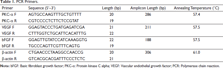
qRT-PCR was used to quantify the expression of genes at the mRNA level. Total RNA was extracted from the ear tissues using the ultrapure RNA extraction kit according to the manufacturer’s instructions, quantified using a Nanodrop spectrophotometer (NanoDrop Technologies, USA), and reversely transcripted into cDNA using the HiFiScript first strand cDNA synthesis kit according to the manufacturer’s recommendations. qRT-PCR was run with UltraSYBR Mixture on an Applied Bio-Rad CFX96 instrument using primers listed in Table 1. The relative mRNA levels were determined using the 2−∇∇Ct method after normalization with rabbit β-actin as the internal reference. 21 The polymerase chain reaction (PCR) was carried out in a reaction system containing 1 µl of diluted and pre-amplified cDNA, 10 µl of TaqMan Gene Expression Master Mix, and 1.5 µl of each fluorescence TaqMan probe. The cycling conditions were 51°C for 2 min, 94°C for 10 min followed by 40 cycles, each for 15 s at 96°C and 1 min at 58°C. The samples were run in triplicate and the mean value was calculated for each case.
PCR Primers.
Western Blot Analysis
The ear tissues were lysed with radioimmunoprecipitation assay (RIPA) buffer (Applygen Tech Inc., Beijing) that contains a protease inhibitors cocktail and quantified using bicinchoninic acid (BCA) kit (CWBIO, Beijing) according to the manufacturer’s instructions. After denaturation, the proteins were separated on sodium dodecyl sulfate-polyacrylamide gel electrophoresis (SDS-PAGE) (MilliporeSigma Corp., MO, USA), transferred to polyvinylidene fluoride (PVDF) membrane (MilliporeSigma Corp., MO, USA), and then detected by the corresponding primary and secondary antibodies before visualization with a chemiluminescence kit. The gray values of reactive bands were analyzed by Quantity One software (BioRad Lab, Inc., CA, USA).
Statistical Analysis
Data were expressed as means ± standard deviation (SD) obtained from at least three independent experiments and analyzed by SPSS version 19.0 for Windows (IBM Corp., Armonk, NY, USA). For normally distributed continuous variables, the means were compared using a student’s t-test or one-way analysis of variance (ANOVA) with the Tukey post hoc test. p <.05 was considered statistically significant.
Results
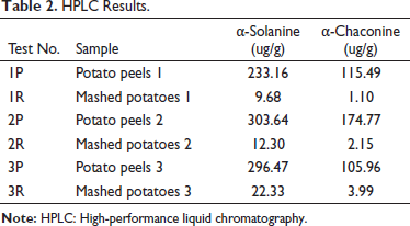
HPLC Results
Both potato peels and mashed potato contained two major alkaloids, α-solanine and α-chaconine, and their contents were very different. The mean content of α-solanine in the potato peels and mashed potato was 277.76 ug/g and 14.77 ug/g, respectively. The mean content of α-chaconine in the potato peels and mashed potatoes was 132.07 ug/g and 2.41 ug/g, respectively. The content of both alkaloids in the potato peel was greater than that in mashed potatoes (Table 2).
HPLC Results.
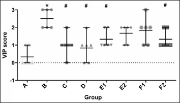
Phlebitis Score
Phlebitis was evaluated using the VIP scale 2 days after treatment and the results are shown in Figure 1. Untreated normal rabbits had the lowest score with no visible phlebitis. Rabbit models treated with Vaseline had the highest score. The number of rabbits in the mashed potato and magnesium gauze groups with scores between 0 and 1 was more than in other groups, indicating that mashed potato and magnesium gauze have better therapeutic effects as compared with alkaloid groups. For alkaloid treatments, there were fewer rabbits with a score of 1 in the 200 mg/kg than in the 100 mg/kg α-solanine cream group, and the results were reversed in the α-chaconine cream group.
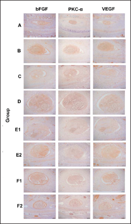
Immunohistochemical Expression of bFGF, PKC-α, and VEGF
The expression patterns of bFGF, PKC-α, and VEGF in the venous vessels and adjacent tissues at the puncture site were examined using immunohistochemical methods. The results showed that the vascular endothelial structure of the ear vein in the control (group A) rabbit was clear and intact, and the lumen was spindle-shaped. After infusion with vincristine (group B), the vascular endothelium was damaged or layered, and the lumen was swollen and deformed to an oval or round shape. bFGF, and to some extent VEGF, was enriched in the vascular endothelium (Figure 2).
mRNA and Protein Levels of bFGF, PKC-α, and VEGF
The effect on the bFGF, PKC-α, and VEGF expression was further quantified with PCR and Western blot. The mRNA expression of bFGF, PKC-α, and VEGF is shown in Figure 3A. Compared with the control (group A), the expression of bFGF and VEGF in the phlebitis model (group B) was significantly decreased (p <.05, both). Compared with group B, the expression of bFGF, PKC-α, and VEGF in group C was significantly increased (p <.05, all). Compared with group B, the expression of bFGF after treatments with 100 and 200 mg/kg α-chaconine (groups F1 and F2) was significantly increased (p <.05, both), the expression of PKC-α after treatment with 200 mg/kg α-solanine (group E2) was significantly increased (p <.05), and the expression of VEGF after treatment with 100 and 200 mg/kg α-solanine and α-chaconine (100 mg/kg) (groups E1, E2, and F1) was significantly increased (p <.05).
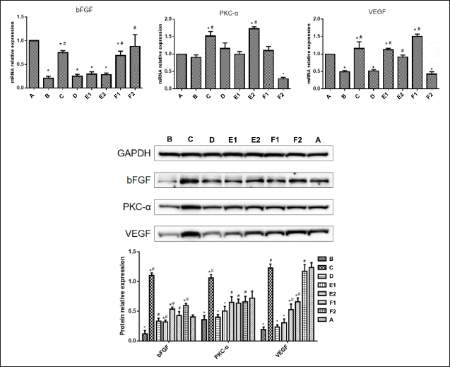
The protein expression levels of bFGF, PKC-α, and VEGF are shown in Figure 3B. Compared with group A, the expression of bFGF, PKC-α, and VEGF in group B was significantly decreased (p <.05). Compared with group B, the expression of bFGF, PKC-α, and VEGF in group C was significantly increased (p <.05, all). Compared with group B, the expression of bFGF in groups D, E1, E2, F1, and F2 was significantly increased (p <.05, all), the expression of PKC-α in groups E2, F1, and F2 was significantly increased (p <.05, all), and the expression of VEGF in groups E2, F1 and F2 was significantly increased (p <.05, all).
Discussion
The starch content in potatoes has a hypertonic effect, which can relieve local swelling and prevent drug extravasation. The glycoalkaloids in potatoes can stimulate smooth muscle and accelerate blood circulation, relieve spasms and reduce exudation. 12 This mainly includes α, β, γ-solanine and α, β, γ-chaconine, of which α-solanine and α-caconine account for more than 90% of the total content. 22 The results of HPLC in this study showed that the potato was rich in α-solanine and α-chaconine, indicating that these two alkaloids may play an important role in the repair of phlebitis. By observing the slices of venous vessels and surrounding tissues, it can be found that phlebitis modeling mainly causes swelling and deformation of venous vessels and damage to the vascular endothelium. It can be speculated that the repair of the venous vascular endothelium is very important for the treatment of phlebitis.
It is generally believed that the blood vessels with chemotherapy-induced phlebitis are accompanied by the release of various biological lineages in the process of trauma and repair. bFGF and VEGF are the key growth factors for angiogenesis. bFGF is one of the most studied fibroblast growth factors in the FGF family. It is a strong angiogenic factor and can promote angiogenesis in damaged tissue by providing nutrition for tissue growth and repair. It also enhances the proliferation, migration, and survival of endothelial cells.23, 24 VEGF is a member of the platelet-derived growth factor family, which has a strong promoting activity for vascular growth. It stimulates the synthesis of protease to enhance the degradation of the extracellular matrix, leading to increased microvascular permeability, extravasation of the plasma proteins, induction of proliferation and metastasis of the vascular endothelial cells, and subsequent formation of the new vascular lumen.25, 26
The receptors of bFGF and VEGF can activate a variety of downstream pathways, including the phosphatidylinositol-3-kinase (PI3K)/Akt pathway and rapidly accelerated fibrosarcoma (Raf)/mitogen-activated protein kinase (MAPK) pathway. Although these two pathways have overlapping effects, it is generally believed that the PI3K/Akt pathway mainly regulates cell survival and migration, while the Raf/MAPK pathway mainly regulates cell proliferation and osmotic balance.27–29 The receptor of bFGF and VEGF activates the Raf/MAPK pathway via Raf and PKC-α. 30 In this study, it was found that bFGF in the vascular endothelium and adjacent tissues was significantly enriched in the vascular endothelium, and VEGF also had this phenomenon, but it was not as obvious as bFGF. It can be seen that bFGF and VEGF are involved in the recovery process of vascular endothelium after phlebitis.
We further detected the expression of bFGF, PKC-α, and VEGF in the vein vessels and adjacent tissues at the puncture site and found that compared with the normal group, phlebitis modeling reduced the mRNA and protein expressions of bFGF and VEGF. While the external application of magnesium sulfate wet gauze could largely restore the expression of bFGF and VEGF, the external application of mashed potato has no significant effect. The increase of bFGF and VEGF content in the venous blood vessels where phlebitis occurs may help in the recovery of venous blood vessels, and the external application of magnesium sulfate wet gauze can provide this effect. Although treatment with mashed potato shows little effect on the bFGF and VEGF expression, it can still produce a good therapeutic effect, suggesting that there are other principles involved in the treatment of phlebitis with potato, such as the reported anti-inflammatory function. 3 And at the same time, this study also found that the potato alkaloids α-solanine and α-chaconine can promote the expression of bFGF and VEGF in venous blood vessels and adjacent tissues. From the results of the protein expression detection, the promoting effect of high-concentration cream is stronger than that of low-concentration cream, and the promoting effect of α-chaconine is higher than that of α-solanine. This also corresponds to other studies on the therapeutic effect of potato alkaloids on phlebitis.13, 14
Conclusion
In conclusion, this study found that the application of mashed potato, α-solanine, and α-chaconine cream has a certain curative effect on phlebitis, and can significantly improve the local swelling, pain, vascular sclerosis, and other symptoms of phlebitis. The potato alkaloid α-solanine and α-chaconine cream can promote the expression of angiogenesis growth factors bFGF and VEGF. Its anti-inflammatory effects have been gradually studied and investigated, which may provide good guidance for the development and exploration of the new usages of natural drugs.
Footnotes
Abbreviations
bFGF: Basic fibroblast growth factor;
VEGF: Vascular endothelial growth factor;
GAPDH: Glyceraldehyde 3-phosphate dehydrogenase;
DAB: 3,3′-Diaminobenzidine;
HPLC: High-performance liquid chromatography;
VIP: Visual infusion phlebitis;
qRT-PCR: Real-time quantitative reverse transcription polymerase chain reaction;
RIPA: Radioimmunoprecipitation assay;
BCA: Bicinchoninic acid;
SDS-PAGE: Sodium dodecyl sulfate–polyacrylamide gel electrophoresis;
PVDF: Polyvinylidene fluoride;
PI3K: Phosphatidylinositol-3-kinase;
Raf: Rapidly accelerated fibrosarcoma;
MAPK: Mitogen-activated protein kinase.
Acknowledgment
We would like to thank the funders for their financial support.
Declaration Of Conflicting Interests
The authors declared no potential conflicts of interest with respect to the research, authorship, and/or publication of this article.
Funding
This study was supported by the Hangzhou Medical and Health Science and Technology Plan (Grant no. 0020190874) and the Science and Technology Projects of Xiaoshan District, Hangzhou (Grant No. 20180218). Statement of Informed Consent and Ethical Approval All animal studies were approved by the Institute of Animal Care and Use Committee of Jiangnan Hospital Affiliated to Zhejiang University of Traditional Chinese Medicine.
Summary
The application of mashed potato, α-solanine, and α-chaconine cream has a certain curative effect on phlebitis.
The potato alkaloid α-solanine and α-chaconine cream can promote the expression of angiogenesis growth factors bFGF and VEGF.