
Abstract
Background:
Obesity is because of excessive fat accumulation that affects health adversely in the form of various diseases such as diabetes, hypertension, cardiovascular diseases, and many other disorders. Our Indian diet is rich in carbohydrates, and hence the sucrose-induced obesity is an apt model to mimic this. Ventromedial hypothalamus (VMH) is linked to the regulation of food intake in animals as well as humans.
Purpose:
To understand the role of VMHin sucrose-induced obesity on metabolic parameters.
Methods:
A total of 24 adult rats were made obese by feeding them on a 32% sucrose solution for 10 weeks. The VMH nucleus was ablated in the experimental group and sham lesions were made in the control group. Food intake, body weight, and biochemical parameters were compared before and after the lesion.
Results:
Male rats had a significant weight gain along with hyperphagia, whereas female rats did not have a significant weight gain inspite of hyperphagia. Insulin resistance and dyslipidemia were seen in both the experimental and control groups.
Conclusion:
A sucrose diet produces obesity which is similar to the metabolic syndrome with insulin resistance and dyslipidemia, and a VMH lesion further exaggerates it. Males are more prone to this exaggeration.
Introduction
Body weight is determined by an interaction between genetic, environmental, and psychosocial factors. 1 Physiologic studies had previously suggested that weight and energy stores are homeostatically regulated, with either weight loss or weight gain producing concerted changes in energy intake and expenditure that resist the obesity initial perturbation. 2 Obesity is because of excessive fat accumulation that may impair health. 3 It adversely affects nearly all physiological functions of the body and poses a significant public health threat. It increases the risk for developing multiple disease conditions, such as diabetes mellitus, 3, 4 cardiovascular disease, 4, 5 several types of cancers, 6 musculoskeletal disorders, 7 and poor mental health, 8 all of which have negative effects on the quality of life, work productivity, and healthcare costs.
Diet is one of the risk factors for obesity. The modern-day energy-dense diet may be the reason for the increasing prevalence of obesity. Li et al. have documented that a diet-induced animal model is the apt model to study obesity in the general population. 9 Various diet models have been studied so far.10, 11Studies have shown a significant difference in glucose intolerance between high-carbohydrate-diet-induced obesity and high-fat-diet-induced obesity. 12 Our Indian diet is rich in carbohydrates, and hence we chose to study the high-sucrose-induced obesity.
Ventromedial hypothalamus (VMH) is designated as the principal satiety center governing feeding behavior. 13 VMH is linked to the regulation of food intake and body weight in animals as well as humans. 14 A lesion of VMH is found to cause obesity. 13 Although there are many studies on hypothalamic obesity (created by an ablation of VMH) and diet-induced obesity, there is very less data available till date about the role of VMH in sucrose-induced obesity on metabolic parameters such as insulin, thyroid profile, lipid profile, and glucose. Hence, the present study was conceived.
Methods
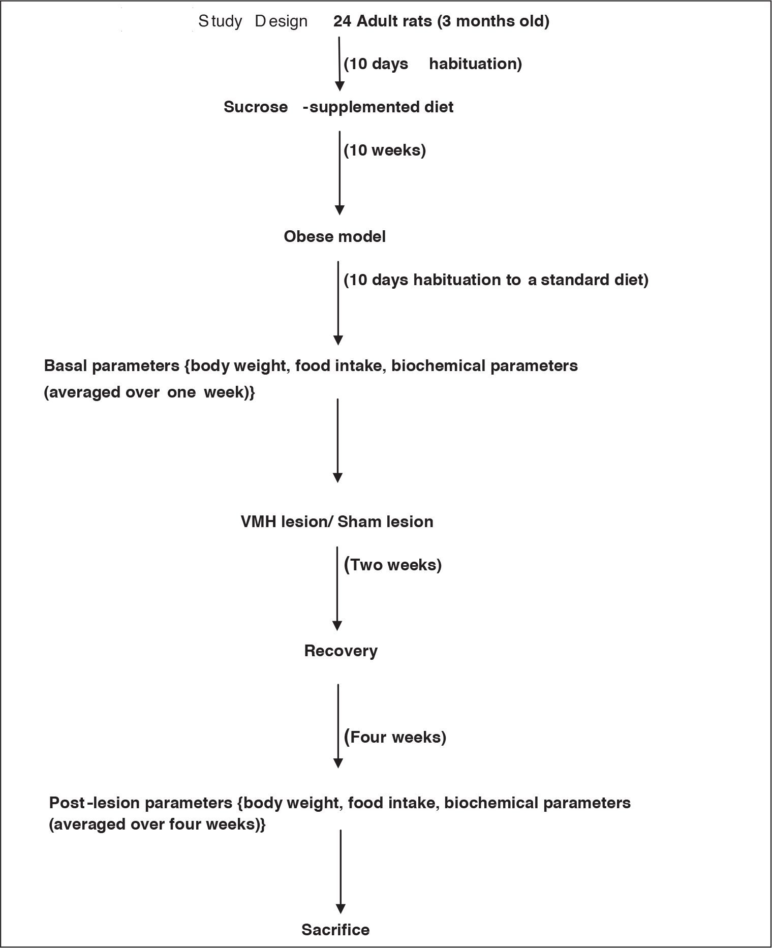
This is an experimental animal study done in the Department of Physiology, Jawaharlal Institute of Postgraduate Medical Education and Research, Puducherry. We commenced the study after obtaining the approval from both the institute scientific advisory committee and animal ethics committee. The guidelines of the Committee for the Purpose of Control and Supervision of Experiments on Animals were diligently followed in the study. A total of 24 (12 males and 12 females) institute-bred healthy adult albino rats of Wistar strain weighing between 150 and 250 g were used for the study. The rats were housed in individual plastic cages with wire lids. A layer of husk was spread on the floor of the cages. A 12-h light–dark cycle was maintained. They were fed on standard rat chow and allowed to habituate for 10 days.
After a habituation period of 10 days, the rats were fed on standard rodent chow supplemented with a 32% sucrose solution 15 and normal tap water. Diet and water were provided adlibitum for a period of 10 weeks to produce the sucrose-induced obese model of rats. Once obesity was attained, they were shifted to standard rodent chow. After 10 days of habituation, 40 g of standard rodent chow and 100 mL of fresh tap water were provided ad libitum every day. Daily food intake and body weight were measured for one week to determine the mean 24-h basal recordings, and pre-lesion blood was collected. The rats were divided randomly into two groups: one serving as the control group and the other as the experimental group. The sample size in each group was 12 (6 males and 6 females). In the experimental group, a lesion was made bilaterally in the VMH nucleus, and the control group included weight- and gender-matched rats, for which sham lesions were made.
Blood Collection
Blood samples were collected after seven days of baseline recordings from the jugular vein for a biochemical analysis under mild anesthesia (ether). A quantification of the thyroid hormone profile—plasmathyroid stimulating hormone[TSH; Human TSH chemiluminescencekit, Siemens, USA (110732)], total triiodothyronine[T3;Human TT3 RIA kit, Immunotech, Czech(119780)], and total thyroxine[T4;Human TT4 RIA kit, Immunotech, Czech (06490092)]—and the lipid profile (chemiluminescence, Siemens, USA)was carried out using the isolated serum as per the manufacturer’s guidelines. Blood glucose was measured using the glucose oxidase and peroxidase method. Insulin concentration was measured using the enzyme-linked immunosorbentassay kit (Millipore, USA). Insulin resistance was calculated using the standard formulae for the homeostatic model assessment of insulin resistance (HOMA-IR). For post-lesion values, 5 mL of rat blood was collected under anesthesia by cardiac puncture before sacrificing. We administered two-fold increased amount of ketamine intraperitoneally before sacrificing the animal. 16
Lesion
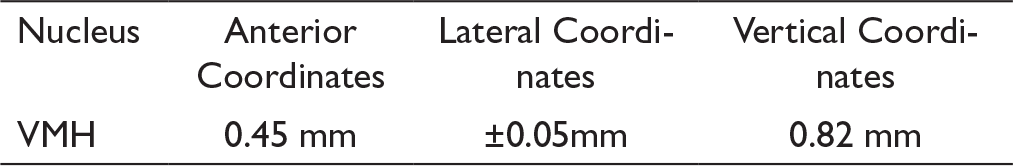
A lesion of the VMH was made according to the coordinates provided from the stereotaxic atlas for rat brain by König and Klippelin 1974. 17
In the experimental rats, the electrodes were passed bilaterally and a mild shock was given for an electrolytic ablation of the VMH nucleus. In the control rats, a sham lesion was made by placing electrodes near the VMH, but without shock to undergo the same level of stress as the experimental rats. After the lesion, the rats were accommodated to their cages with standard rodent chow and water for a fortnight, and we monitored them for bleeding and distress till their recovery. We recorded the post-lesion variables ensuring the complete recovery of the rats from the surgical procedure.
Statistical Analysis
All the data were analyzed and expressed in mean ± SD. Unpaired t-test was done between the groups and paired t-test was done before and after the lesion. All the data analysis was carried out in the IBM SPSS statistics software (version 20, New York, USA). The significance was set atthe P-value <.05.
Study Design 24 Adult rats (3 months old)
Results
Comparison of Body Weight and Food Intake in Female Rats
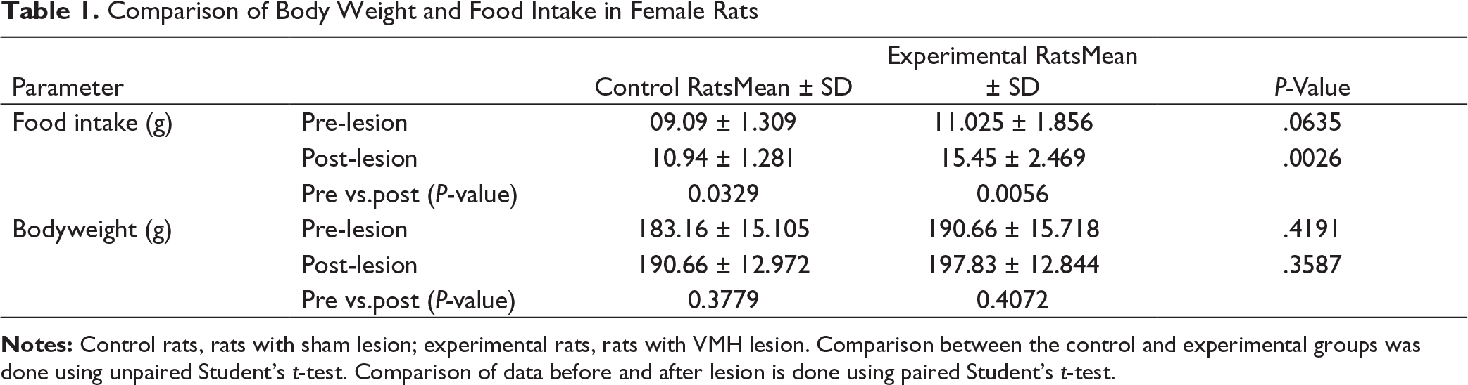
Notes: Control rats, rats with sham lesion; experimental rats, rats with VMH lesion. Comparison between the control and experimental groups was done using unpaired Student’s t-test. Comparison of data before and after lesion is done using paired Student’s t-test.
Comparison of Body Weight and Food Intake in Male Rats
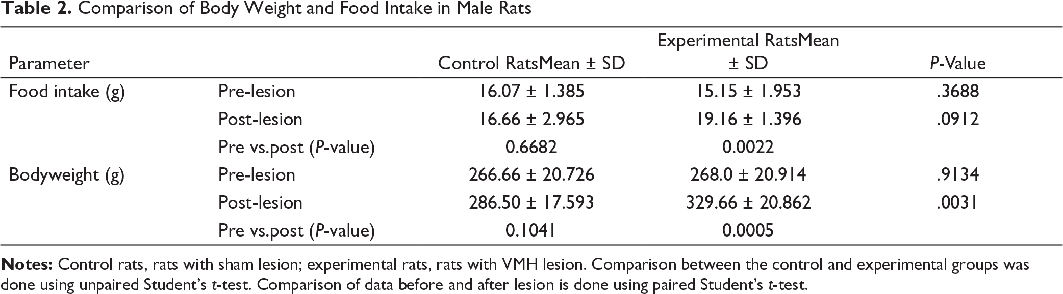
Notes: Control rats, rats with sham lesion; experimental rats, rats with VMH lesion. Comparison between the control and experimental groups was done using unpaired Student’s t-test. Comparison of data before and after lesion is done using paired Student’s t-test.
Males showed that pre-lesion values were comparable among the groups.
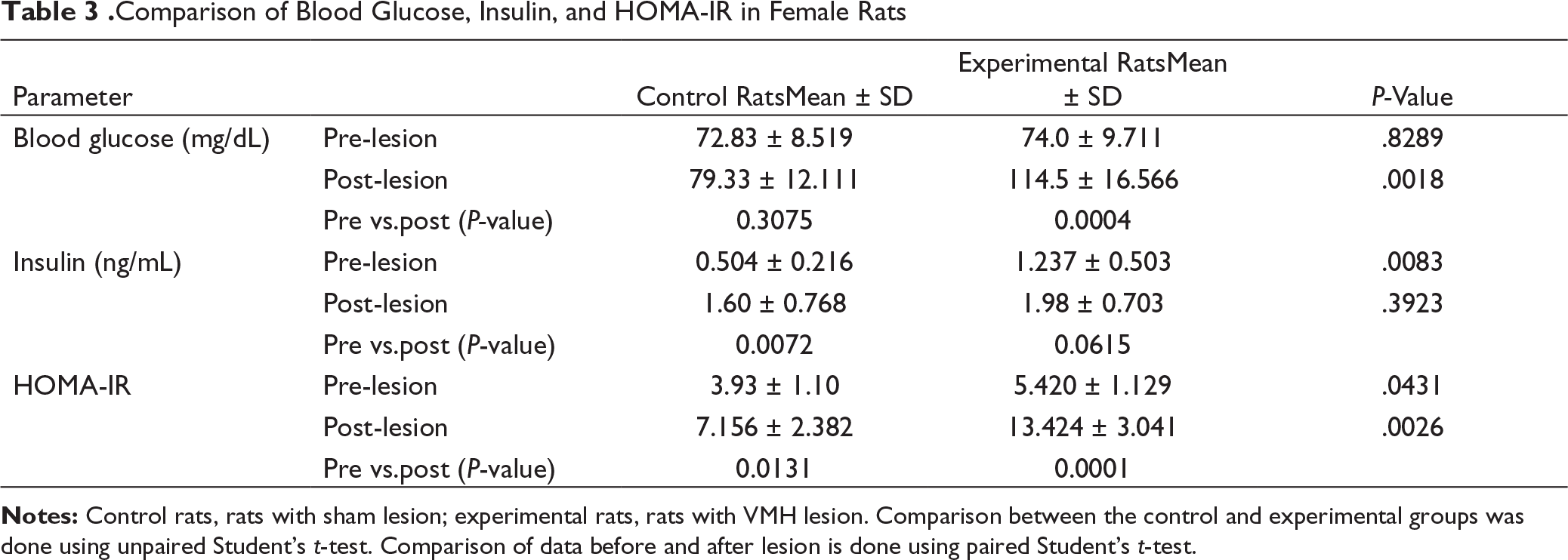
.Comparison of Blood Glucose, Insulin, and HOMA-IR in Female Rats
Notes: Control rats, rats with sham lesion; experimental rats, rats with VMH lesion. Comparison between the control and experimental groups was done using unpaired Student’s t-test. Comparison of data before and after lesion is done using paired Student’s t-test.
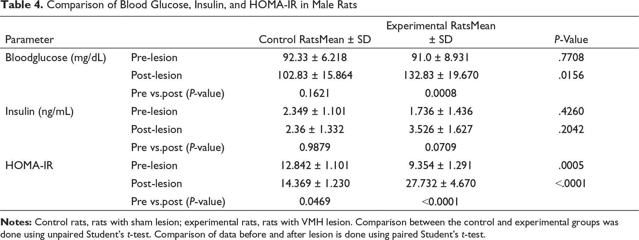
Comparison of Blood Glucose, Insulin, and HOMA-IR in Male Rats
Notes: Control rats, rats with sham lesion; experimental rats, rats with VMH lesion. Comparison between the control and experimental groups was done using unpaired Student’s t-test. Comparison of data before and after lesion is done using paired Student’s t-test.
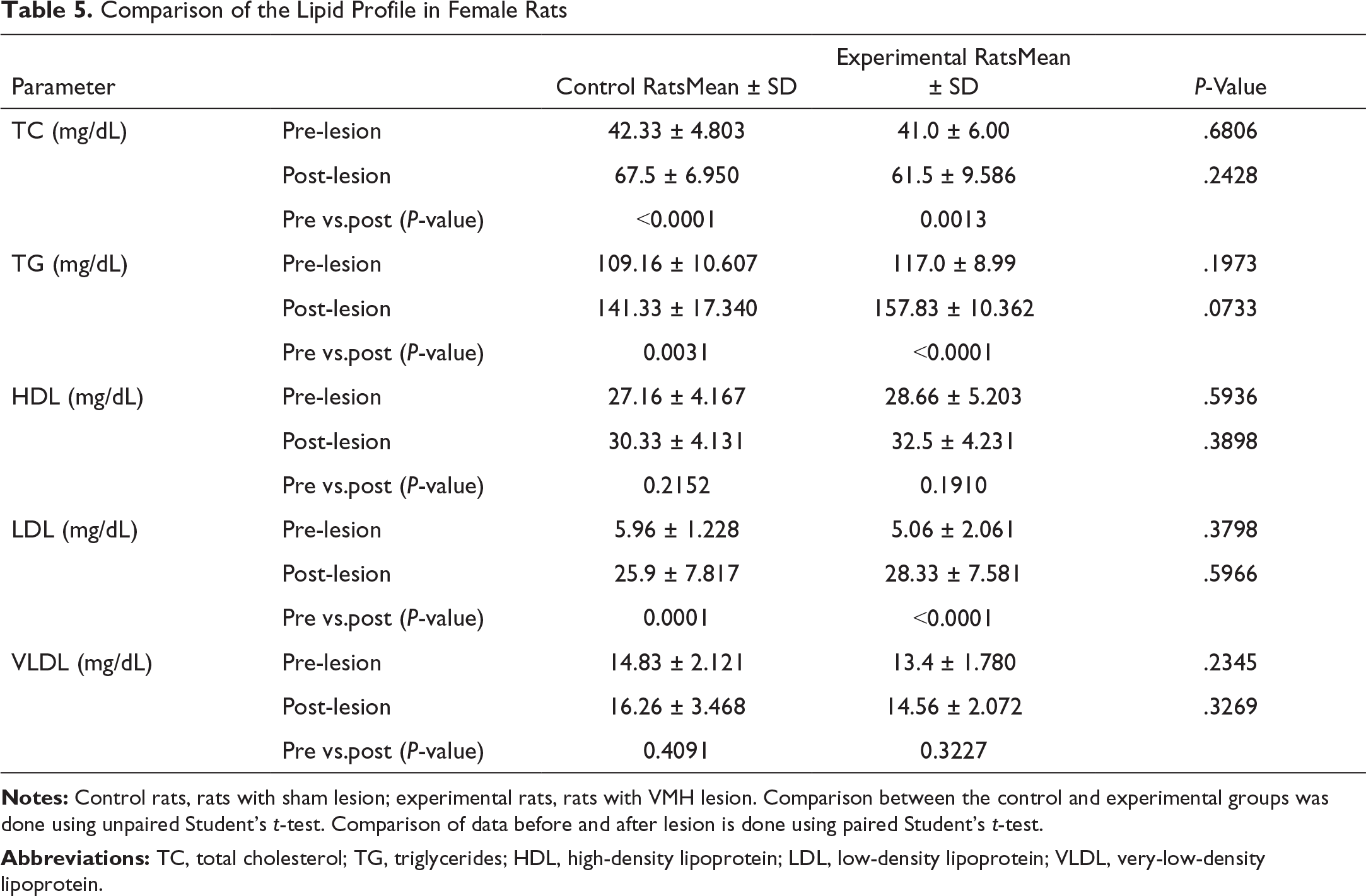
Comparison of the Lipid Profile in Female Rats
Notes: Control rats, rats with sham lesion; experimental rats, rats with VMH lesion. Comparison between the control and experimental groups was done using unpaired Student’s t-test. Comparison of data before and after lesion is done using paired Student’s t-test.
Abbreviations: TC, total cholesterol; TG, triglycerides; HDL, high-density lipoprotein; LDL, low-density lipoprotein; VLDL, very-low-density lipoprotein.
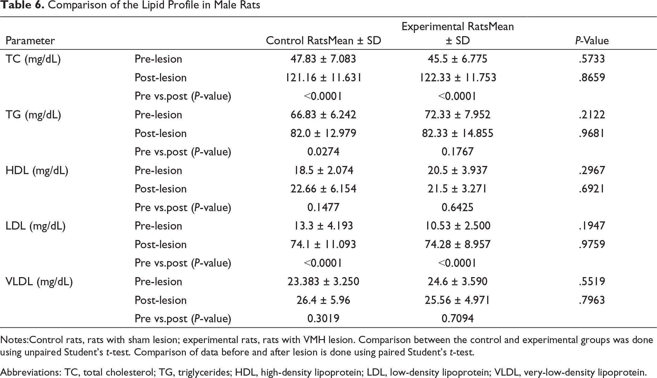
Comparison of the Lipid Profile in Male Rats
Notes:Control rats, rats with sham lesion; experimental rats, rats with VMH lesion. Comparison between the control and experimental groups was done using unpaired Student’s t-test. Comparison of data before and after lesion is done using paired Student’s t-test.
Abbreviations: TC, total cholesterol; TG, triglycerides; HDL, high-density lipoprotein; LDL, low-density lipoprotein; VLDL, very-low-density lipoprotein.
Comparison of Thyroid Profile in Female Rats
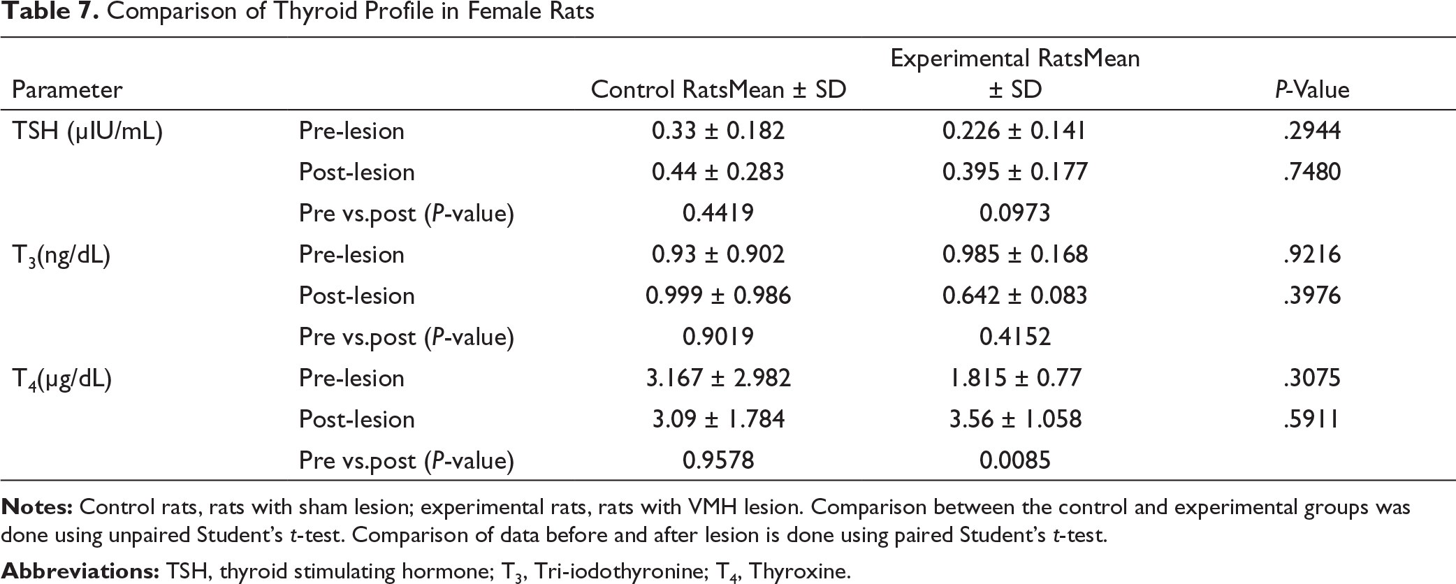
Notes: Control rats, rats with sham lesion; experimental rats, rats with VMH lesion. Comparison between the control and experimental groups was done using unpaired Student’s t-test. Comparison of data before and after lesion is done using paired Student’s t-test.
Comparison of Thyroid Profile in Male Rats
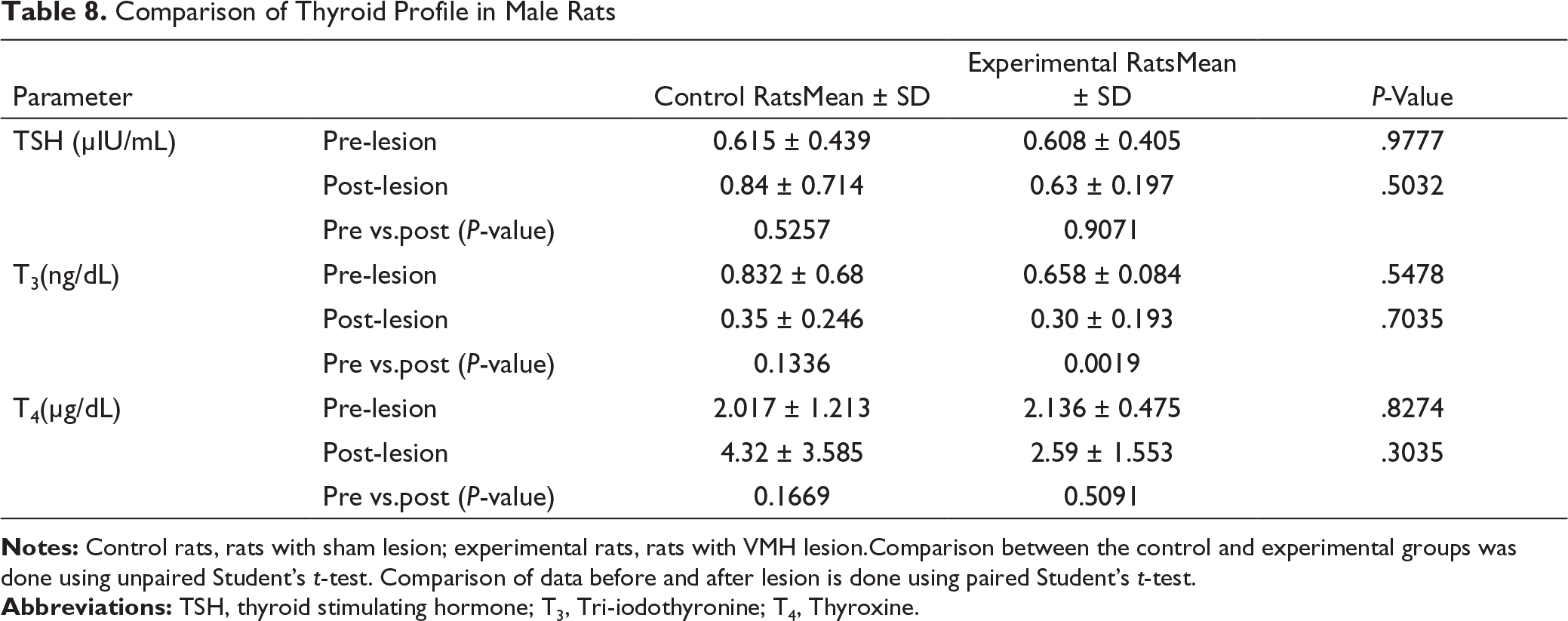
Notes: Control rats, rats with sham lesion; experimental rats, rats with VMH lesion.Comparison between the control and experimental groups was done using unpaired Student’s t-test. Comparison of data before and after lesion is done using paired Student’s t-test.
T3 significantly decreased in the experimental group(Tables 7 and 8).
Discussion
VMH is designated as the principal satiety center governing feeding behavior. 13 Established pathways involving orexigenic neuropeptide Y and agouti-related polypeptide, as well as the anorexigenic pro-opiomelanocortin and cocaine- and amphetamine-regulated transcript neurons project from the arcuate nucleus to other important hypothalamic nuclei, including the paraventricularnucleus, dorsomedial nucleus, VMH, and lateral hypothalamus nuclei. 18 Apart from these, there are also projections to and from other brain stem areas, cortical areas, and reward pathways.
This study was conducted to assess the role of VMH in already-obese rats. The rats were made obese by providing a sucrose solution, and then a lesion was made in the VMH nucleus. We observed a varied difference in male and female rats. The female rats showed an increase in food intake after the lesion in comparison to the control rats. But this did not result in a weight gain in them. However, in male rats, there was a significant weight gain along with hyperphagia. This is in confirmation with our previous study by Dev et al., where there was an increase in body weight in both male and female rats compared to their own control rats following the VMH lesion; the increase was significant in male rats and not significant in female rats. 16 Hence, the females are protected from hyperphagic obesity to some extent in comparison to males. This indicates a dissociation of the mechanism controlled by VMH regulation in different genders. On the contrary, Coxet al. observed that extensive bilateral VMH damage resulted in a diminished rate of weight gain inspite of an increased food intake in both the genders. 19 They compared the rate of weight gain instead of the overall weight gain, which was not assessed in our study. Another study by Sclafani et al. showed that vagotomy suppresses hyperphagia in rats on a chow diet and sucrose solution when VMH was damaged, but not on a palatable mixed diet. 20 This suggests that VMH is not involved in the regulation of feeding when a palatable diet is given. Hence, VMH is not the final common pathway for the regulation of feeding.
The important parameters of energy homeostasis are blood glucose and insulin levels. The blood glucose levels significantly increased after the lesion in both males and females, and a corresponding increase in insulin levels also observed in both the groups, though statistically not significant. But the HOMA-IR values were quite significant in both males and females. The control rats also had a significant increase in insulin which may be, to maintain homeostasis for the increase in blood glucose, an effect of the sucrose diet. Therefore, the sucrose diet by itself can cause a diabetes-like condition where there is an increase in blood glucose, insulin, and insulin resistance, and the VMH lesion further exaggerates this. In a study by Cao et al. the abdominally obese and normal-weight rats, which were created by giving a modified sucrose diet, showed a significantly reduced glucose-to-insulin ratio, demonstrating a decreased overall capability of disposing of ectogenic glucose. 21 Another study by Yang et al. described that a sucrose-rich diet can cause a change in insulin, signaling by the downregulation of genes involved in the insulin secretion. 22
There was a significant change in post-lesion values of TC, TG, and LDL and an insignificant rise in HDL and VLDL in both the control and experimental groups in females. Males also showed similar results, except for TG which was higher in females. This suggests that the VMH lesion did not produce these effects, but may be because of a sucrose diet there are higher values in both the control and experimental groups. This is in confirmation with a study by Yang et al., where the measurement of hepatic TG clearly indicated an increased hepatic lipid accumulation in response to the high-fat and high-sucrose diet as early as two weeks. This may be explained by the upregulation of genes involved in lipid metabolism and inflammation. 22 The high triglyceridemia in the high-sucrose diet was because of an increased hepatic triacylglycerol secretion and a decreased removal of triacylglycerol from the plasma in contrast to the high-fat-diet-induced triglyceridemia, which is because of a decreased removal of triacylglycerol alone. 23 Another study by Cao et al. observed that a modified high-sucrose diet produces hepatic lipidosis and hepatocyte mitochondrial swelling. 21 To infer that sucrose diet creates a model of dyslipidemia.
Thyroid hormone is a major regulator of energy metabolism and food intake, greatly influencing the energy homeostasis of the body. In our study, the experimental group of females showed a significant rise in T4 values, whereasthe experimental group of males showed a decrease in T3 values.The increasedT4 and decreased T3values in the experimental group of both the genders after lesion, may be because of the reduced conversion of T4 to T3. The VMH lesion might have resulted in the deficiency of type II deiodinase enzyme, which is primarily localized in the brain and pituitarygland. 24 Previous studies have found that T3 has a direct influence on feeding; T3 directly injected into the ventromedial nucleus increased the food intake by four times 25 and the inhibition of thyroid hormone receptors in VMH reverses the weight loss observed in hyperthyroidism, 26 interpreting that the thyroid hormone regulates the food intake and body weight via VMH through the hypothalamus–pituitary–thyroid axis. However, at this stage, we are not clear whether the VMH lesion resulted in a deficiency of type II deiodinase or there was a decreased expression of receptors, which needs further evaluation.
This study was done only with biochemical parameters; this is a major limitation of the study. Other parameters like fat % and other metabolic and inflammatory changes were not measured. A future study with fat % and other metabolic and inflammatory changes associated with obesity could be planned. Since aVMH lesion exaggerates obesity, the role of drugs and stem cell therapy, which enhance the neurological recovery, may be explored in the treatment of obesity. 27
Conclusion
A sucrose diet produces obesity, which is similar to the metabolic syndrome with insulin resistance and dyslipidemia, and aVMH lesion further exaggerates it. Males are more prone to this exaggeration. Females seem to be protected to some extent which may be because of the effect estrogen, which needs further analysis.
Footnotes
Acknowledgment
We thank JIPMER for providing the intramural research grant for this study.
Author Contribution
All authors have equally contributed.
Ethical Statement
Ethical clearance wasobtained before the start of experiment and all procedures followed the CPCSEA guidelines.
Declaration of Conflicting Interests
Funding
The conduct of this project was funded by the JIPMER intramural research fund.