
Abstract
Identification of an individual plays a critical role in scenarios involving mass disasters, missing persons, and unidentified bodies. Various factors such as race, sex, stature, and age are key parameters considered during the identification process. Among these factors, estimating stature stands out as a crucial element in the overall identification process. Here, we conducted a study in a tertiary care center in South India that was dedicated to formulating a standardized method for determining stature in patients. The study focused on utilizing the length of the first metatarsal from radiographic images as a key indicator for accurately estimating stature. Utilizing the advanced digital X-ray equipment available at the hospital, we meticulously measured the length of the first metatarsal in either foot of a sample consisting of 161 individuals. Through a detailed analysis of the data collected, the researchers discovered a more pronounced relationship between stature and the length of the first metatarsal in females compared to males. The study’s outcomes led to the formulation of regression equations specifically for this demographic group. For males, the regression equation was computed as H = 134.8 + 4.8 M1, while for females, it was determined as H = 133.4 + 3.86 M1. These significant findings highlight the practicality of utilizing the regression equations derived from this study to estimate stature within the South Indian population. The regression equation established through this research offers a dependable and precise approach for determining stature in this specific demographic group by solely considering the length of the first metatarsal bone.
Introduction
Identification is the crucial process of determining an individual’s identity, regardless of their status or circumstances. This task has become even more significant in light of the recent surge in mortality rates, especially in cases involving severed body parts, decomposed bodies, or mass disasters.1, 2 In situations where remains have been extensively decomposed, burned, or disfigured, such as in explosions, earthquakes, or plane accidents, where only fragments of bones or sections of bodies may be recovered, employing multiple identification methods becomes imperative. There are primarily two types of identification methodologies: Absolute and partial. Absolute identification refers to correctly identifying a person with 100% accuracy, irrespective of whether they are alive or deceased, while partial identification is less precise in comparison.
Both natural and man-made catastrophic incidents can be attributed to factors such as rapid population growth, industrialization, advancements in civilization, and climate change. It is under these challenging circumstances that establishing the identity of individuals becomes not just important but also a task for forensic investigators. Determining the stature or height of individuals is a crucial aspect when analyzing unidentified human remains since height serves as a distinctive biological characteristic.2–4 This analysis is rooted in the understanding that there exists a direct linear correlation between stature and the length of various skeletal elements such as vertebrae, long bones, metacarpals, metatarsals, pelvis, scapula, calcaneus, talus, and skull.
Although various bones can be utilized, the long bones of the limbs are commonly employed to estimate an individual’s stature.2, 3 Different methods tailored to specific ethnic and geographical populations can be utilized to calculate stature from these long bones.3, 4 Numerous research studies have been conducted to develop regression formulas customized to the stature patterns of distinct population groups. However, the utility of long bones is hindered by the challenges posed by fragmentation, leading to inaccuracies in measurements. Consequently, smaller bones such as foot bones can also be considered when determining the stature of an adult. 4
Studies focusing on measuring ulnar length, humerus length, and percutaneous tibia length have been undertaken to assess stature in the South Indian population. Conversely, there is also research on estimating stature from smaller bones, such as metatarsals, in populations such as Koreans, Egyptians, and Spaniards. This highlights the need for further research and exploration in forensic anthropology to enhance identification methods, particularly in scenarios involving mass disasters and challenging environmental conditions.5–8
This study aims to estimate the stature of individuals from the first metatarsal length of adults in tertiary care centers. The objective of the study is to determine the first metatarsal length from the radiograph, to correlate the first metatarsal length with the actual height, and to derive a regression formula to apply for the South Indian population.
Material and Methods
Ethics Approval
The research was conducted after getting ethical approval from the Institutional Ethics Committee of Sri Ramachandra Medical College. The ethical reference number is CSP-MED/22/DEC/81/162.
Study Design
This is a cross-sectional study which is conducted on 161 selected subjects (86 males and 75 females). All individuals aged between 18 and 60 years are included in the study. Subjects with congenital deformities, disease, or fractures of the first metatarsal bones, aged less than 18 and more than 60 years, are excluded from the study.
Methods
After getting consent from the subject, the height of the subject was measured with the help of the standard height measuring tool. These were collected from the individuals who attended the outpatient department with inclusion criteria and who were advised to undergo an X-ray of either foot at the Department of Radiology, Sri Ramachandra Higher Education and Research Institute, Porur, Chennai.
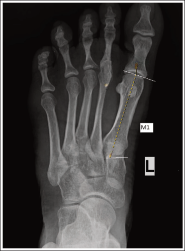
The subject was asked to stand in an upright position. The subject was allowed to stand erect with his/her back resting against a scaling instrument and the arms held at the sides of the body. Distance from the vertex to the foot is measured in an erect posture. The first metatarsal length of the subjects was measured from the dorso-plantar digital X-ray using a digital image viewer. By taking previous studies as reference, the definition of metatarsal length measurement was as follows: M1–the maximum length of the first metatarsal–the distance between the tip of the tuberosity and the most distal point of the head (Figure 1).
First Metatarsal Length.
The measurement obtained was statistically analyzed by using the Statistical Package for the Social Sciences (SPSS) program for statistics. The statistics techniques include mean, median, standard deviation, Pearson correlation coefficient, p value, linear regression analysis, and scatter diagram for correlation coefficient.
Results
The average age of study participants (n = 161) was 37.5 years, with a standard deviation of 10.9. The average age of females (n = 75) is 39.2 years, with a standard deviation of 11.9, and for males (n = 86) is 36.1 years, with a standard deviation of 9.8. Table 1 shows the descriptive statistics of both sexes. The results of an independent t test showed that height significantly differed among males and females (p = .001). The average height of males was found to be higher than that of females.
The Details on the Descriptive Statistics of Male and Female Measurements.

Table 2 shows the relationship between height and M1. The correlation values of M1 are found to be significant (p < .05). The height was correlated with the size of the metatarsal bone in both genders. Figure 2 shows the relationship between height and M1. Stronger correlations are detected between the height and length of the M1 metatarsal bone (M1; R = 0.540) in females.
Correlation of Height with M1.

Relationship Between Height and Length of First Metatarsal Bones.

In univariate correlation analysis, height was found to be correlated with the first metatarsal bone. Regression analysis was performed to fit a model to predict the height based on the length of the first metatarsal bone. The regression equation derived for females was H = 133.4 + 3.86 M1, and for males, H = 134.8 + 4.8 M1. The details are depicted in Table 3.
Regression of Length of First Metatarsal Bone with Height According to Gender.

Discussion
When estimating height, it is crucial to consider various factors, with age being a significant determinant.9–11 In our study, we specifically focused on individuals aged 18 years and above, as this age signifies the completion of epiphyseal fusion. Our research involved participants within the age range of 18–60 years, encompassing a diverse demographic. The primary objective of our study was to develop a regression equation for our specific population, enabling us to accurately predict and estimate heights based on relevant variables. By analyzing data across different age groups and incorporating age-related factors, we aimed to create a robust model that can assist in height estimation for individuals within our target age group.
In a study conducted by Park et al. researchers looked into the correlation between height and metatarsal measurements by utilizing a specialized regression equation to account for the unique characteristics of the population. The study sample comprised 200 adults aged between 20 and 86 years. To see accurate measurements, the researchers employed distal X-rays to assess the lengths of the first and second metatarsals of the foot. The analysis of the collected data revealed a statistical correlation, with a correlation coefficient (R value) of 0.468 (M1), 0.4312 (M2) for males and 0.475 (M1), 0.4773 (M2) for females, indicating a moderate positive relationship between foot bone dimensions and height within the studied population. 5 These findings shed light on the potential association between metatarsal measurements and stature. This study has M1 values lower than our study, which implies the racial difference, and it shows that males have a better association with M1 in contrast to females, which is associated with M2. This is against our study, where both males and females are associated with M1.
Ibrahim et al. conducted a study in the Egyptian population to analyze metatarsal length using radiography for a sample of 220 individuals aged between 21 and 62 years. They employed a regression analysis and found that the correlation coefficients (R) were 0.8904 (M1) and 0.8878 (M2) for men, and 0.8818 (M1) and 0.8808 (M2) for women. Interestingly, the research highlighted that the first metatarsal bone demonstrated a stronger correlation with stature. 6 The values are higher than those in our study, and they showed that the stature of both males and females showed a stronger association with M1, which is similar to our study.
Rodríguez et al. conducted a thorough investigation into adult stature among the Spanish population, utilizing a sample group comprising 228 individuals aged between 19 and 82 years. Their study focused on assessing adult stature by precisely measuring metatarsal length using radiography. Through the meticulous analysis of all gathered measurements, the research team was able to craft a comprehensive formula. This formula, when applied, yielded correlation coefficient values denoted as R for both male and female subjects. Specifically, they found correlation coefficient values of 0.783 (M1) and 0.705 (M2) for males, while for females, the correlation coefficients were calculated to be 0.731 (M1) and 0.711 (M2). The results of this study provide valuable insights into the relationship between metatarsal length and adult stature within the Spanish demographic, shedding light on the factors influencing physical characteristics among adults in this population segment. 7 Both the Spanish study and Egyptian study have shown M1 is better associated than M2 in both sexes. Both these study values are higher than our population, which repeatedly emphasizes how physical and racial variation influence the stature.
The cadaveric investigation conducted by Park et al. aimed to establish a specific regression equation for the Korean population to estimate stature based on metatarsal bones. For their study, a total of 31 female and 50 male adult formalin-fixed cadavers were utilized. Through their research, a statistically significant correlation was identified between stature and metatarsal length, yielding a correlation coefficient of M1 of 0.460 in males and 0.510 in females. These are higher than the M2 value. This finding tells us the potential utility of metatarsal bones in estimating stature within the Korean demographic, offering valuable insights for forensic investigations and anthropological studies in the region. 8 We could also able to see that the M1 value derived in cadavers is higher in females when compared to the radiographic value of females in the same Korean population. This denotes there could be variation between radiography and bony values because the position of the foot during X-ray examination can also account for variation in values.5, 8
Sharma et al. (2024) demonstrated a strong correlation between humeral length and stature, reinforcing the reliability of long bones as primary indicators. However, compared to their higher correlation values, our study using first metatarsal length showed moderate correlation (R ≈ 0.53–0.54), highlighting that although small bones are useful, they are relatively less accurate than long bones. 12
Similarly, Chawla et al. (2024) reported a significant correlation using upper limb measurements with regression analysis. Their findings align with our study in demonstrating statistically significant relationships (p < .001), although their correlation strength was comparatively higher, possibly due to the inclusion of larger limb segments. 13
Kumar et al. (2023), in a South Indian population, evaluated stature from foot length and reported a significant positive correlation. Their findings closely support our results, as both studies are region-specific and emphasize the utility of foot-related measurements. However, our study specifically focuses on the first metatarsal, which provides a more localized skeletal parameter. 14
Lomi et al. (2023) used odontometric and skull parameters and found comparatively variable correlation values. This contrasts with our study, where metatarsal length showed a more consistent linear relationship, suggesting that appendicular skeletal elements may provide better predictive accuracy than cranial parameters. 15
Singh et al. (2023) demonstrated a strong correlation using percutaneous lower limb measurements, which is in agreement with the general principle that lower limb bones contribute significantly to stature. However, their higher correlation values compared to our study further emphasize that long bones and limb segments are superior predictors when available. 16
Meena et al. (2022) reported reliable stature estimation using ulna length. Their findings are comparable to our study in terms of statistical significance, though ulna (a long bone) exhibited better predictive accuracy than metatarsal measurements. 17
Patil et al. (2023) utilized arm span and its segments and found a strong correlation with stature. While their results support the concept of proportional body measurements, the correlation observed in our study is slightly lower, which can be attributed to the smaller size and variability of metatarsal bones. 18
Sinha et al. (2022) evaluated lower limb measurements and found strong correlation coefficients, which are higher than those observed in our study. This again reinforces that larger skeletal elements yield more accurate stature estimation compared to smaller bones. 19
Gupta et al. (2023) demonstrated a significant correlation using tibial length, with better predictive value than smaller bones. This is consistent with our findings, where metatarsal length, although useful, shows comparatively moderate correlation. 20
Das et al. (2022) studied hand length and breadth and found a significant association with stature. Their results are comparable to our findings, as both studies utilize smaller skeletal elements, and both demonstrate moderate correlation with stature. 21
Within the scope of this research, analysis revealed that the average height of the participants was 162.1 cm, with a standard deviation of 6.7. When it comes to the average length of the M1 metatarsal bone, it was documented at 6.4 cm with a standard deviation of 0.6. Particularly noteworthy findings emerged when investigating the relationship between height and M1 length in female subjects, showing a correlation coefficient of 0.540. This correlation shows a significant connection between an individual’s stature and the specific elongation of the M1 metatarsal bone, shedding light on potential anatomical and physiological implications that warrant further exploration and analysis. The limitation of the study is that it was done only in one institution and only for males and females. So, this study should be extended to include the unknown sex.
Conclusion
The first metatarsal bone can be a crucial element in stature estimation when combined with other bones, especially long bones. Despite its diminutive size in comparison to long bones, the first metatarsal exhibits a lower susceptibility to fragmentation, making it particularly useful in scenarios involving mass disasters. Thus, the aim of the study has been achieved by formulating the regression equation for the first metatarsal. To effectively apply the regression formula for stature estimation, further research studies on the South Indian population are essential. By conducting multiple similar studies within this demographic, the accuracy and applicability of the regression formula in estimating stature can be significantly enhanced.
Footnotes
Acknowledgment
I would like to extend my thanks to the X-ray technician, Mahalakshmi, for helping me to collect the samples from the Radiology Department.
Declaration of Conflicting Interests
The authors declared no potential conflicts of interest with respect to the research, authorship, and/or publication of this article.
Ethical Approval
Ethical approval was obtained from the Institutional Ethics Committee.
Funding
The authors received no financial support for the research, authorship, and/or publication of this article.
Informed Consent
Informed consent was obtained from the participants for conducting the study.