
Abstract
Fingerprint evidence is one of the most reliable biometric tools in forensic identification due to its uniqueness and permanence. Fingerprints are frequently encountered at crime scenes in both partial and complete forms. An emerging morphological feature of interest is fingerprint ridge density, defined as the number of ridges within a specified area of the fingerprint. The present study aimed to evaluate sex-based differences in fingerprint ridge density across three anatomical regions of the fingerprint: Radial (R), ulnar (U), and lower (L) in the Indian Tamil population. The study was conducted on 274 individuals (137 males and 137 females) aged between 18 to 60 years. Fingerprints were collected from all 10 fingers of each participant. Ridge density was measured in three defined regions of the fingerprint: R, U, and L areas. Statistical analysis was carried out using the independent t-test to compare ridge density values between males and females in each region. The findings revealed that females exhibited significantly higher ridge density than males across all three regions of the fingerprint. Among the studied regions, the L area demonstrated the highest mean ridge density, followed by the R and U regions. These differences were consistent across both hands and all fingers, although slight variations were observed in a few anatomical areas. Overall, the results strongly indicated the presence of sexual dimorphism in fingerprint ridge density. The study concludes that fingerprint ridge density is a reliable morphological parameter for sex differentiation in the Indian Tamil population, with females consistently showing higher ridge density than males. The L region of the fingerprint was found to be the most dependable area for distinguishing sex.
Keywords
Introduction
Dermatoglyphics has been used for a very long time as an important morphological trait for the purpose of individual identification. It is based on the study of the patterns on our hands and feet, which are unique, hereditary, and stable throughout life, making them extremely useful for both forensic and medical fields. 1 Fingerprints are formed by ridges and furrows during the intrauterine period of fetal development. Fingerprints are unique in nature for each individual and are permanent throughout human life. They are considered the most potent evidence encountered in all types of crimes. 2 Dactyloscopy is the study of fingerprint ridges, types, and patterns to identify an individual. 3 Law enforcement has been utilizing fingerprints as conclusive evidence for individual recognition for more than a century. Ridge counting and ridge characteristics are usually studied by fingerprint examiners during the comparison of fingerprints obtained from suspects with questioned prints recovered from crime scenes. Therefore, these characteristics of fingerprints have been widely studied by researchers and analysts for personal identification. The ridges of the fingerprint also play a vital role in the identification of gender based on ridge density. 4
Ridge density, or the number of ridges occurring in a specific area, is the most predominant constituent of epidermal ridge detail in the fingerprint. 5 The ridge density of fingerprints can be influenced by two factors: The width of the ridges and the distance between them.6, 7 Sex determination plays a vital role during crime investigation, so ridge density from fingerprints aids in possible identification.8, 9 A unique aspect of ridge density analysis is that investigators can determine an individual’s sex even from partial fingerprints, which are the most frequently encountered at crime scenes.10–12 The variations between genders are primarily attributed to genetic influences on ridge formation during embryonic development. 13 The width of ridges determines the number of ridges that can be present within a specific area of a fingerprint. 14 The present study focused on determining whether the ridge density of a single fingerprint can be used to identify the gender of an individual, and to assess the variation in ridge count across different fingers, between the left and right hands, and among the designated areas (lower [L], radial [R], and ulnar [U]) within a single fingerprint. This study will be valuable in forensic investigations, as it demonstrates that even a single fingerprint, commonly encountered at crime scenes, can provide reliable clues for gender identification.
Methodology
This cross-sectional study was conducted at Sri Ramachandra Institute of Higher Education and Research. The ethical clearance was obtained from the Institutional Ethics Committee (IEC-NI/24/DEC/99/175). A total of 274 adult participants (137 females and 137 males), aged 18–60 years, and belonging to the Indian Tamil ethnic group, were enrolled. Rolled impressions from all 10 fingers of each participant were collected on a fingerprint sheet using the inking method. Ridge counts were performed diagonally within a 5 mm × 5 mm square, focusing on the R and U regions of the central core, as well as the L area adjacent to the flexion crease of the terminal phalanx on each fingerprint obtained from both hands, following the procedures outlined by Gutiérrez-Redomero et al. 15 Ridge density was calculated for each area across all 10 fingers separately for males and females. Statistical analyses were conducted to determine significant differences between genders.
Criteria for Sample Selection
Inclusion Criteria
The study includes only adult participants aged between 18 to 60 years who can provide complete rolled patent fingerprints showing both the core and delta clearly. To maintain cultural and genetic homogeneity, only individuals from the Indian Tamil population will be included, as this can influence fingerprint ridge density. Participants must be free from skin diseases or conditions, such as eczema or psoriasis, that could affect the quality of fingerprints and introduce confounding variables. Both male and female participants will be clearly defined to facilitate an accurate analysis of sexual dimorphism in fingerprint ridge density.
Exclusion Criteria
Participants with plain, partial, smudged, or unclear fingerprints will be excluded, as these may not provide reliable data. Individuals with skin conditions, diseases, or injuries affecting the fingers or hands that could compromise fingerprint quality will also be excluded. Furthermore, participants who do not belong to the Indian Tamil population will be excluded to maintain cultural and genetic homogeneity in the study.
Result
Statistical Analysis
Data were analyzed using R version 4.4.1. The dataset consisted of ridge density values measured on the R, U, and L areas of each finger of both hands. Descriptive statistics, including the mean and standard deviation, were calculated for fingerprint ridge density across each specified area for all 10 fingers, segmented by sex, presented in Tables 1–4, along with the corresponding p values obtained from independent sample t-tests (Table 5). Significant gender differences (p < .05) were observed in 27 out of 30 variables after confirming approximate normality through visual inspection of histograms. No correction for multiple comparisons was applied, given the exploratory nature of the analysis. However, highly significant p values (<.001) were observed in the majority of variables, supporting robustness of the findings.
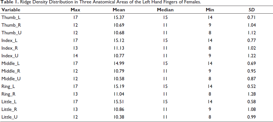
Ridge Density Distribution in Three Anatomical Areas of the Left Hand Fingers of Females.
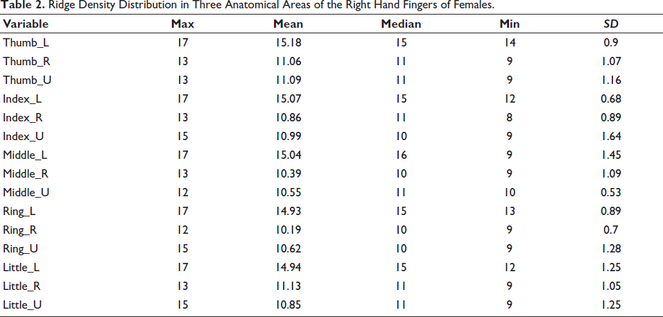
Ridge Density Distribution in Three Anatomical Areas of the Right Hand Fingers of Females.
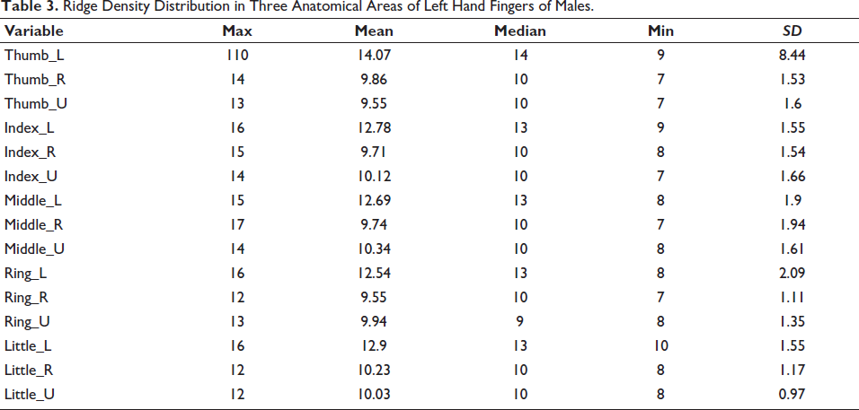
Ridge Density Distribution in Three Anatomical Areas of Left Hand Fingers of Males.
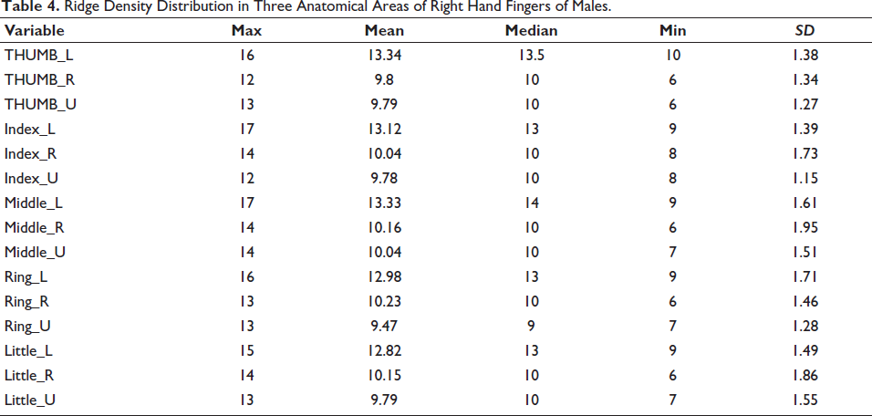
Ridge Density Distribution in Three Anatomical Areas of Right Hand Fingers of Males.
Descriptive Statistics
As shown in Tables 1–4, the data indicate that mean ridge density is higher and more consistent in the lower area across all fingers in females (left and right hands) than in males.
Independent Sample t-tests
Gender Based Comparison of Fingerprint Ridge Density (mean ± SD) Across Digits of the Right and Left Hands.
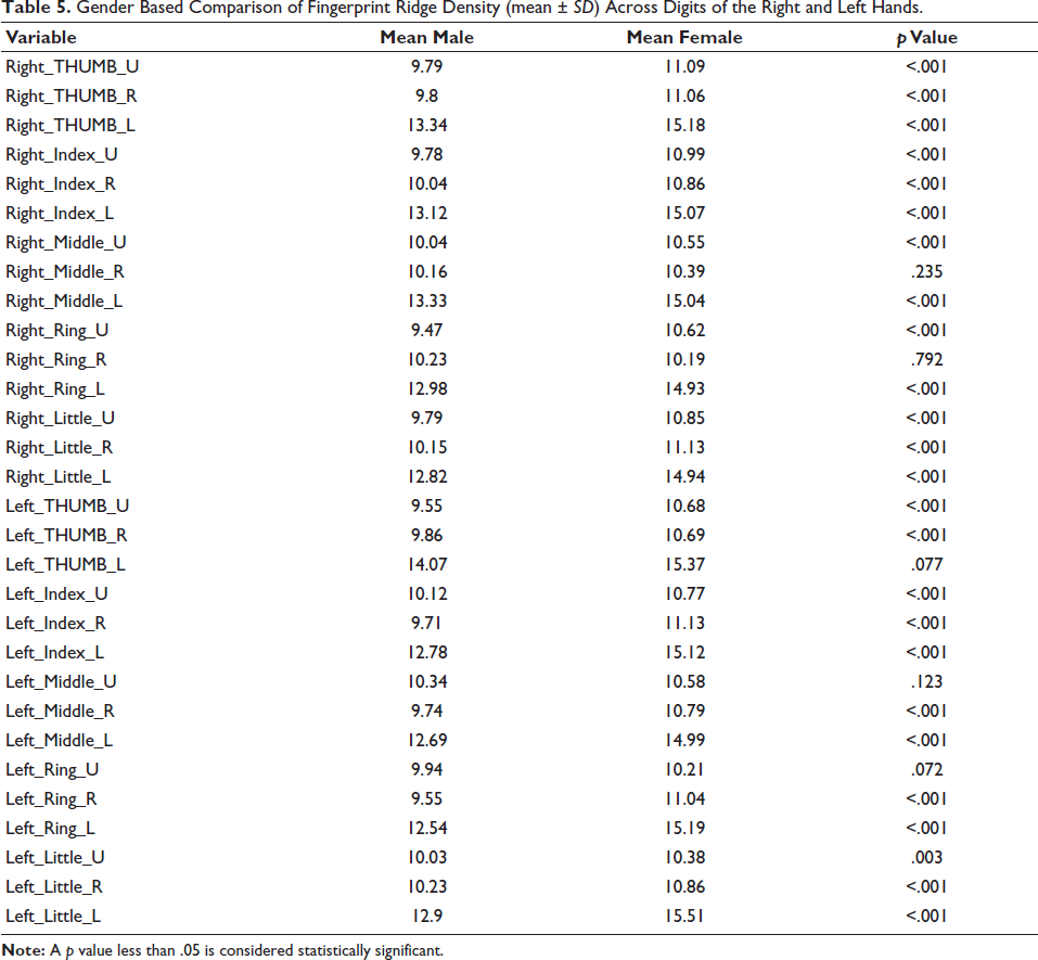
As shown in Table 5, the results of independent sample t-tests comparing male and female ridge density measurements across all 10 digits (thumb to little finger) of the right and left hands. Ridge density was significantly higher in females for most finger segments, especially in the left and right thumb, index, and little fingers, as evidenced by extremely small p values. A few segments did not show statistically significant differences.
Discussion
In the present study, fingerprint ridge density measurements covering R, U, and L areas of all fingers on both hands were analyzed for 274 participants (137 males and 137 females) to explore sexual dimorphism. The results indicate that females consistently exhibit higher ridge density than males across most fingers and regions. This finding is consistent with Gutiérrez-Redomero et al. 16 who reported that female fingerprints were characterized by finer ridges and greater ridge counts within the same unit area compared to males. The current study findings further reveal that the L area showed the most pronounced differences. Specifically, the mean ridge density in females’ L (proximal) areas ranged between 14 and 17 ridges/25 mm² on the left hand and 9–17 ridges/25 mm² on the right hand, whereas males exhibited a L and slightly more variable range of 8–16 ridges/25 mm² on the left hand and 9–17 ridges/25 mm² on the right hand. In the R region, females showed densities of 8–13 ridges/25 mm² on the left hand and 8–13 ridges/25 mm² on the right hand, whereas males ranged between 7–17 ridges/25 mm² on the left hand and 6–14 ridges/25 mm² on the right hand. In the U region, females exhibited densities of 8–14 ridges/25 mm² on the left hand and 9–15 ridges/25 mm² on the right hand, while males showed 7–14 ridges/25 mm² on the left hand and 6–14 ridges/25 mm² on the right hand. Similar patterns were noted in the R and U regions, where females exhibited narrower but consistently higher ridge densities than males. These results are in agreement with previous studies, which consistently reported finer ridge structures and greater ridge density in females across diverse populations.17–19 These quantitative differences reinforce the observation that female fingerprints are generally finer, with a higher number of ridges per unit area, particularly in the L regions, compared to males. The variation observed in males, especially in the R and U areas, indicates a coarser and more heterogeneous ridge pattern, consistent with previously reported sexual dimorphism in fingerprint ridge density.20, 21
The sexual dimorphism observed in ridge density in the present study can be explained by anatomical differences in finger size and ridge spacing. Males generally possess larger finger pads, which accommodate broader ridge breadths and consequently result in fewer ridges per unit area. In contrast, females tend to have smaller fingers with finer, more closely spaced ridges, thereby producing higher ridge density values. Our findings are consistent with previous studies,22–25 which similarly reported that ridge density differences between sexes are largely attributable to variations in ridge width and finger pad dimensions rather than differences in ridge formation processes. This supports the notion that ridge density is an indirect reflection of sexual dimorphism in finger morphology and is therefore a reliable criterion for sex estimation across populations.
Our findings also align with a Sudanese study, 26 which reported higher ridge density in females and emphasized the L areas as most reliable for sex estimation. However, unlike their observation of consistently higher densities in the left hand and some regions lacking significant sex differences, our results showed more uniform dimorphism across both hands and regions, suggesting possible population-specific variability.
When compared with previous studies, several similarities and differences emerge. Gutiérrez-Redomero et al. 27 observed differences in ridge density across U, R, and lateral regions, consistent with the current study showing the highest densities in the L areas. Krishan et al. 28 reported that in the North Indian population, R and U regions had higher ridge density than L areas, whereas in the Indian Tamil population, the present study shows that the L areas consistently exhibit higher ridge density, indicating population-specific variation. Similarly, Nayak et al. 29 reported that females generally have ridge density greater than 13 ridges/25 mm² while males have fewer than 12 ridges/25 mm². The current study corroborates this trend with mean values in females ranging from 14 to 17 ridges/25 mm² and males showing L ranges, highlighting a consistent gender difference. Unlike prior studies on European and sub-Saharan populations, where right-hand ridges were coarser, the Indian Tamil participants displayed higher ridge density in the left hand, suggesting potential genetic or developmental influences unique to this population. Additionally, recent studies30, 31 emphasize the forensic utility of establishing population-specific thresholds, as ridge density values can vary not only between genders but also across ethnic and regional groups. Incorporating such standards could greatly improve the accuracy of forensic applications in diverse populations.
Female fingerprints are finer and show higher ridge density primarily because of smaller finger size, and the thinner epidermis compresses the ridges per unit area. Additionally, sex-specific hormonal influences during fetal development, such as L testosterone and higher estrogen exposure in females, lead to more closely spaced ridges, whereas males develop coarser, more widely spaced ridges. Overall, the study confirms that fingerprint ridge density is sexually dimorphic in the Indian Tamil population, with females showing finer and denser ridges than males, particularly in the L areas. The quantitative data provided strengthen the basis for using ridge density as a reliable parameter for gender determination in forensic investigations, highlighting specific anatomical regions that are most informative.
Conclusion
The present findings affirm the utility of fingerprint ridge density as a reliable, non-invasive, and cost-effective indicator for gender estimation. The strong statistical differentiation in 27 of 30 areas across multiple fingers and fingerprint regions underscores its forensic value. Moreover, the study emphasizes the importance of standardized, area-specific sampling and bilateral data acquisition to enhance the robustness of ridge density-based identification. This study is particularly useful in the case of partial or distorted fingerprints collected from the crime scene. The inclusion of data from the Indian Tamil population also helps bridge a critical gap in population-specific forensic research. Future research should further investigate the role of genetic, environmental, and age-related factors in influencing ridge density and explore machine learning integration for automated gender classification using ridge-based parameters.
Footnotes
Declaration of Conflicting Interests
The authors declared no potential conflicts of interest with respect to the research, authorship, and/or publication of this article.
Ethical Approval
Ethical clearance was obtained from the Institutional Ethics Committee (Ref: IEC-NI/24/DEC/99/175).
Funding
The authors received no financial support for the research, authorship, and/or publication of this article.
Informed Consent
Written informed consent was obtained from all individual participants included in the study.