
Abstract
Seventy years ago, two medical art students painted a group portrait of the staff of the anatomy department in the University of Manchester Medical School. The painting is an unusual allegorical portrayal of the staff as pantomime characters. This paper asks: who were they and what were their subsequent careers? Does this picture tell us anything about the role of anatomy in medical education in the 1950s?
Introduction
The painting (Figure 1), entitled, Acta Anatomica − Christmas 1951, was intended to celebrate the award in 1950 of a DSc by Aberdeen University to George Archibald Grant Mitchell (1906–1993), professor of anatomy at Manchester, for his research on the autonomic nervous system (ANS). 1 The framed picture (1.5ms square) depicts the staff of the anatomy department as a chaotic group of random pantomime characters. It was painted by medical art students, Marjorie Beck and Shirley Manley, and hung in the staff room of the old Manchester Medical School on Coupland Street until 1973, when it was transferred to the then new Stopford Building Medical School on Oxford Road. The painting was put into a storeroom and screwed face-forward onto the wall for protection; there it remained until it was rediscovered by workmen in 2004.
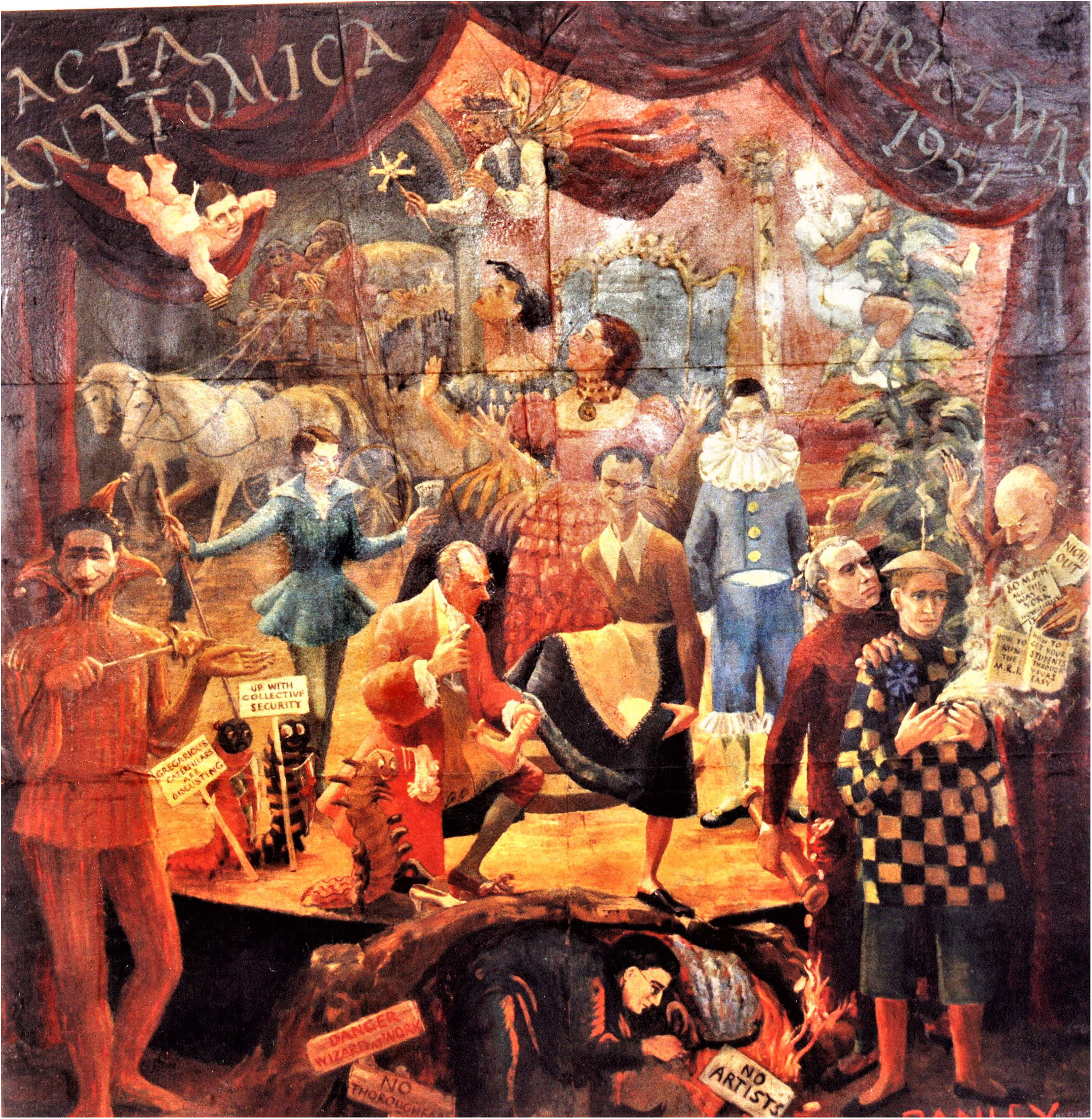
Acta Anatomica - Christmas 1951. Allegorical painting of the staff of the department of anatomy, University of Manchester, 1951.
At that time, the author, then honorary curator of the Medical School Museum of Medicine and Health (MMH), investigate the history of the painting. Most of the information was gleaned from the former executive dean, Dr Frederic Beswick (1925–2019), who had been lecturer in physiology during the 1950s. 2 His wife, Mrs Charlotte Beswick (1926–2013), who was the first honorary curator of the MMH (1980–95), had made some notes about the identity of the characters in the painting, later updated by Dr Beswick. 3 Contact was also made with retired surgeon Hanuš Weisl (1923–2007), the only surviving character whom could be located at that time (2004); he had been an anatomy demonstrator in the department and was able to provide a good account of the painting and the staff during 1950-51. 4 Three of the characters, Miss Dorothy Davison, Professor GAG Mitchell and Dr Eugenia Cooper, had been the subject of previous articles in the Journal of Medical Biography, and further information was collected from obituaries and the Medical Directories. 5 A brief article in the Faculty Newsletter (2004) reported the discovery and the painting was then hung in the dissecting room, where it remains today. 6
It is seventy years since its creation and a review might ask: do the pantomime characters reflect the personality of the depicted staff members? What was the relationship between their work in anatomy and their subsequent careers? What, if anything, does this ‘pantomime’ tell us about an anatomy department in the mid-twentieth century. This paper first identifies the characters, followed by an interpretation and discussion of the painting.
The dramatis personae
This section lists the members of staff of the University of Manchester department of anatomy in 1951, their qualifications, life-dates, position in the department and the pantomime character that represents them in the painting. They are identified in the annotated picture (Figure 2).
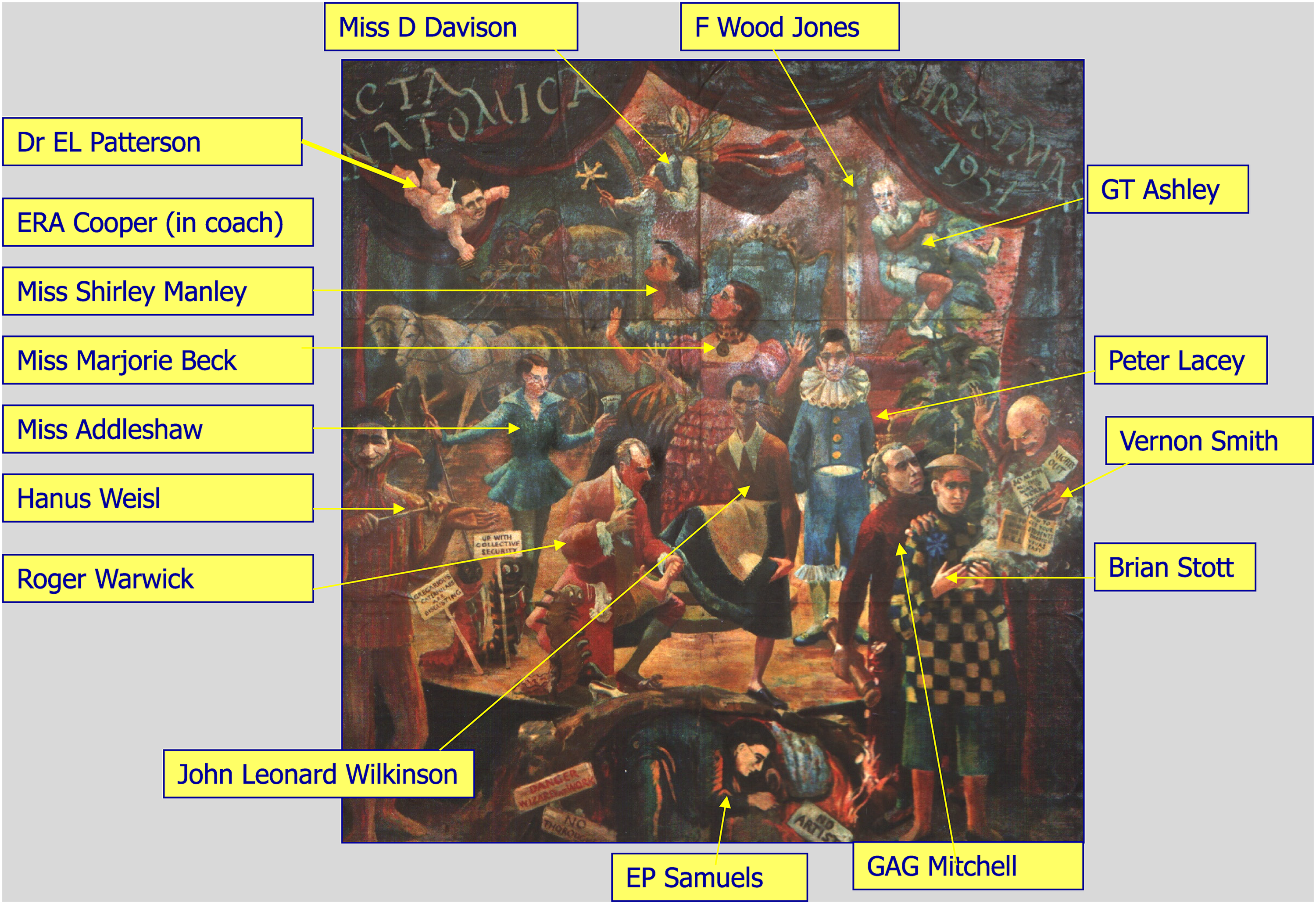
Names of the staff depicted in the painting.
Commentary
There is nothing in the painting to suggest an actual anatomy department or dissecting room; the pantomime is established by the costumes and is set on a theatre stage, the curtain is inscribed, ‘ACTA ANATOMICA CHRISTMAS 1951’. The characters appear to be distributed at random, either alone or in small groups, which coveys a sense of chaos. Miss Dorothy Davison, the senior medical artist, is the Fairy Godmother at the top of the painting. Her connections with the Anatomy Department and the Manchester Royal Infirmary (MRI) dated back to the 1920s. She illustrated many anatomical, anthropological and surgical publications and was one of the founders of the Medical Artists Association (MAA) in 1949. 7 Below her, at the centre of the picture, are the medical art students, Marjorie Beck and Shirley Manley, the Wicked Sisters, who under Davison's tuition painted the picture. They were known as ‘M&B’, a pun on Mitchell's work during the War on ‘May & Baker’ sulphonamide drugs. Marjorie was a member of the MAA and later worked at the Department of Anaesthetics, University of Oxford. Shirley worked as a freelance medical artist, but little is known about their later careers. 8 All three women are looking towards the coach and horses in the left upper corner; Dr Eugenia Cooper, reader in histology, was Cinderella inside the coach, perhaps taking her away? She had had a long and successful career in the department since 1921, and aspired to a professorship, however she felt badly let-down when Professor Mitchell advised her not to apply for the chair of anatomy in Liverpool. Her image has been painted-out; perhaps, because she had taken a long leave-of-absence during 1951 or maybe she just thought the painting was undignified. 9 The lady in blue with glasses is Miss Addleshaw, a secretary; possibly a new recruit to the department, she is depicted as Buttons and holding a note, ‘on approval’, the stick she is holding is a book-grab, presumably she also looked after the departmental library. In the corner, hovering over this group of ladies is an Angel, Dr Patterson, senior lecture; perhaps the ladies are looking to this ‘Guardian Angel’ for support? He was regarded as a ‘gentleman’, calm and a source of good counsel. As the author's anatomy tutor in 1967, he was always helpful and gave good advice based on his encyclopaedic knowledge of anatomy and the blood supply of the nervous system. 10
Both Dr Cooper and Roger Warwick used monkeys in their research, so the two monkeys driving the coach were probably included as a recognition of their sacrifice? Dr Warwick, reader in anatomy, is shown as a courtly-dressed Prince Charming, kneeling to try the slipper on the foot of the Wicked Stepmother. His study of the topographical anatomy of the third cranial nerve nucleus was an important advance in neuroanatomy. 11 He became professor of anatomy at Guy's Hospital Medical School in 1955 and a celebrated editor of Gray's Anatomy (1973–90). 12 Dr John Leonard Wilkinson, an anatomy demonstrator studying for his Fellowship of the Royal College of Surgeons (FRCS), is depicted as the Wicked Stepmother with an evil grin! A reputation, presumably reversed by the award of his OBE in 1967 for his medical work as a Primitive Methodist missionary work in Sierra Leone (1955–71), from where he published papers on tropical disease. After his return, he was appointed senior lecturer in anatomy at University College, Cardiff, and wrote Neuroanatomy for Medical Students in 1986. 13
Another demonstrator, Dr Hanu
Professor Mitchell is depicted as the powerful magician, Abanazar, attired in his dark-red doctoral gown (DSc Aberdeen) and holding a rolling pin or perhaps his doctoral scroll? He qualified at Aberdeen University (1929), where he continued as lecturer in anatomy and surgery. During the War he was a Lt. Colonel surgeon in Cairo, involved in the early trials of penicillin. In 1943 he was made ‘adviser on penicillin therapy for 21 Army Group’ for the invasion of Europe. After the War, the University of Manchester vice chancellor JSB Stopford (1888–1961) appointed him to the chair of anatomy in 1946. 16
Mitchell's hand is on the shoulder of Dr Stott, dressed as Aladdin wearing an oriental hat, he looks worried, he had failed his FRCS examination, and he is throwing away papers: ‘how to run the MRI’ and ‘how to get your students through vivas easily’. Dr Beswick remembered that Mitchell wanted him to stay in the department, but Stott decided to abandon his surgical ambitions and joined a general practice in Cannock. 17 The Genie arising from the smoke of Aladdin's lamp is Dr Vernon Smith, a demonstrator, described by Beswick as a ‘wild card’ who is trying to lead Stott astray with his signs for a ‘night out’ at ‘80 MPH all the way to York’. Smith eventually became an ophthalmic surgeon in Birmingham. 18 Dr Samuel, a reclusive researcher, is the Wizard working under the stage, protected by the signs ‘danger wizard at work’ and ‘no artists’! Note the money falling from Mitchell's pocket towards him; he worked in the Medical School basement producing expensive silver-stain slides of the nervous system. After his MD he settled on a successful career in general practice, unfortunately cut short by a serious illness, aged 41. 19
High up on the right, is Thomas Ashley, dressed in shorts; he was in the Royal Army Medical Corp in the Far East (1942–45) before his appointment as lecturer in embryology (1946), but was seconded to Uganda in 1948 to teach anatomy. He was back in the department in 1950, probably full of tales of foreign adventures, and a good character for the painting! He was a keen sportsman, so appropriately shown as Jack at the top of the beanstalk in his jungle attire. 20 Finally, just visible on the left of Ashley, is the Ghost of Frederic Wood Jones, the former professor of anatomy (1938–45), still haunting the department; he is perched on top of Cleopatra's Needle ─ a reference to his anthropological work in Egypt. 21 The Caterpillars represent the medical students; Dr Beswick though that the sign, ‘gregarious caterpillars are disgusting’ was a reflection of the feelings of the two art students towards the medical students!
Discussion
There are several well-known paintings of groups of anatomists, for example: Rembrandt's Anatomy Lesson (1632), William Cheselden giving an anatomical demonstration to six spectators in the anatomy-theatre of the Barber-Surgeons’ Company (1730), John Cook's painting of Arthur Keith examining the Piltdown skull (1915) and many others. Apart from been great works of art, they are a record of the history, status and fashion of the anatomists and their working environments in past times. Acta Anatomica is not a great painting, nor does it reveal anything about the anatomy department; any historical analysis has to be based on the players in the pantomime, that is the staff members of the department, and their part in the wider history of medical education.
In 1951 the department was based in the University of Manchester Medical School (MMS) on Coupland Street. When the MMS building opened in 1873 as part of Owens College (the forerunner of the University), it was almost entirely devoted to anatomy with a large dissecting room, histology laboratory, lecture theatres, museum and library. 22 An extension for physiology was added in the 1880s with a teaching laboratory and lecture theatre, however the anatomy department continued to occupy most of the building, adding new facilities such as histochemistry and radiological anatomy. By 1951, under Professor Mitchell guidance, the department had recovered from its wartime dereliction: student numbers, research publications, BSc and MD degrees, and demonstrators studying for their FRCS were all well established.
Acta Anatomica has fortuitously captured the anatomy department at what was probably its peak. ‘M&B’ have cleverly caught the strengths, and weaknesses of the characters. The apparent chaos is striking; Dr Beswick described the picture as showing ‘a very disparate group of characters who never had the ethos to do something collectively’. Weisl attributed the anarchy to Mitchell: ‘he lacked the vision to weld his team into a single unit and everyone was left to do their own thing’. However, it is more likely this ‘anarchy’ was illusionary, in part a reaction to the stress and restrictions of the War years; the Medical School, indeed the whole University had been severely disrupted by air raids, conscription of staff, compression of the curriculum etc. Professor Wood Jones, due for retirement, struggled with reduced staff and a shortened curriculum to maintain academic standards. The vice-chancellor, JSB Stopford, a former professor of anatomy, specifically appointed Mitchell in 1946 to restore normality to the department and to prepare for an expansion of the number of medical students. 23 Mitchell had to quickly recruit sufficient staff to restart a normal teaching schedule; also, he was busy re-establishing his own research – during 1946–51 he wrote nine papers on the ANS and later published two monographs (all illustrated by Miss Davison). 24
It was not chaos, but it was a group of individuals, some of whom had served in the armed services, focused on establishing or re-establishing their career aspirations during a period of post-war austerity and reconstruction. All the senior staff and researchers were interested in some aspect of neuroanatomy, and Roger Warwick and John Wilkinson went on to successful careers in anatomy. The post-war department was male dominated; Eugenia Cooper was already well established with an excellent track record in research but was disappointed not to be appointed as professor, nevertheless she wrote two textbooks and continued with successful collaborative research projects in endocrinology, neuropathology, neurophysiology until her retirement. Her work on the origin and function of ‘oligocytes’ (oligodendrocytes) is especially noteworthy. 25
The demonstrators, who were employed to supervise the students in the dissection room, were also busy preparing for their FRCS examination: Drs Smith and Weisl passed and pursued careers in surgery, while Stott, Lacy and later, Dr Samuel, decided on careers in general practice. Even with a ‘FRCS’, not everyone was able to gain a foothold on the surgical career ladder; the new National Health Service had increased the number of salaried hospital registrar posts, but this also increased the number of applicants, on the other hand, general practice, funded by the NHS per capita payment scheme, was an attractive option. Mitchell also encourage promising students to stay an extra year to do an intercalated BSc degree, and also supported a number of MD research thesis.
Dan Hoyte (1923–2004), a career anatomist (he later became professor of anatomy at the University of the West Indies) joined the department as a researcher in 1952. He found Professor Mitchell friendly and supportive and the ‘staff of high calibre, a melange of personalities, ideas, of wit, of friendship’, and commented on Mitchell: ‘it is a mark of his great stature that he was able to run a productive department, oversee the teaching of 150 medical and dental students, encourage excellent youngsters to spend an Honours year with us, take an interest in our research [and] promote one of the foremost neuroanatomical departments in the world.’ 26
During the 1960s genetics and electron microscopy were added to the department, which by then was a smooth running machine, processing an increasing number of pre-clinical students. 27 However, the traditional ‘pre-clinical’ model of medical teaching was increasingly seen as old-fashioned and out of step with modern medical sciences. During the 1960s Mitchell and Beswick were part of the team that planned the move to the new Stopford Building Medical School and the implementation of a new integrated medical curriculum in 1973. This was part of a national reform of medical education; anatomy (including dissection) was still important, but it was now just one of several medical sciences, integrated around clinical problems and centred on ‘problem based learning’. 28
The painting does highlight the increasing importance of the work and professionalisation of the medical artist during the twentieth century. The contribution of Miss Davison to the foundation of the MAA and medical illustration has been previously discussed, and her department had close links with anatomy and neurosurgery. 7 She had worked as an independent medical illustrator since the 1920s and after 1945, when employed by the University, she started a course in medical illustration for art students such as misses Manley and Beck. After Miss Davison retired in 1953 the medical art department continued under Zita Stead Blackburn (1904–1986), also a founder member of the MAA, and who had been a medical artist at St Bartholomew's Hospital. 29 When artist Richard Neave was appointed as director in 1959, he expanded the services to include facial reconstruction, and in 1973 organised the move of the department to a spacious studio in the new Stopford Building Medical School. 30 For many years the unit gained in reputation and was busy supporting medical education, research, and training medical art students. However, after the millennium a period of decline set in; the medical art unit came under increasing competition from other departments for resources, and the demand for bespoke illustrations, lecture slides etc. declined. The department closed in 2008, and the staff members moved to other universities or used their skills to diversify into other areas. 31
Summary
Acta Anatomica was just an amusing interlude, part of the anatomy department's celebration of Professor Mitchell's DSc. The painting also inadvertently caught the department at its zenith; a slow decline in status and influence was brought about by advances in other medical sciences, imaging, genetics, pharmacology and surgery. Dr Beswick remembered the picture as a focus of interest and amusing comment for new staff members and visitors to the staff room. During the 1970s the reorganisation of the medical curriculum and problem-based learning broke-down the traditional barrier between pre-clinical and clinical (hospital) teaching. Memorising the minutia of regional anatomy gave way to a more functional, biological approach. Anatomy no longer dominated the curriculum, though dissection remains an important part of the course. The painting still hangs on the wall of the dissecting room, from where the old anatomists can watch the students; what would they think? They would notice some technical improvements, especially the use of computerised images and the training area for ‘keyhole surgery’, but what of the present-day medical students? Perhaps they might say ‘little has changed’ − just a group of serious, hard-working caterpillars? What a pantomime!
Footnotes
Acknowledgements
I owe thanks to Stephanie Seville, University of Manchester MMH Heritage Officer for access to MMH information files and Ingrid Gouldsborough, Professor of Anatomy, for permission to photograph the painting. Acta Anatomica is copyright of the artists, Marjorie Beck and Shirley Manley. I have tried to contact the artists for permission but have failed to locate either of them; the author would be grateful to hear from anyone with information about them. The painting belongs to the University of Manchester Medical School in the Faculty of Biology, Medicine and Health.
Declaration of conflicting interests
The author(s) declared no potential conflicts of interest with respect to the research, authorship, and/or publication of this article.
Funding
The author(s) received no financial support for the research, authorship, and/or publication of this article.