
Abstract
The emphasis of biocompatible polymer applications in medical sciences and biotechnology has remarkably increased. Developing new low-cost, low-toxicity and lightweight composite forms of biopolymers has become even more attractive since the addition of new species into polymer matrices assist to improve biomedical activities of such materials to a higher extend. Developments in nanoscience and nanotechnology recently contribute to controlled fabrication and ultraprecise diagnosis of such materials. This study concerns the observation of solution processing effects in the fabrication of porous PLA/AGNWs bionanocomposite coatings using electron and ion processing based serial cross-sectioning and high-resolution imaging. The nanostructuring and characterization were both performed in a focused ion-beam-scanning electron microscope (FIB-SEM) platform. HR-SEM imaging was conducted on-site to track solvent based morphological property alterations of PLA and PLA/AgNWs structures. Simultaneous SEM-EDS analyses revealed the elemental distribution and the chemical composition along the cross-sectioned regions of the samples. Accordingly, it was observed that, in case of acetone dissolved materials, both pristine PLA and PLA/AgNWs samples sustained their foamy structure. When chloroform was used as the solvent, the porosity of the polymer matrices was less and the resulting structure was found to be denser than samples dissolved in acetone with a lower surface area ratio inside the material. This can be attributed to the rapid volatilization of acetone compared to chloroform, and hence the formation of interconnected pore network. For both nanocomposite biopolymers dissolved in acetone and chloroform, silver nanowires were homogeneously distributed throughout PLA matrices.
Keywords
Introduction
Biodegradable and biocompatible polymer composites offer numerous benefits due to the modification flexibility of their production processes and characteristic properties and their multifunctionality. 1 During the last decades, these composites in particular have effectively been employed in medical sciences and biotechnology due to their improved mechanical strength and bioactivity, which makes them good candidates for drug delivery and tissue engineering applications. Among all, poly-lactic acid (PLA) has received significant attention, as from its different forms three-dimensional porous scaffolds can be manufactured. PLA is immunologically inert and biodegradable through hydrolytic and enzymatic pathways and hence frequently takes part in numerous biomedical applications such as wound healing, implants, resorbable surgical sutures and controlled drug release systems.2,3 More importantly, because PLA is an immunologically inert synthetic polymer, it is preferred for designing a composition of tissue engineering scaffold.4,5 Both biocompatibility and dissolvability of PLA in the body makes it a good candidate for several medical applications. As the main degradation mechanism, hydrolysis of the ester backbone of PLA eventually produces non-harmful and non-toxic compounds as ultimate products. The biocompatibility and antibacterial activity of PLA can be enriched by forming nanocomposite structures via introducing nanomaterials into such material systems. On the other hand, the PLA has poor mechanical properties for tissue engineering applications such as bone scaffolds. 6 Hence, biocompatible polymers are often doped with nanomaterials, e.g. silver nanoparticle, boron nitride in order to improve their morphological, mechanical and thermal properties.7-10
Silver is known to possess good antibacterial activity and low toxicity in animal cells. 11 In particular, one-dimensional structure and higher aspect ratios of silver nanowires (AgNWs) allow for displaying different physicochemical properties when compared to other forms of silver at the nanoscale. 12 Antibacterial activity of biopolymers is considerably enhanced when metal nanoparticles, which act as antimicrobial agents are added to form new polymer composites. 13 Accordingly, doping of biocompatible and biodegradable polymers with silver nanowires would be of interest for advanced applications where antibacterial activity is also required in combination to polymers’ expected biological and medical properties. Micro/nano-porous biocomposite polymer systems are highly preferred for pharmaceutical and cosmetic applications due to their utility in controlled release systems where diffusion of drug or active substances through the pores is of interest. 14
Over the last decades, nanoscience and nanotechnology have been actively contributing to the developments in bioengineering where the characteristics of biomaterials can be defined, tuned and analyzed in high precision. Especially electron microscopy science opens many ways for tailoring and structuring of a large spectrum of materials besides providing physical, chemical and morphological information down to sub-nanometer scale. FIB instruments, when coupled to a SEM can handle various tasks including micro/nano-machining, site specific analysis and 3D tomography applications in combination to high-resolution imaging and structural/elemental analyses. 15
In this study, PLA/AgNWs biocomposite coatings were fabricated by using different solvents, such as acetone and chloroform. The materials were then characterized at the nanoscale in order to observe the inner-porosity morphology and the distribution of nanospecies throughout the polymer matrices in correlation to the solvent used during processing. The biocomposite materials were drop-coated on silicon wafer substrates, and this was followed by simultaneous ion-beam cross-sectioning and high-resolution imaging in a FIB-SEM multi-beam platform. The cross-sections revealed the inner morphology of the samples, hence he degree of porosity in PLA nanocomposites determines the potential of these materials to be used for biopackaging, controlled drug delivery and wound healing applications.
Materials and methods
Silver nanowires having an average diameter of 50 nm and length of 5–10 µm were dispersed in IPA solution. Silicon wafer slides (1 × 1 cm2) were sonicated in acetone and ethanol for 15 min and then they were dried under N2 flow.
Samples dissolved in acetone
The PLA solution (100 mg/ml, in acetone) was drop-coated on the silicon wafer substrates and dried under atmospheric conditions. The PLA/AgNWs composite solution was prepared using 0.5 ml PLA (200 mg/ml, in acetone) and 0.5 ml AgNWs (5 mg/ml in IPA), then drop-coated onto silicon wafers and left for drying under ambient conditions.
Samples dissolved in chloroform
The PLA solution (100 mg/ml, in chloroform) was drop-coated on the silicon wafer substrates and dried under atmospheric conditions. The PLA/AgNWs composite solution was prepared using 0.5 ml PLA (200 mg/ml, in chloroform) and 0.5 ml AgNWs (5 mg/ml in chloroform), then drop-coated onto silicon wafers and left for drying under ambient conditions.
The experimental study has been carried out at the JEOL JIB 4601F Multi-Beam FIB-SEM platform equipped with a FEG-SEM column, a gallium-ion column, gas injection system for local carbon deposition. Prior to FIB processes, all samples were coated with thin Au/Pd films using sputter coater, in order to avoid surface-charging effects. Before FIB processing, a local carbon protective layer was deposited on the regions of interest in order to protect the materials from the irradiation damage that may be caused by ion and electron bombardment. To view the sub-surface, cross-sections of the PLA and PLA/AgNWs structures, samples were ion-sectioned and simultaneously observed using HR-SEM imaging. Ion milling was performed using 30 keV ions over a range of currents tuned for precise control over the beam-specimen interactions. For trench milling, relatively higher ion currents (1–5 nA) were applied, whereas polishing of side-walls was performed using moderate ion current values (0.3–1 nA). The cross-sections were observed using secondary electron (SE) at 5 keV electron energy and backscattered electron (BSE) imaging modes for identifying the morphological properties of PLA and PLA/AgNWs composite structures. For the chemical compositional analysis, energy dispersive X-ray analysis (EDS) measurements were performed using an Oxford X-MaxN EDS detector attached onto the FIB-SEM platform. EDS mapping was performed using 15 keV electron energy.
Results and discussion
The drop-coated samples were examined by FIB-SEM platforms in order to observe the alterations in the microstructure and morphology by the solvent before and after the addition of silver nanowires into the PLA. Figures 1 and 2 show the SEM-SE (a) and SEM-BSE (b) images taken from the inner microstructure of the acetone and chloroform dissolved pristine PLA samples after FIB cross-sectioning. According to the cross-sectional analysis, it can be monitored that the samples that were dissolved in acetone ended up in highly interconnected porous network, compared to the PLA samples those were solution processed in chloroform. This can be attributed to the highly volatile nature of acetone, which creates pores when escaping from the materials very fast during drying.

The FIB cross-sections of pristine PLA dissolved in acetone: (a) SEM-Secondary Electron image; (b) SEM-Back Scattered Electron image.

The FIB cross-sections of pristine PLA dissolved in chloroform: (a) SEM-Secondary Electron image; (b) SEM-Back Scattered Electron image.
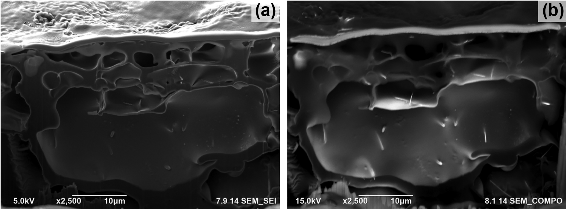
A further study was performed on the silver nanowire doped PLA samples, in order to monitor the microstructure and morphology of the nanocomposite forms. Besides porosity, the distribution of the nanowires throughout the polymer matrix was also examined in the case of using different solvents. Figure 3 shows the cross-sections of PLA/AgNWs sample dissolved in acetone. SE image in Figure 3(a) reveals the interconnected porous network, whereas BSE image in Figure 3(b) shows the distribution of silver nanowires along the polymer structure. Also an EDS mapping study was conducted on the same cross-sectional surfaces in order to indicate the silver nanowires that are present inside the nanocomposite that is dissolved in acetone. The EDS maps are given in Figure 4.

The FIB cross-sections of PLA/AgNWs dissolved in acetone: (a) SEM-Secondary Electron image; (b) SEM-Back Scattered Electron image.

SEM-EDS mapping analysis performed at the FIB cross-sections of PLA/AgNWs dissolved in acetone: multilayered map, C map (red), O map (green) and Ag map (yellow).
A similar work was conducted for the cross-sectional analysis of PLA/AgNWs nanocomposites dissolved in chloroform. Figure 5(a) shows SE images for the morphology and porosity characteristics, while BSE image in Figure 5(b) reveal the silver nanowire distribution along the polymer structure in the case of using chloroform as the solvent. Again the EDS maps in Figure 6 helped for the elemental analysis of cross-sections by which it is easy to observe the presence and dispersion of silver nanowires.
The FIB cross-sections of PLA/AgNWs dissolved in chloroform: (a) SEM-Secondary Electron image; (b) SEM-Back Scattered Electron image.

SEM-EDS mapping analysis performed at the FIB cross-sections of PLA/AgNWs dissolved in chloroform: multilayered map, C map (red), O map (green) and Ag map (yellow).
The cross-sectional SEM-SE and -BSE images in Figure 1 clearly demonstrate the existence of micron and sub-micron-sized pores which represent the foam-like structure of acetone dissolved PLA materials without any addition of nanowires into the matrix. On the other hand, cross-sections of the PLA samples which were prepared using the same conditions but are dissolved in chloroform are given in Figure 2. Morphologically, due to their high porosity fraction and hence large surface areas, these biopolymers can be considered to have high degree of bioactivity when interacted with surrounding environment. However in the case of using acetone as the solvent, the interconnected porous network is found to be dominating when compared to chloroform, due to the fast volatilization properties and hence resulting in foamy and spongy inner structures in drop-coated PLA samples.
Acetone dissolved PLA/AgNWs nanocomposites’ cross-sectional images are given in Figure 3, which show both porosity Figure 3(a) and nanowire Figure 3(b) distribution along the nanocomposites. After the addition of silver nanowires, the polymer sustained its foamy structure, while it can be observed that the dispersion of the nanowires were homogenous and uniform. The SEM-EDS maps in Figure 4. Also confirm these findings by elemental indication on the corresponding cross-sections.
Similarly, chloroform dissolved PLA/AgNWs nanocomposites were cross-sectioned and the porous network as well as the silver nanowire distribution were investigated. The results in Figure 5 showed that these samples are consisting of pores, however they found to be less foamy and the interconnection of the holes are limited when compared to that of acetone dissolved nanocomposites. However again the dispersion of silver nanowires were uniform for the chloroform dissolved samples as well. The EDS maps present the homogenous elemental distribution of nanowires from the cross-sectional view in Figure 6.
Considering all results, the effect of using different solvents during solution processing of PLA and PLA/AgNWs can clearly be observed. Due to the differences in volatilization regimes of acetone and chloroform, similar polymeric solutions generated different morphologies and variations in evaluation of micro/nano-pores and porous network. However, the addition of silver nanowires for the enhancement of bioactivity properties were successful for both solvents and the resultant nanobiocomposites were structurally and chemically uniform.
Conclusion
In this study, advanced processing capabilities of FIB-SEM platforms were used for serial cross-sectioning and high-resolution imaging for investigating the inner structure of the materials in terms of porosity and matrix-dopant distribution, which then can be attributed for sustaining homogenous antibacterial and bioactivity properties. It was observed that PLA matrix had a high degree of micro/nano-porous network and dispersion of AgNWs within the matrix was quite effective for both acetone- and chloroform-dissolved samples and the addition of silver nanowires did not make any alterations in the porous nature of the matrices which is needed for increased bioactivity. Consequently, the effective use of FIB-SEM platforms enabled to observe the porous microstructure and the nanocomposite characteristics of novel PLA/AgNWs nanocomposites, which may be offered as good candidates for a variety of biomedical applications including scaffolds, medicine and packing.
Footnotes
Declaration of conflicting interests
The author(s) declared no potential conflicts of interest with respect to the research, authorship, and/or publication of this article.
Funding
The author(s) received no financial support for the research, authorship, and/or publication of this article.