
Abstract
A study was recently published that sought to develop an in vivo model of facioscapulohumeral muscular dystrophy by transplanting muscle precursor cells from a patient into immunodeficient mice. The study largely applied the methodology used by our team in a study published more than two decades ago with a similar objective, albeit for another muscular dystrophy. However, our study is not cited, leaving the wrong idea that the concept, methodology, and part of the results are original to this recent study. Although the recent study is of interest, the omission of our publication, as well as other relevant references, deprives it of an adequate scientific context. We, therefore, want to point out the importance of a careful bibliographic search in any scientific work.
Recently, Mueller et al. 1 have published a study in which they attempted to develop an in vivo model of facioscapulohumeral muscular myopathy (FSHMD). To this end they transplanted muscle precursor cells (MPCs) from a patient with FSHMD into immunodeficient mice. At first, we were enthusiastic to learn that another research group took up an idea that we had more than two decades ago and that we published in 1999 2 . At the time, we sought to develop an in vivo model of another muscular dystrophy, namely myotonic dystrophy (MD). In our study, MPCs from five adult MD patients as well as MPCs from a fetus with the congenital form of MD were transplanted into severe combined immunodeficiency (SCID) mice, with MPCs of other origins serving as controls (Table 1). In the following 1 to 4 mo, we observed human-derived myofibers in all mouse muscles grafted with human MPCs. Mouse muscles grafted with human MD cells had the same genetic abnormalities as the donors, and we found electromyographic abnormalities characteristic of MD in some of them 2 .
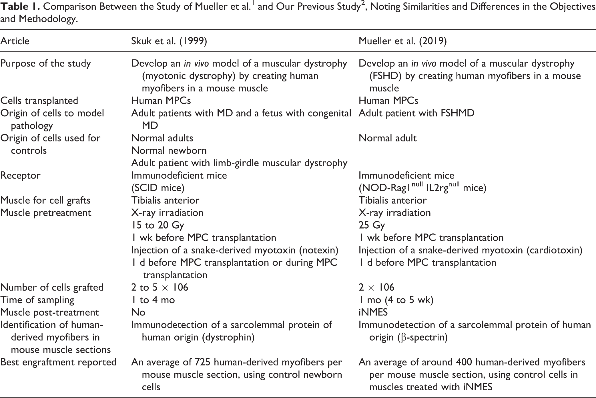
FSHMD: facioscapulohumeral muscular myopathy; iNMES: intermittent neuromuscular electrical stimulation; MD: myotonic dystrophy; MPC: muscle precursor cell; SCID: severe combined immunodeficiency.
Since the study published by Mueller et al. 1 reproduced much of our previous methodology and had an objective similar to that of our 1999 study 2 (Table 1), it is understandable that our enthusiasm turned into disappointment as we noted that the recent manuscript did not mention our work as a precedent in terms of concept and methodology.
Referencing is an important part of any scientific work. Quoting an editorial from B. Penders on the rules for responsible referencing 3 , citations in a scientific article “demonstrate the foundation upon which our studies rest as well as how they are different from previous work” 3 . As this editorial pointed out, the omission of essential citations “can wrongfully suggest that your own publication is the origin of an idea, a question, a method, or a critique, thereby illegitimately appropriating them” 3 . In this sense, the recent report published by Mueller et al. 1 may impart on readers the impression that their study is the origin of an idea and a methodology that are, in effect, over two decades old.
Furthermore, we noted other omissions or citation errors in Mueller et al.
1
, which we point out below: Papers published by our group in 2007
4
and 2012
5,6
were quoted to say that in earlier studies of human MPC transplantation in the muscles of immunodeficient mice, human-derived myofibers were partially “contaminated” with mouse myonuclei. This mention was used in Mueller et al.
1
to explain that the authors decided to treat the mouse muscles with high doses of X-rays prior to the MPC graft. Expressed in this way and without more appropriate references, this might lead readers to believe that muscle irradiation before MPC transplantation is a novelty of the recent study. However, the suppression of the proliferative capacity of endogenous satellite cells to favor the regeneration of myofibers by the grafted MPCs using high doses of gamma or X-rays is a standard technique whose first use dates back almost three decades
7,8
. In fact, a careful reading of the 2007 study
4
would have shown that we did indeed irradiate the host muscles previous to the human MPC transplantation and there was no mention of mouse myonuclei contamination in the human-derived myofibers
4
. On the other hand, our first studies regarding human MPC transplantation in immunodeficient mice are much earlier, having been published in 1993 and 1994
9
–11
, and one of them
10
used high-dose irradiation (20 Gy) of the muscles prior to the transplantation of human MPCs. In this study, an analysis of whether the human-derived myofibers had mouse myonuclei was made, and we reported that the majority were completely of human origin
10
. Therefore, the protocol we used in 1994 already minimized the contamination of human-derived myofibers by mouse myonuclei. This protocol afforded our team a very efficient engraftment, i.e., we reported SCID mouse muscles almost entirely composed by human-derived myofibers (92.8% ± 6.5%) in 1994
10
, and up to 725 ± 329 human-derived myofibers per mouse muscle in 1999
2
. MPCs from FSHMD patients have previously been transplanted into immunodeficient mice and compared with MPCs of normal donors in a study published by Vilquin et al.
12
. Although the objective of the study was not to develop an in vivo model of FSHMD, they reported that the engraftment of FSHMD cells was similar to those from normal individuals. As this finding was similar to that observed by Mueller et al.
1
, proper etiquette dictates that it ought to have been cited
3
. The fact that some MPCs (from mouse and human origin) give rise to satellite cells once grafted into mice has already been sufficiently established to be “intriguing” (reviewed in reference 13). In fact, one of the experiments proposed by Mueller et al.
1
, i.e., treating the muscles that have human-derived myofibers with a myotoxin (which allows to observe if the satellite cells derived from the graft can participate in new muscle regeneration cycles), has already been carried out in studies with murine
14
and human
13
MPCs. Therefore, these publications should be cited at least in future studies if that methodology is used.
Leaving aside these shortcomings in referencing, we found that the study of Mueller et al. 1 is interesting and surely relevant for the study of FSHMD. From the specific point of view of our expertise with MPC xenografts in immunodeficient mice (which includes not only MPCs of human origin, but also from dogs 15 and monkeys 4,16 ), we found the intermittent neuromuscular electrical stimulation (iNMES) of the peroneal nerve innovative. It is interesting that the authors reported a substantial increase in the number of human-derived myofibers when they used iNMES to induce repeated maximal contractions of the muscle transplanted with human MPCs 1 . This good engraftment was achieved, according to what we interpret, with only one longitudinal injection of cells, i.e., without the need to distribute the MPCs throughout the muscle by multiple microinjections, as we usually do in our studies 2,10 . Importantly, with this technique Mueller et al. 1 reported an increase in the average diameter of the human-derived myofibers, close to 30 µm according to figure 1c in that article, while in our earlier papers we had reported 22.9 ± 12.4 µm in the best case 2 . They also reported that iNMES enhanced the differentiation of the human-derived myofibers, as shown by a reduction in the levels of embryonic myosin when compared to muscles that were not treated with iNMES 1 . Therefore, iNMES could improve the protocols previously developed to create human “mini-muscles” in immunodeficient mice. These updated protocols could more effectively model some human genetic myopathies and help researchers gain more reliable data. Finally, we hope that the references mentioned here will serve to better contextualize future studies in this field.
Footnotes
Acknowledgments
The authors wish to express their gratitude to Gabriel Lamothe for correcting the text as well as for his helpful suggestions.
Declaration of Conflicting Interests
The author(s) declared no potential conflicts of interest with respect to the research, authorship, and/or publication of this article.
Funding
The author(s) received no financial support for the research, authorship, and/or publication of this article.