
Abstract
Sendai virus (SeV) vectors have potential clinical applications because they can efficiently introduce foreign genes without toxicity into various organs. A recent study reported the green fluorescent protein (GFP) gene transfer to adipose tissue-derived stem cells (ASCs) with SeV vectors results in more efficient expression of GFP than AdV and identified the preservation of the multilineage potential of ASCs transfected with SeV vectors. This study assessed the gene transfer efficiency to floating ASCs with SeV vectors. Although a slight cytotoxicity was observed, the efficiency of gene transfer to cells in the floating state was much higher at all times and all concentrations at MOIs of 2, 10, and 20 than in the adhesion state. Moreover, ASCs transfected with SeV vectors in floating state have the same potential for their differentiation into specific tissues, such as adipocytes and osteocytes, as untransfected ASCs. These data suggest that SeV transfection to ASCs in the floating state could therefore be useful for gene transfer technology.
Keywords
Introduction
Adipose tissue has a multipotent population of cells capable of differentiating along a number of mesenchymal pathways (19,21). These cells are termed adipose tissue-derived stem cells (ASCs) (3). ASCs have characteristics similar to bone marrow-derived stem cells (BMSCs) and skeletal muscle-derived stem cells (2,10,11,26,35) and can be induced to differentiate into various mesenchymal tissues, including chondrocytes, adipocytes, osteoblasts, myocytes, and endothelial cells (6,15,16,21,34). However, in comparison to BMSCs or other MSCs, ASCs are easy to obtain, carry relatively lower donor site morbidity, and can be harvested in large numbers (9,12,31). In addition, ASCs possess a long culture period and high proliferation capacity (10). Therefore, adipose tissue may be an ideal source of large numbers of autologous stem cells attainable by a less invasive method and ASCs may be more expected to have clinical applications than other stem cells. Therefore, efficient, stable, and low toxicity gene transfer into ASCs would also be useful.
F (envelope fusion protein)-defective Sendai virus (SeV) vectors were previously developed which are nontransmissible (20). SeV vectors can efficiently introduce foreign genes without toxicity into various organs, including airway epithelial cells (29), vascular tissue (13), skeletal muscle (23), synovial cells (21), retinal tissue (7), and hematopoietic progenitor cells (8,30). Because SeV vectors use a cytoplasmic transfection system, they can mediate gene transfer to a cytoplasmic location (18). They replicate in the form of negative-sense single-stranded RNA in the cytoplasm of infected cells without a DNA phase and theoretically avoid vector integration-related adverse events (20). There are technical advantages in the use of the recombinant SeV vectors for gene therapy (14,24). First, the vector particles are stable and can be easily concentrated to high titers unlike retroviral vectors. Second, the modalities of target cell processing and viral transfection are technically uncomplicated and possible in clinical situations that require transduction into large numbers of target cells (8).
A recent study reported on the use of SeV vectors for gene transfer to ASCs. Gene transfection to ASCs with SeV vectors resulted in more efficient expression of the green fluorescent protein (GFP) gene than AdV vectors. In addition, ASCs transfected with SeV vectors maintained their multilineage potential (31).
Generally, ASCs are incubated in the adhesion state. However, when ASCs are used for cell transplantation, they are trypsinized and the number is counted in a floating state. Therefore, gene transfection into ASCs with SeV vectors in the floating state is more important for clinical application. In this study, for the sake of clinical applications, GFP gene transfection efficiency into ASCs with SeV vectors was verified and compared in both an adhesion and floating state.
Materials and Methods
Animals
C57BL/6 mice were purchased from SLC Japan. The mice were housed in a controlled environment (12-h light/dark cycles at 21°C) with free access to water and a standard chow diet before killing. All conditions and handing of the animals in this study were conducted with protocols approved by the Nagoya University Committee on Animal Use and Care.
Isolation and Culture of Mouse Adipose Tissue-Derived Stem Cells (ASCs)
Adipose tissue-derived stem cells (ASCs) were collected from 7–14-month-old female C57BL/6 mice. The mice were killed by cervical dislocation and the adipose tissues in the inguinal groove were isolated and washed extensively with Hank's balanced salt solution or phosphate-buffered saline (PBS) to remove blood cells. The isolated adipose tissues were cut finely and digested with 1 ml of 1 mg/ml type I collagenase (Collagenase Type I, 274 U/mg, Koken Co., Ltd., Tokyo, Japan) at 37°C in a shaking water bath for 90 min. Adipose tissue cells were filtered using 250-μl nylon cell strainers (BD Biosciences) and suspended in Dulbecco's modified Eagle's medium (DMEM)/F12 containing 20% fetal bovine serum (FBS; Trace Scientific Ltd., Melbourne, Australia, Uin: 53141, Lot: B01249-500) and 100 U/ml penicillin/streptomycin. The cells were centrifuged at 1,200 rpm for 5 min at room temperature and the pellet contained the ASCs. The cells were washed three times by suspension and centrifugation in the culture medium and incubated overnight at 37°C with 5% CO2 in culture medium (DMEM/F12, 20% FBS, and 100 U/ml penicillin/streptomycin). The primary cells were cultured for 4–5 days until they reached confluence and were defined as passage 0. The cells used in all of the experiments were between passages 2 and 5.
Recombinant Sendai Virus (SeV) Vector
Recombinant Sendai virus (SeV) vectors were constructed as described previously. In brief, the entire cDNA coding jelly fish enhanced green fluorescent protein (GFP: for SeV-GFP) were amplified by PCR, using primers with a NotI site and new sets of SeV E and S signal sequence tags for an exogenous gene and then inserted into the NotI site of the cloned genome. Template SeV genomes with an exogenous gene and plasmids encoding the N, P, and L proteins (plasmids pGEM-N, pGEM-P, and pGEM-L, respectively) were conjugated with commercially available cationic lipids, then cotransfected with UV-inactivated vaccinia virus vT7-3 into LLMCK2 cells. Forty hours later, the cells were disrupted by three cycles of freezing and thawing and injected into the chorioallantic cavity of 10-day-old embryonic chicken eggs. Subsequently, the virus was recovered and the vaccinia virus was eliminated by a second propagation in eggs. The virus titer was determined using chicken RBC in a hemagglutination assay and virus were kept frozen at −80°C until use (22,31).
Gene Transfection to ASCs with SeV Vectors in the Adhesion and Floating State
In the adhesion state, ASCs (1.0 × 10⁵ cells) were seeded in each well of a 12-well plate (BD Biosciences) with 1 ml of culture medium for 2 h and were confirmed to adhere to the flask bottom. The GFP gene transfer was performed by adding SeV vectors at a multiplicity of infections (MOIs) of 2, 10, 20, 50, and 100 to 150 μl of the culture medium and the cells were incubated for 1 h with rocking every 10 min. After 1-h incubation, 1850 μl of the culture medium was added and cultured. After 24-h incubation, the culture medium was removed and washed by PBS and an equal volume of fresh medium was added. After various times of incubation, the GFP expression of ASCs in each well was observed by the fluorescence microscope and was calculated (Fig. 1).
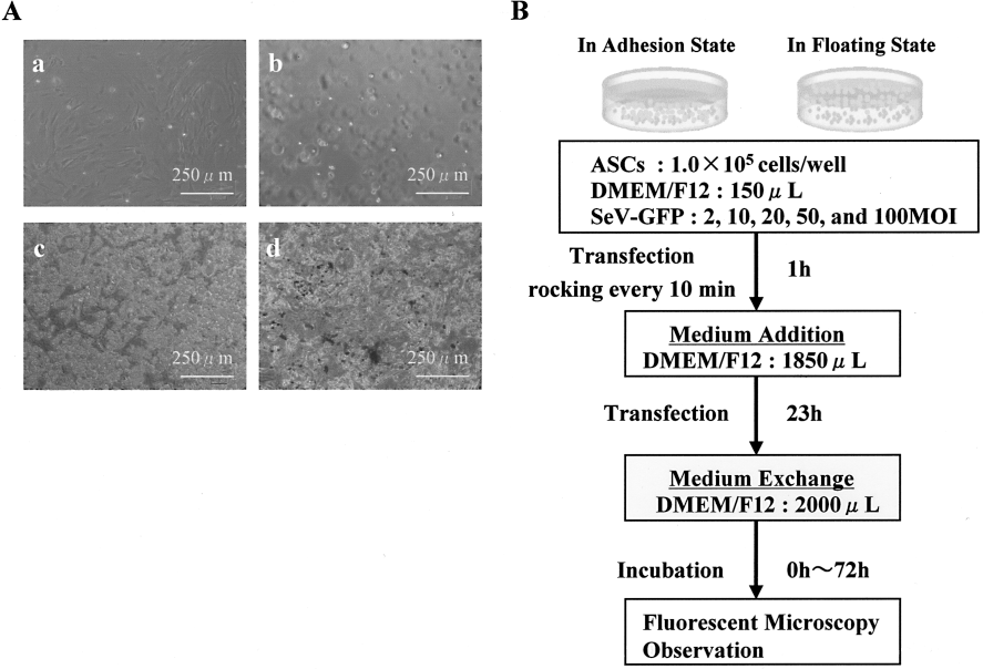
Method of transfection with SeV vectors, and the morphology of adipose tissue-derived stem cells (ASCs) in the adhesion and floating state. (A) Adipogenic differentiation of ASCs confirmed by the presence of intracellular lipid vesicles staining for oil red O (a). Osteogenic differentiation of ASCs confirmed by Von Kossa's staining (b). Morphology of ASCs used in this experiment in the adhesion state (c) and floating state (d). (B) Method of transfection of ASCs with SeV vectors in the adhesion and floating state.
In the floating state, immediately after the seeding of ASCs in each well under the same conditions, the GFP gene transfer was performed by adding SeV vectors at the same MOIs to the culture medium (Fig. 1).
For cytotoxicity, ASCs (1.0 × 10⁴ cells) were seeded in each well of a 96-well plate with 100 μl in the same way. Viable cells were counted using Cell Counting Kit-8 (CCK-8; Dojindo Laboratories, Kumamoto, Japan) after removing the medium. CCK-8 reagent (10 μl) was added into each well and the reaction was allowed to proceed for up to 4 h. The absorbance of the sample at 450 nm was measured against a background control, using a microplate reader. The data were analyzed for statistical significance using the t-test.
Adipogenic Differentiation
Adipogenic differentiation was induced by culturing the cells for 3 days in DMEM (high glucose) containing 100 μM indometacin, 1 μM dexamethasone, 1 μM hydrocortisone, 10 μM insulin (Sigma, I-5500), and 10% FBS. The cells were cultured further in DMEM (high glucose) containing 10% FBS for 2 weeks and the medium was changed every 3 days. Differentiation was confirmed by microscopic observations of intracellular lipid droplets and Oil Red O staining as an indicator of intracellular lipid accumulation. Briefly, the cells were fixed in 10% solution of formaldehyde in PBS for at least 10 min at room temperature and washed with 60% isopropanol. The cells were then stained with 2% (w/v) Oil Red O reagent for 10 min at room temperature followed by repeated washing with distilled water and destaining in 100% isopropanol for 1 min.
Osteogenic Differentiation
Osteogenic differentiation was induced by culturing the cells for 2 weeks in DMEM containing 200 μM dexamethasone, 50 μM ascorbate-2-phosphate (Wako Pure Chemical Industries Ltd., 013-12061), 10 mM α-glycerophosphate (Sigma, G-9891), and 10% FBS and the medium was changed every 3 days. The differentiation was confirmed by an examination of the extracellular matrix calcification using Von Kossa's method. The cells were washed twice with PBS and fixed in 10% formalin for 15 min at room temperature. They were washed and incubated with deionized water for 15 min. Therefore, they were stained with a solution containing naphthol AS MX-PO4, N,N-dimethylformamide, Red Violet LB salt, and Tris-HCl (pH 8.3) for 45 min. Von Kossa staining was carried out with 2.5% silver nitrate solution for 30 min.
Results
Gene Transfer to ASCs with SeV Vectors in the Adhesion and Floating State
ASCs were harvested from adipose tissue in the inguinal region of each mouse. Some cells from adipose tissue adhered to the bottom of the culture flask and exhibited a fibroblastic shape. They proliferated quickly in the culture medium and reached a uniform confluent cell monolayer and differentiated into adipocytes and osteocytes (Fig. 1A-a, A-b). These data suggest that the harvest cells were ASCs.
In the adhesion state (Fig. 1A-c, 1B), ASCs were seeded and confirmed to adhere to the flask bottom for 2 h. The GFP gene transfer was performed by adding SeV vectors at multiplicity of infections (MOIs) of 2, 10, 20, 50, and 100. After 1-h incubation, the culture medium was added and cultured. After 24-h incubation, the culture medium was removed and washed by PBS and fresh medium was added. In the floating state (Fig. 1A-d, 1B), immediately after the seeding of ASCs, the GFP gene transfer was performed at the same MOIs to the culture medium.
Comparison of GFP Expression Efficiency in the Adhesion and Floating State
To evaluate the dose dependency of transfection efficiency of SeV vector to ASCs, ASCs were transfected with SeV vector in the floating or adhesion state at MOIs of 2, 10, 20, 50, and 100 (Fig. 2). The efficiency in the floating state was 17.5 ± 2.2% 2 MOI, 46.1 ± 4.4% 10 MOI, 70.9 ± 9.9% 20 MOI, 78.9 ± 5.8% 50 MOI, 80.5 ± 10.3% 100 MOI (Fig. 2A, B). In the adhesion state, the efficiency was 12.5 ± 1.1% 2 MOI, 18.4 ± 2.3% 10 MOI, 32.2 ± 6.0% 20 MOI, 72.4 ± 10.0% 50 MOI, 76.8 ± 5.8% 100 MOI (Fig. 2A, B). These data suggest that the transfection of ASCs with SeV vectors in the floating state results in more efficient expression of transgene (GFP expression) than in the adhesion state at MOIs of 2, 10, and 20.

Comparison of GFP expression efficiency of ASCs transfected with Sendai virus (SeV) vectors in the adhesion and floating state. (A) The dose-dependent GFP expression levels of ASCs transfected with SeV vectors in the adhesion and floating state after culture for 48 h at MOIs of 2, 10, 20, 50, and 100. The bar graph indicates the transfection of ASCs with SeV vectors results in more efficient expression of the transgene (GFP expression) with transfection in the floating state. The expression efficiency was significantly higher in the floating state at MOIs of 2, 10, and 20. In this graph, the white and black bars show the viability of ASCs transfected with SeV vectors in the adhesion and floating state, respectively. The data, each in triplicate, are shown as the mean ± SD values. *p < 0.05. **p < 0.001. The efficiency almost reached its peak at 50 MOI of SeV vector transfection in both states. (B) Fluorescence photomicrographs of ASCs transfected with SeV vectors in the adhesion and floating state after culture for 48 h at MOIs of 2, 10, 20, 50, and 100.
GFP Expression Time in the Adhesion and Floating State
To evaluate the time course of the GFP expression in ASCs, they were transfected with SeV vectors in the floating state or in the adhesion state at MOIs of 2, 10, 20, 50, and 100 (Fig. 3; 10 MOI and 20 MOI date not shown). The GFP expression was seen from 12 to 96 h and the line graph indicates the efficiency almost reached its peak at 48 h in the adhesion and floating state. These data suggest that the GFP expression efficiency of ASCs transfected with SeV vectors in the floating state was much higher at all times and all concentrations than in the adhesion state. On the other hand, the transfection with SeV vectors in the floating state did not prolong the period of the GFP expression.

GFP expression time of ASCs transfected with SeV vectors in the adhesion and floating state. The time course of the GFP expression of ASCs transfected with SeV vectors in the adhesion and floating state at MOIs of 2, 20, and 100. The GFP expression was observed from 12 to 96 h after SeV vector transfection, and the line graph indicates the efficiency almost reached its peak at 48 h in both states. The data, each in triplicate, are shown as the mean ± SD values.
Cytotoxicity in the Adhesion and Floating State
To investigate the cytotoxicity of SeV vectors to ASCs in the adhesion and floating state, ASCs were treated at MOIs of 2, 10, 20, 50, and 100 of SeV vectors for 48 h. In the adhesion state, the viability of ASCs was not significantly affected at all MOIs (Fig. 4). No cytotoxicity was confirmed in the adhesion state. On the other hand, the viability of ASCs transfected with SeV vectors in the floating state was decreased by approximately 85–95% at all MOIs (Fig. 4). Therefore, the cytotoxicity was slightly confirmed in the floating state, but the level was substantially low and a lot of ASCs were florescent.

Cytotoxicity of ASCs transfected with SeV in the adhesion and floating state. This graph was shows that the cytotoxicity of ASCs transfected with SeV vectors in the adhesion and floating state at MOIs of 0, 2, 10, 20, 50, and 100 of SeV vectors for 48 h. In the graph, the white and black bars show the viability of ASCs transfected with SeV vectors in the adhesion and floating state, respectively. The data, each in triplicate, are shown as the mean ± SD values.
Morphology After Differentiation Into the Adipogenic and Osteogenic Lineage in the Floating State
To ascertain whether ASCs transfected with SeV vectors in the floating state preserve their stem cell function, ASCs transfected with SeV vectors were treated with adipogenic or osteogenic medium. After adipogenic differentiation intracellular lipid droplets were observed (Fig. 5A). Moreover, after osteogenic differentiation by the above-mentioned method, cuboidal shapes and tightly packed arrangements were confirmed (Fig. 5B). These data suggest that ASCs transfected with SeV vectors still possessed multilineage differentiation potential. On the other hand, in the adhesion state, a previous study confirmed that ASCs transfected with SeV vectors maintained their multilineage differentiation potential (30). No significant differences in the differentiation potential were observed between the two states.

Morphology and staining after differentiation into adipocytes and osteocytes of ASCs transfected with SeV vectors in the adhesion and floating state. (A, B) Adipogenic differentiation of ASCs transfected with SeV vectors in the floating state at 100 MOI confirmed by the presence of intracellular lipid vesicles. (C, D) Osteogenic differentiation of ASCs transfected with SeV vectors in the floating state at 100 MOI confirmed by the extracellular matrix calcification. The GFP expression with SeV vectors was observed by fluorescence microscopy after 2 weeks.
Discussion
Adipose tissue can be obtained safely and abundantly from adult donors with minimal morbidity by such techniques as liposuction. The ASCs can be easily obtained from adipose tissue and expanded speedily by culturing (31). Recently, ASCs have been confirmed to differentiate into several cell types, not only in the mesenchymal lineage to produce adipocytes, chondrocytes, myocytes, osteocytes but also hepatocytes (1,15). Therefore, it is expected that the clinical utility of ASCs for gene therapy will increase.
The target cells need to be efficiently transfected with a vector and able to express the desired protein for the clinical application of gene therapy. To date, various vectors have been examined for their efficiency of transfection into ASCs. Adenoviral vectors are able to transduce a variety of cells and tissues (17). Adenoviral vectors applied at high MOIs result in integration and gene expression (4,32). Although the transfection efficiency into ASCs is 61% at MOIs of 100, transfection with adenoviral vectors at high MOIs is cytotoxic (31), as has been reported for other cell types (25,27,33). SeV vectors have several advantages over other vectors. The receptor for SeV vectors is sialic acid bound to gangliosides, present on most cell types. In addition, gene transfection with SeV vectors requires a cytoplasmic location, not a DNA phase. In addition, exogenous gene expression occurs at this site, removing the additional barriers of nuclear import. Finally, the SeV vectors are much less unlikely to generate wild-type virus in vitro or in vivo, because homogeneous recombination between RNA genomes is very rare indeed in negative-stranded RNA viruses (20). The transfection into ASCs with SeV vectors results in efficient expression of a transgene (GFP expression) and much lower cytotoxicity (31).
In this study, for the sake of clinical applications, the GFP gene transfection efficiency into ASCs with SeV vectors was verified and compared in both adhesion and floating state. The expression efficiency was higher in the floating state after 48 h of culture. At MOIs of 50 and 100, the efficiency in the floating state was higher or equal to that observed in adhesion state, but at MOIs of 2, 10, and 20, the expression efficiency was significantly higher in floating state. On the other hand, the cytotoxicity was observed but it was minor and more than 85% ASCs were alive in all MOIs observed. In addition, in both states, the expression time was at least from 12 to 96 h after SeV vector transfection and their efficiency almost reached their peaks at 48 h, suggesting that the expression time in the floating state was not affected. Moreover, the influence of ASCs transfected in the floating state on differentiation into mesodermal lineages, such as adipocytes and osteocytes, was investigated. The ASCs maintained in control medium were used as negative controls for each lineage of differentiation. ASCs transfected with SeV vectors in the floating state showed no differentiation without induction by specific medium. The ASCs after transfection with SeV vectors in the floating state were differentiated into both adipogenic and osteogenic cells with green fluorescence, suggesting that the cells after transfection with SeV vectors retain their potential for their differentiation, as observed in nontransfected ASCs. Therefore, gene transfection into ASCs with SeV vectors in the floating state was more effective than the transfection in the adhesion state.
In undifferentiated ASCs transfected with SeV vectors, the GFP fluorescence intensity is attenuated after 96-h culture. On the other hand, in differentiated ASCs transfected with SeV vectors, the GFP fluorescence intensity was relatively higher than in undifferentiated ASCs (Fig. 5). The observed differences may be due to their proliferative activity (5). Undifferentiated ASCs have a high proliferation, whereas differentiated ASCs reduce their proliferation. Future studies should verify not only the expression period in undifferentiated ASCs transfected with SeV but also in differentiated ASCs.
Footnotes
Acknowledgment
We thank the Kobayashi Pharmaceutical Co., Ltd and Rina Yokota (Nagoya University) for their assistance.