
Abstract
Modern forensics encounters new challenges and demands new analytical methods that would meet variety of prerequisites regarding their accuracy, rapidness, flexibility, and reliability. Vibrational spectroscopic methods, in particular near-infrared spectroscopy, offer such potential and meet an increasing interest in forensics for authentication of various documents. Pittcon 2020 Conference, which took place in Chicago, Illinois, included a Session dedicated to the role of novel tools of investigation in the forensics of tomorrow. This article summarizes and complements the presentation upon how the current state-of-the-art and future prospects of vibrational spectroscopic techniques fits into this role. The application of near-infrared spectroscopy, including the benefits stemming from using novel miniaturized portable instruments, Raman and surface-enhanced Raman scattering techniques, is discussed in detail in the present article.
Keywords
Introduction
Recent decades have witnessed a tremendous increase in importance of modern non-invasive analytical methods in forensic applications.1,2 A number of factors contributed to this occurrence. The world underwent a rapid introduction of printing technologies available to every consumer, while the increasing volume of documentation being in everyday use make the problem of print identification and authentication more challenging. Similar problem can be observed in the identification of print materials used in photography.3,4
Using modern office equipment, one may easily counterfeit the documents that are questioned. The particular reason for this ubiquitous problem lies in the continuous development technology of digital printing. 5 On the one hand, many forgeries are prepared with the use of inkjet printers. Especially, the most popular black-on-white documents may be difficult to be differentiated from their counterfeited copies. On the other hand, there is a lack of efficient and non-destructive methods for authentication of documents. Countering this type of crimes becomes more difficult and requires time-consuming verification procedures.
Fortunately, new technologies also deliver novel, potent tools for qualitative analysis that are highly useful for such scenarios. This trend was well-reflected by the Organized Session “Innovations in Technology to Advance Forensic Science” chaired by the National Institute of Justice (NIJ), at Pittcon Conference held in Chicago, Illinois, in 2020. The present article will shortly summarize the opening lecture given at the Session, in which the current and future potential of vibrational spectroscopic techniques, as the reliable and cost-effective methods of non-destructive analysis, was discussed. The value of these technologies for forensic analysis in the discussed area will be presented on the examples of print material identification and authentication for documents and photography, using near-infrared (NIR) spectroscopy, compared with Raman and surface-enhanced Raman scattering (SERS) techniques. This investigation, performed in the Institute of Analytical Chemistry and Radiochemistry at the University of Innsbruck, Austria, is one of the promising research directions performed in our Institute, where the forensic applications of these techniques benefit from being incorporated into a wider research horizon of vibrational spectroscopy and analytical chemistry. 6
Practical application potential of NIR spectroscopy compared with other vibrational spectroscopic techniques in print classification and photography identification
Classification of prints
The first case study aimed at the forensic distinction of black inkjet on the documents in question. 7 Non-destructive analysis was based on NIR spectroscopy combined with supervised classification methods; discriminant analysis (DA), linear discriminant analysis (LDA), and quadratic discriminant analysis (QDA). The case study took into examination 22 different prints of the three most common brands of office printers offered at retail market; carbon black and black colorant prints were used. All samples were printed on the same type of office paper. The spectra were recorded on a NIRFlex N-500 FT-NIR spectrometer (Büchi Labortechnik AG, Flawil, Switzerland) equipped with the fiber probe unit (i.e. the Büchi cell “Fiber Optic Solids”). The instrument operated in the spectral range of 10,000–4000 cm−1. There spectra were recorded 45 times, and acquired from the three spots designated at the sample surface by printed squares at each sample. Thus, the experiment collected 990 spectra for the subsequent analysis. The NIR spectra of the printed squares were divided into calibration and test sets, based on which the classification accuracy (CA) of unknown samples was evaluated. In order to increase the robustness of the developed method, three different combinations of calibration and test sets were implemented and compared. The performance of three different methods of discriminatory models (DA, LDA and QDA) in classification of the samples was directly compared. Additionally, the CA of each of the calibrated models was also tested using varying numbers (1–5) of principal components (PC) to build the appropriate DA model. Two groups of models were also constructed, one using raw spectra and the other with the spectra being first pre-treated with standard normal variate algorithm; the performance of classification in these two cases is presented in Table 1. The results showed that the Euclidean method yielded the highest accuracy in predicting independent test samples and was clearly superior to the QDA and Mahalanobis algorithms. It was also found the analysis of the carbon black print was embarked with a higher CA value than that of the black colorant. This work demonstrated a full suitability of NIR spectroscopy, combined with supervised DA methods, to perform fast and accurate analysis of scanning of inkjet-printed documents in a non-destructive manner. 7
Comparison of DA methods in classification (percentage of correct classification) of carbon black and black colorant using various numbers of PCs in regard to the average of Unscrambler computed accuracy (UA), classification accuracy (CA), Unscrambler computed accuracy of SNV spectra pre-treatment (UA SNV), and Classification accuracy of SNV-treated sample spectra (CA SNV).
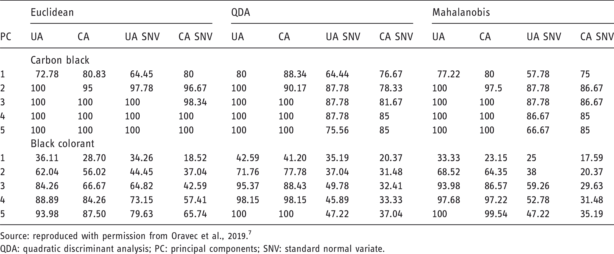
Source: reproduced with permission from Oravec et al., 2019. 7
QDA: quadratic discriminant analysis; PC: principal components; SNV: standard normal variate.
The feasibility of using other vibrational spectroscopic techniques for analysis of black inkjet and laser prints for forensic analysis of the documents was evaluated as well. The aim of the second case study was to investigate the spectral properties and to compare the suitability of applying SERS with Fourier transform Raman (FT-Raman) to identify the spectra of prints. 8 Further specific goals included the establishment of an SERS substrate for this purpose, as well as to select a well-performing spectra processing and chemometric algorithms for future document analysis with the use of the developed method. The sample set included the printouts of eight laser and four inkjet printers available at retail market. Samples were measured with two Raman dispersive spectrometers and an FT-Raman instrument. The former type of devices were DXR Raman microscope with 532 nm excitation line and Foram 685-2 spectrometer with 685 nm excitation laser. The FT-Raman instrument used in this study was Bruker MultiRAM spectrometer with 1064 nm excitation line. Silver nanoparticle colloid (AgNP) from the SERS experiment was synthesized and verified by UV-Vis spectroscopy and scanning electron microscopy (SEM). Significant differences due to silver colloid spin were observed only on SEM images. The main contribution of the work was the developed new successful approach to yield SERS signal of an adequate intensity and quality for the reliable analysis of black prints obtained by both laser and inkjet printers. In addition, the established method is based on only one metal colloid and the analysis can be performed in situ; that is, directly on the surface of the printed sample. SERS delivered much improved quality of spectra, bearing significantly higher levels of absolute sensitivity and selectivity toward the print materials than the FT-Raman (Figure 1). This investigation delivered evidence that SERS can be promising and universal for forensic analysis of printed documents when traditional methods are not effective.
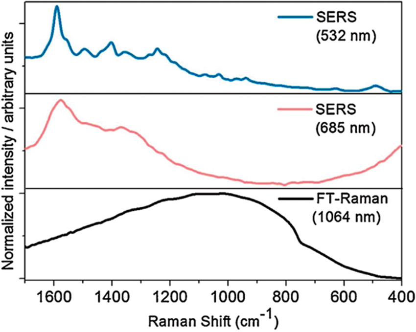
The comparison of the SERS and FT-Raman spectra of a selected exemplary inkjet sample from Oravec et al. 8 (Sample V). The spectra measured using different instruments, two dispersive and one FT-Raman. The first spectrum represents sample treated by poly-L-lysine with AgNPs measured using DXR Raman microscope with 532 nm laser (blue). The second spectrum represents sample treated only by AgNPs measured by Foram 685-2 instrument with excitation line 685 nm (red) and the third spectrum represents sample without treating measured by FT-Raman with excitation line 1064 nm (black). Source: reproduced with permission from Oravec et al., 2018. 8
Identification of print materials in historic photography
Identification and authentication of historical photographs is a challenging task. In recent years, there has been a significant increase in interest in old photography, especially among the general public, from conservatives, photographers, collectors, archives, and merchants to amateurs looking to preserve valuable family albums. 9 Special photographic techniques require different storage conditions, recovery and preservation methods, e.g. water treatment, is not suitable for gelatin binder due to swelling. Material identification and characterization of its structural arrangement in the photography substrate (i.e. paper) is an essential and critical stage to develop an appropriate approach to the process of recovery of an old photography, its subsequent maintenance, and damage prevention. A successful analysis of the mentioned properties of a photography dictates one’s ability to assume an appropriate way to storage and conservation of photographs. 10 Historical photographic materials are combinations of inorganic materials, e.g. silver or baryte, and organic polymeric materials, e.g. paper, albumen, gelatin, or collodion. The chemical composition of these materials, e.g. albumen constituting proteins, vitamins, and minerals that are dispensed in an aquatic environment and their structural deposition in the substrate, create a challenging and complicated sample for non-destructive analysis.
Although both gelatin and albumen contain proteins and feature very similar infrared (IR) spectra, attenuated total reflection Fourier transform IR (ATR-FTIR) was demonstrated as a promising technique, capable of providing rapid and highly reliable ways for characterization and identification of photographs.11–13 The FTIR method is widely used to assist conservation of historical photography. Because collodion and gelatin are absorbed at specific frequencies, identification by IR spectroscopy is relatively more straightforward. However, finer differences between albumen and gelatin appear in IR spectra. Both are proteins with strong amide I and II amide bands appearing in the same spectral range. Furthermore, the processes used to prepare the emulsion can significantly affect the measured spectra. Therefore, robust analysis requires taking into account examination of several forms of the sample originating from different sources. 11 A significant disadvantage of the ATR-FTIR method is that the spectra acquisition spot at the sample surface in relation to the size of the analyzed photo is limited (Figure 2(a)). Additionally, a relatively high pressure on the sample needs to be imposed to secure an adequate contact surface between the sample and the internal reflective element (i.e. ATR crystal) resulting in the danger of scratching of the photo surface. In addition, the presence of coatings, lacquer layers (Figure 2(b)), or the use of alternative fabrication methods may make the interpretation of IR spectra even more difficult. 10
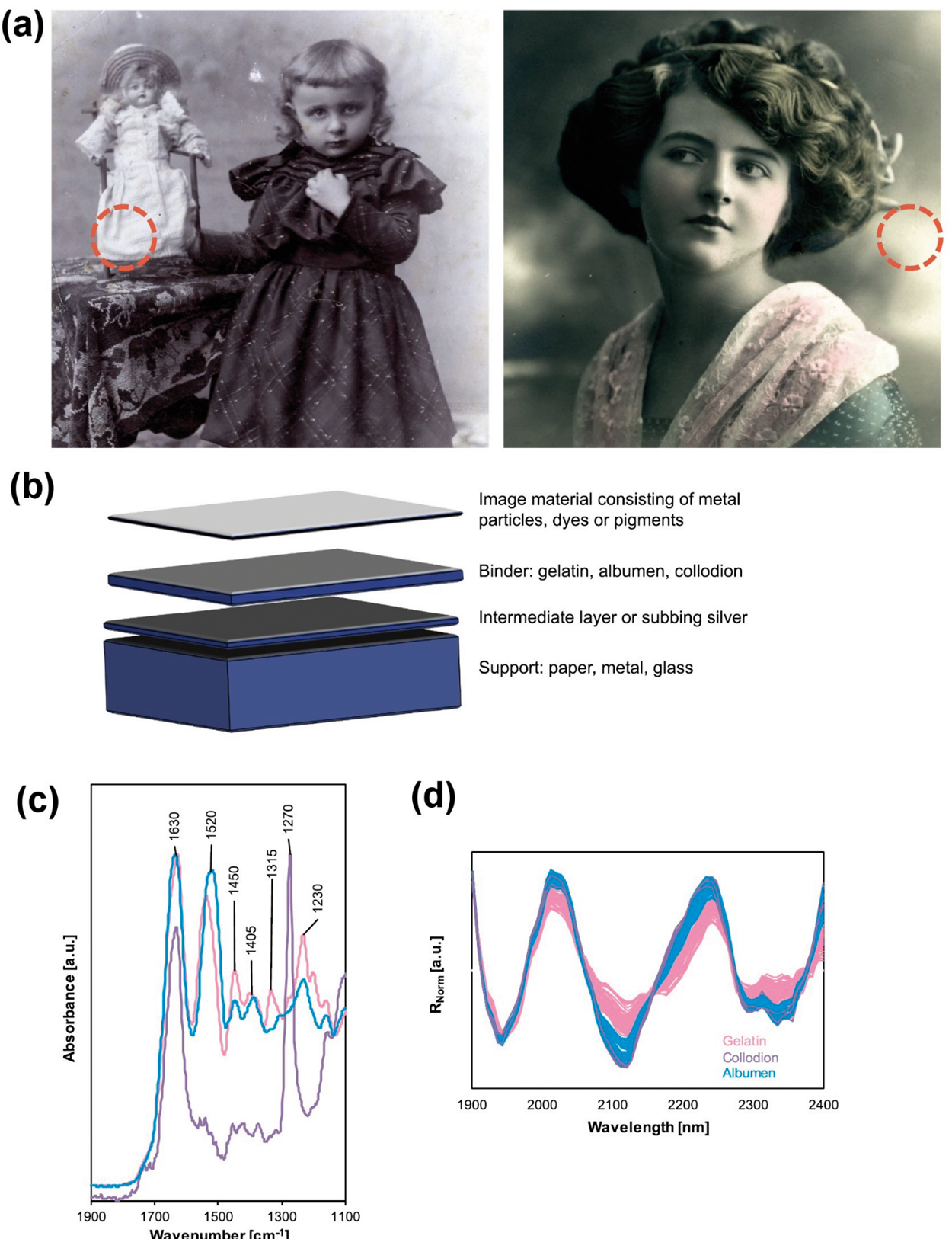
(a) The sampling points were focused on lightest areas of the image (red dashed circles); (b) the schematic cross-section of photographs; (c) the detail of a typical ATR-FTIR normalized spectral pattern of a gelatin (magenta), collodion (violet), and albumen (cyan) prints; (d) The NIR spectra comparison of all samples. Raw spectra of whole acquired in the spectral range of 1900–2400 nm.
Because the coatings and lacquer layers overlap the IR spectrum of the proper pigment layer, ATR-FTIR technique may be more susceptible for misidentification of the photography. Compared to ATR-FTIR, NIR spectroscopy is characterized by a significantly higher penetration depth, which allows you to get information better averaged across the sample depth (Figure 2(b)) and the local (i.e. across different spots in the single sample) and between-sample variation in the analyzed layers of photography is less detrimental for reliability of identification. In the third of the reviewed case studies, we focused on identifying and classifying a data set of 98 historic old photographs and then classified them according to the character of their binder. The developed method offered practical gains in the increased reliability in a non-destructive identification of the binder material in old photographs. The method compared NIR (Figure 2(c)) and ATR-FTIR (Figure 2(d)) techniques, both combined with Principal Component Analysis (PCA), in the explored scenario and presented a very promising potential of NIR spectroscopy. NIR spectroscopy very well discriminates between gelatin and the other two constituents (albumen and collodion), as concluded from the sample grouping along the first Principal Component (PC-1) that explains 94% of the variance in the spectral dataset (Figure 3(a)). The discrimination between albumen and collodion was possible with the third component (PC-3) as presented on the respective PCA score plot (Figure 3(b)). This analytical method offers practical advantages in selecting the appropriate condition for storage, maintenance, and refurbishment procedures of old and valuable pieces of photography.
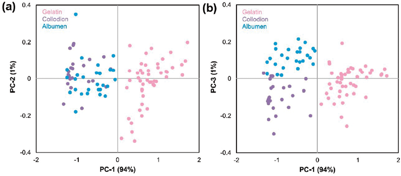
(a) PCA biplot of PC-1 and PC-2, and (b) PCA biplot of PC-1 and PC-3.
Document examination in forensic applications
In the fourth of the case studies discussed here, we examined whether handheld NIR spectroscopy can be successfully used to identify paper and colorants as a rapid screening method in document examination. 14 Portable NIR spectroscopy is an extremely potent tool as it combines the typical advantages of this technique with on-site capability. 15 The technology of miniaturized spectrometers rapidly progresses in NIR spectroscopy, more so than witnessed for most other techniques. 16 In the mentioned case study, we checked whether the samples, which are difficult to distinguish by naked eye approach, can be discriminated based on their NIR spectra by unsupervised PCA and supervised classification method, DA. 14 Eight types of printer paper (average spectra resented in Figure 4(a)) and 10 types of colorants (average spectra resented in Figure 4(b)) were included in the experimental set. A portable NIR spectrometer (microPHAZIR, Thermo Scientific) was used for spectral measurements. The spectra were analyzed first by PCA and then by DA. The robustness of the classification model was evaluated by a five-step cross-validation. All paper samples were properly distinguished in the primary step, the analysis of the variance by means of PCA (Figure 4(c) to (e)). Accurate discrimination of black colorants was achieved in the secondary step of supervised classification DA, using the first major PCs (Figure 4(d) to (f)). Our results showed that miniaturized NIR spectroscopy can be successfully used to identify paper and black colorants in documents in a non-destructive manner directly on-site.
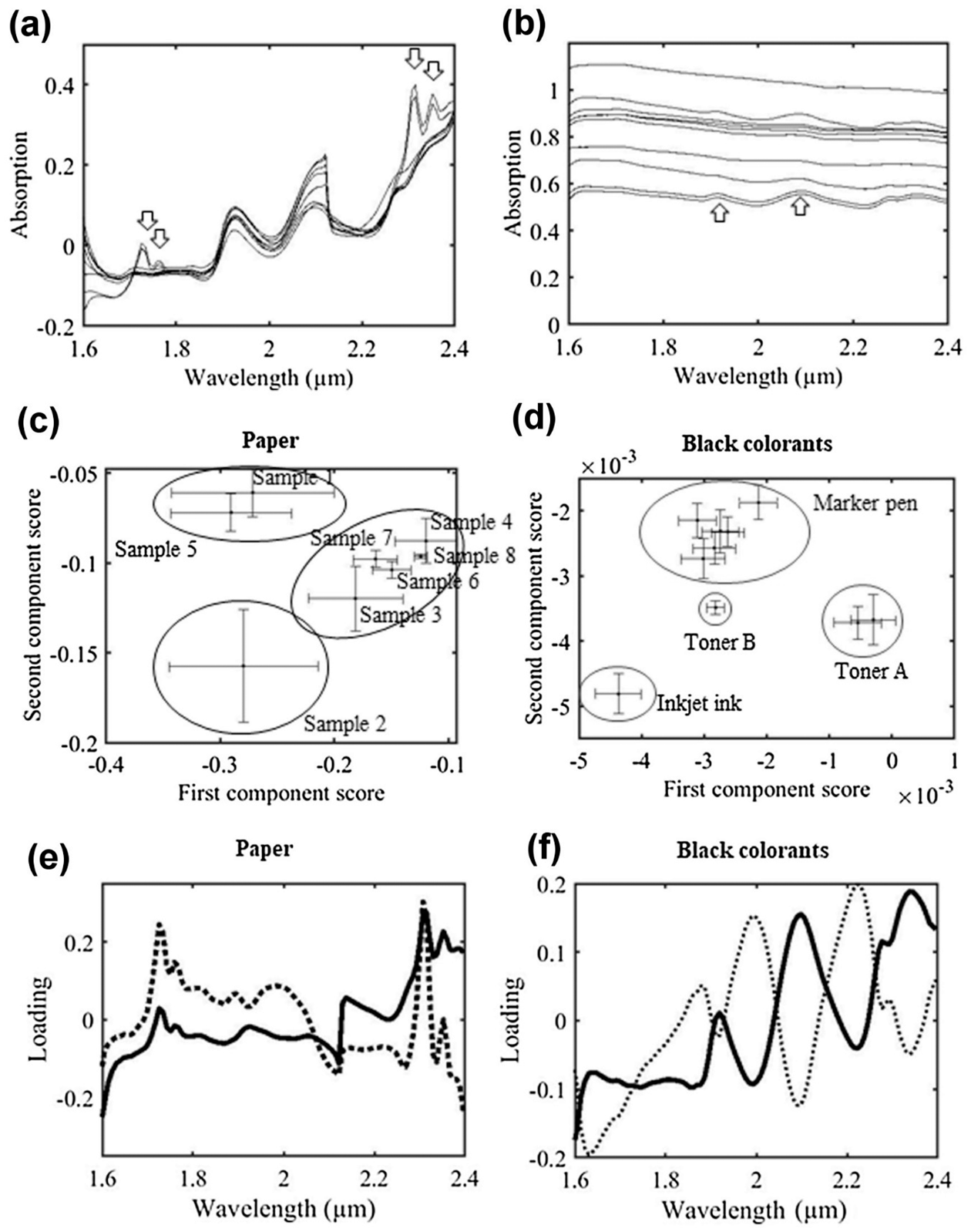
(a) Average spectra of paper samples; (b) average spectra of black colorant samples; (c) scatter plots of principal component scores of paper samples; (d) scatter plots of principal component scores of black colorant samples; (e) loading plots of principal component spectra of paper samples; and (f) loading plots of principal component spectra of black colorant samples. Solid lines show the first components and dotted lines show the second components.
Summary
The challenges of today’s analytical chemistry in the forensic examination of documents attract critical attention as illustrated by the scope of the Organized Session “Innovations in Technology to Advance Forensic Science” chaired by the NIJ, at Pittcon 2020 Conference held in Chicago, Illinois. The importance of establishing novel technologies in this type of analysis has worldwide importance, given the currently insufficient accuracy, reliability, and rapidness of the conventional methods used for detecting forged documents. The lecture opening the Session pointed the attention to the great potential of vibrational spectroscopic techniques used in this role. The presentation summarized the most recent advances accomplished in this field, and evaluated the current trends in the development of spectroscopic methods. Particularly, NIR spectroscopy offers there the necessary performance in the analysis, combined with practical advantages as well as competitive miniaturized spectrometer technology, resulting in much desired on-site capability. On the other hand, SERS techniques yield unparalleled sensitivity in the scenarios where this element of the performance is prioritized. Similar problems and the scope for analysis are observed in the area of the identification of old photographs and using the information available from spectroscopic analysis for optimizing the renovation, storage, and conservation of old pieces of valuable photography.
Footnotes
Declaration of conflicting interests
The author(s) declared no potential conflicts of interest with respect to the research, authorship, and/or publication of this article.
Funding
The author(s) received no financial support for the research, authorship, and/or publication of this article.