
Abstract
1-Bromopropane (1-BP) is an increasingly used chemical solvent for cleaning metals and gradually replacing spraying adhesives. Workers exposed to 1-BP (WBP) showed nervous system dysfunction and other symptoms. This study focused on the proteomic change between healthy individuals (HIs), WBP and poisoned patients with 1-BP (PBP). Total proteins from serum samples were isolated, and high-abundance proteins were filtered out. Large-scale label-free proteomics platform was utilized for protein identification and quantitative comparison, followed by biological function analysis by bioinformatics tools. Compared to HI, 99 proteins were up-regulated and 55 proteins were down-regulated in WBP; 59 proteins were up-regulated and 94 proteins were down-regulated in PBP. With WBP as control, 63 proteins were up-regulated and 127 proteins were down-regulated in PBP. These differently expressed proteins were mainly involved in the immune response, neuron system regulation, blood coagulation, wound healing, endopeptidase activity, lipid metabolic process and apoptosis. The proteomic profiling change of HI, WBP and PBP provides a comprehensive view on 1-BP poison through immune response, signal transduction, metabolism, coagulation and response to stress. This study expanded our understanding on early development and maintenance and provided more potential protein markers for diagnosis of 1-BP poisoning.
Introduction
1-Bromopropane (1-BP) is an increasingly used organic solvent in many manufacturing process, such as immersion degreasing and metal cleaning, as it is less dangerous to the depletion of the ozone layer. 1 Workers exposed to 1-BP (WBP) are facing increased occupational health risk. It is reported that the symptom from 1-BP exposure included numbness, vibration sense losses in lower extremities, ataxic gait, weakness and other nervous system impair. 2 –5 Animal experiments showed that 1-BP induced central nervous system (CNS) toxicity, microglial changes, oxidative stress, reproductive toxicity and liver toxicity. 6 –8 The pathogenesis and maintenance mechanism of 1-BP neurotoxicity is far from lucid. It is reported that the 1-BP may deplete glutathione (GSH) in the liver and brain 9,10 and metabolism of 1-BP via oxidation of cytochrome P450IIE1. 6
The proteomic mechanism of 1-BP toxicity has not been well studied. We collected the serum of the healthy individuals (HIs), WBP and poisoned patients with 1-BP (PBP), and the large-scale label-free proteomics technology was used to identify involved proteins and perform quantitative comparison. Differently expressed proteins (DEPs) were screened and analysed by bioinformatics tools in order to extend our understanding on proteomic change during the development of illness associated with 1-BP exposure and provide potential protein markers for poisoning diagnosis.
Materials and methods
Peripheral blood of each 2 ml was collected from three groups of workers in the same enterprise, six HIs, six WBP and three PBP. Information and cases were provided by Baoan Center for Disease Control and Prevention (Shenzhen, Guangdong Province), and all objects were male (Table 1). The work of both WBP and PBP was metal cleaning. For the WBP and PBP, the periods of exposure to 1-BP were 10.50 ± 2.07 months and 10.00 ± 0.58 months, respectively. All PBP were hospitalized for lower limb weakness and numbness. The information of three poisoned patients is shown in Table 1. The patients were diagnosed according to the National diagnostic criteria of occupational disease, occupational bromopropane poisoning. Other diseases such as cerebral cardiovascular disease, immune-mediated disease and other reagent poisoning diseases were excluded. All collected blood samples were placed in refrigerator at 4°C for 24 h, followed by centrifuging at 4000 rpm for 10 min. All samples were collected with written informed consent using protocols that comply with the Declaration of Helsinki Principles. Permission for these studies was obtained from the medical ethics committee (The 8th People’s Hospital of Wuxi).
Demographic information of three poisoned patients.
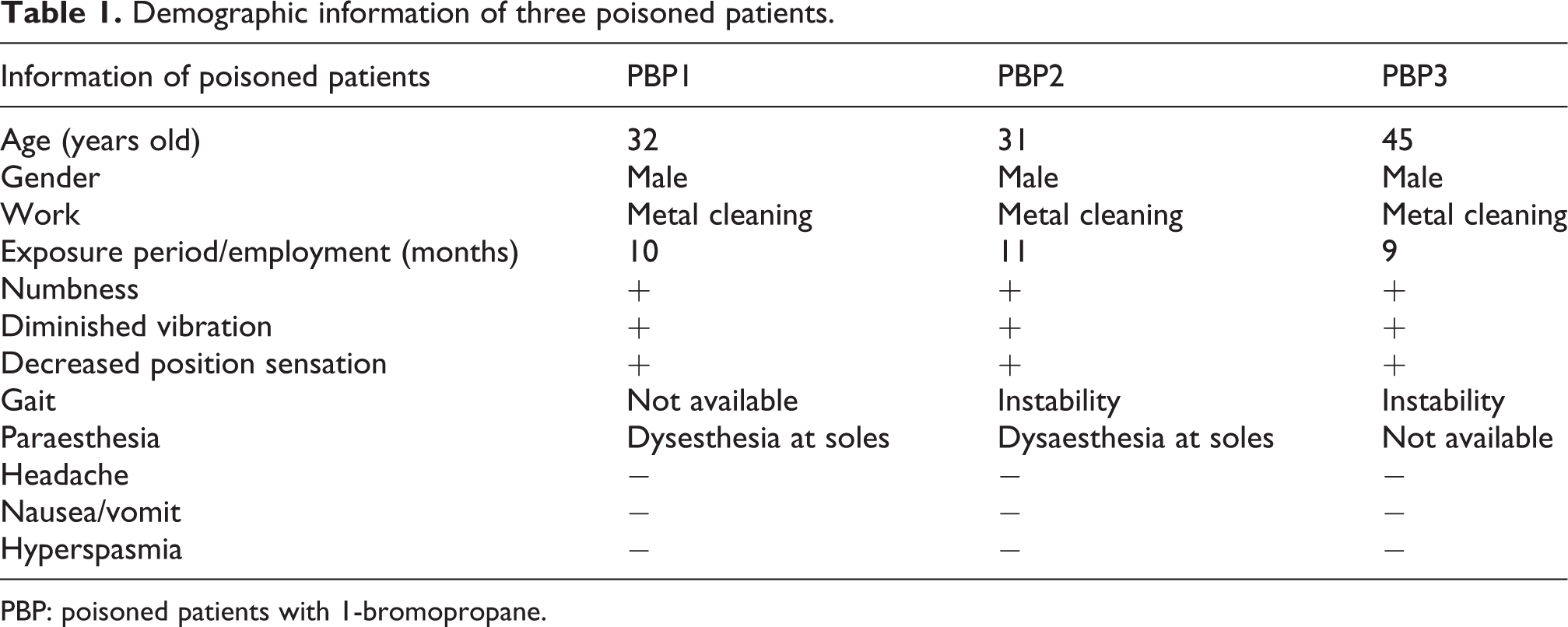
PBP: poisoned patients with 1-bromopropane.
Protein extraction and preparation
In order to increase reliability, sample pooling method was performed. Within the WBP group, on average two samples were pooled into a new sample and three repeated samples were generated. Total protein was extracted by optimized phenol extraction method. 11 High-abundance proteins that remove reagent (Proteoprep Blue Albumin and IgG Depletion Kit; Sigma-Aldrich, St Louis, Missouri, USA) were used to enrich the low-abundance proteins in the serum for next protein analysis.
For each 80 µl serum sample, 200 µl 8 mol/L urea was added and centrifuged at 14,000 g for 4 min. Precipitated proteins were transferred to 200 µl 0.1mol/L tris-HCl/8 M (hydrochloric acid) urea (pH 8.5) buffer and centrifuged. For reductive alkylation, 10 µl of 1 mol/L iodoacetamide (Sigma-Aldrich) is dissolved in 0.1 mol/L tris-HCl/8 mol/L urea and centrifuged, followed by dissolving in 100 µl 0.1 M tris-HCl/8 M urea and centrifugation. The aforementioned process was performed twice. By 1:5 ratio, trypsin (Promega, Madison, Wisconsin, USA) was added at 37°C for 16 h, followed by freeze-drying. Peptides were desalinated (Waters, Milford, Massachusetts, USA) before MS/MS (Mass Spectrometry) was performed. Protein concentration was determined by the Bradford protein assay method (Thermo Scientific, Waltham, MA, USA).
Label-free proteomic analysis
LTQ orbitrap Elite Liquid mass spectrometry with EASY-nLC1000 system (Thermo Fisher Scientific, Odense, Denmark) was employed for protein analysis followed by manufacturer’s instruction. Afflux sample of 10 µl passed through protect column (C18 PepMap 100, 300 μm × 1 mm, 5 μm, 100 Å) and analysis column (Acclaim PepMap C18, 15 cm × 75 μm, 2 µm, 100 Å; Dionex, Sunnyvale, California, USA), respectively. The assay was performed with the following gradient 5–45% (water A and acetonitrile B, both with 0.1% formic acid) for 190 min, followed by another gradient of 95% B for 10 min. Then, the gradient of 5% was set to re-equilibrate the column for the next injection. Eluting peptides from the column were analysed by MS/MS spectrometer, and top-15 method was used for data collection. The parameter of spectrum was as follows: cation model, crash model, resolution 70,000 and mass range 350–1800.
Data analysis
Thermo Proteome Discovert (1.4.0.288) software with Mascot tool was used for label-free data analysis against the reference protein database of Human Uniprot-2015 at an false discovery rate (FDR) <1%. The searches were performed with fixed parent ion mass error ±10 ppm, ion mass error ±0.05 Da, least match one unique peptide and tryptic specificity allowing a maximum of two missed cleavages. The DEPs over 1.5 fold change between HI, WBP and PBP were analysed by t-test with SPSS software (V20). The accession numbers of proteins were inputted into Blast2GO (V2.8.0), 12 David Bioinformatics tool (david.ncifcrf.gov) 13 and KOBAS (2.0) 14 to determine their biological function.
Results
Proteomic expression profiling in HI, WBP and PBP
Using label-free LC-MS/MS technology, 497 unique proteins were identified. In detail, 348, 363 and 340 unique proteins were identified in HI, WBP and PBP (Table 2), respectively. Compared to HI, 99 proteins were up re-regulated and 55 proteins were down-regulated over 1.5 fold change in WBP (Table 3). In PBP, 59 proteins were up-regulated and 94 proteins were down-regulated (Table 3). With HI as control, 32 up-accumulated proteins and 24 down-accumulated proteins displayed similar tendency in WBP and PBP (Figure 1, Table 4). With WBP as control, 63 proteins were up-regulated and 127 proteins were down-regulated in PBP (Table 3).
Identified proteins information by label-free proteomic technology.

PBP: poisoned patients with 1-bromopropane; WBP: workers exposed to 1-bromopropane; HI: healthy individual.
Differently expressed proteins between WBP, PBP and HI.

PBP: poisoned patients with 1-bromopropane; WBP: workers exposed to 1-bromopropane; HI: healthy individual.

Venn diagram displaying the coexpressed proteins with similar tendency in WBP and PBP with HI as control. The number of up-regulated and down-regulated proteins in two groups is indicated in the intersections. WBP: workers exposed to 1-bromopropane; PBP: poisoned patients with 1-bromopropane; HI: healthy individual
Coexpressed proteins with similar tendency in WBP and PBP with HI as control.

PBP: poisoned patients with 1-bromopropane; WBP: workers exposed to 1-bromopropane.
Protein function analysis on WBP and HI
Bioinformatics tools were utilized for protein function analysis on gene ontology (GO Consortium, http://geneontology.org/) and pathway. Compared to HI, the DEPs in WBP were mainly divided into two levels of biological process and molecular function (Figures 2(a) and (b)) according to GO enrichment analysis. For molecular function, DEPs were mainly involved in enzyme regulator activity, ion binding, hydrolase activity, protein binding, lipid binding and transmembrane transporter activity (Figure 1(a)). In view of biological process classification, the top nine enrichment terms covered establishment of localization, response to stress, single-organism cellular process, single organism signalling, regulation of biological process, cellular response to stimulus, organic substance metabolic process and primary metabolic process (Figure 1(b)).

GO enrichment of DEPs between WBP, PBP and HI. (a) Molecular function enrichment of DEP between WBP and HI. (b) The enrichment of biological process in DEP of WBP and HI. (c) The molecular function analysis of DEP between PBP and HI. (d) The biological process analysis of DEP from PBP and HI. (e) The enrichment of molecular function of DEP of PBP and WBP. (f) The biological process analysis of DEP between PBP and WBP. GO: gene ontology; WBP: workers exposed to 1-bromopropane; PBP: poisoned patients with 1-bromopropane; HI: healthy individual; DEP: differently expressed protein.
By pathway analysis, 11 proteins (accession nos.: P02671, P01031, Q59GS8, P11226, P02747, P02745, P08697, P13671, P00748, P00747, P12259 and P0C0L5) were involved in the pathway of complement and coagulation cascades. Three proteins (Accession nos.: P02671, P00748 and P12259) were involved in the intrinsic prothrombin activation pathway. Four DEPs (P01031, Q59GS8, P11226, P13671 and P0C0L5) were involved in the lectin-induced complement pathway. Fibrinogen alpha chain and plasminogen (P02671 and P00747) were involved in the fibrinolysis pathway. Mannose-binding protein C (P11226) was involved in the inflammation of the CNS (neurotic disorder). Pigment epithelium-derived factor (P36955) annotated to the regulation of neurogenesis (neurogenesis) and neuron projection development. Five DEPs that include serotransferrin (P02787) and others (P11226, Q7Z7Q0, P00747 and P01011) were involved in the biologic process of neurology.
Function analysis of PBP and HI
The DEPs between PBP and HI were analysed by GO enrichment. Viewed from molecular function classification, it was mainly divided into ion binding, hydrolase activity, enzyme regulator activity, lipid binding, protein binding, oxidoreductase activity and signal transducer activity (Figure 2(c)). For biological process level, the top 10 annotated terms included response to stress, establishment of localization, single-organism cellular process, single organism signalling, cellular response to stimulus, regulation of biological process, primary metabolic process, organic substance metabolic process, anatomical structure development and single-organism developmental process (Figure 2(d)).
According to Kyoto Encyclopedia of Genes and Genomes pathway analysis, five DEPs (accession nos: P01031, Q59GS8, P11226, P02745, P13671 and P0C0L5) were involved in the complement pathway. Eleven proteins that included fibrinogen alpha chain, complement C5, complement component 5 variant and so on (P02671, P01031, Q59GS8, P11226, P02747, P02745, P08697, P13671, P00748, P00747, P12259 and P0C0L5) were involved in the pathway of complement and coagulation cascades. Prothrombin (P00734) is involved in the pathway of neuroactive ligand–receptor interaction and regulation of neurogenesis. Fibrinogen alpha chain (P02671) is involved in the neurodegenerative diseases. Serum paraoxonase/arylesterase 1 (P27169) was annotated to neurological process. Serum paraoxonase/arylesterase 1 (P27169) and apolipoprotein M (O95445) were annotated to the process of response to toxic substance.
Function analysis of PBP and WBP
By level of molecular function, ion binding, hydrolase activity, enzyme regulator activity, lipid binding, protein binding, oxidoreductase activity, signal transducer activity and transmembrane transporter activity were mainly involved in this classification (Figure 2(e)). Viewed from the biological process classification, the top 10 annotated terms included were single-organism cellular process, establishment of localization, response to stress, single organism signalling, cellular response to stimulus, regulation of biological process, primary metabolic process, organic substance metabolic process, anatomical structure development and cellular component organization (Figure 2(f)). Coagulation factor X, plasma kallikrein and prothrombin (P00742, P03952, P00734 and Q16519) were mainly involved in the pathway of prothrombin activation pathway. Ten proteins (accession nos: P02787, P00734, P02042, P03952, P05546, P08697, B7ZLE5, P00742, P68871 and P62328) were involved in the function of haemostasis. The protein of HCG40889, isoform CRA_b (A0A024R962), was involved in the nervous system diseases. Pigment epithelium-derived factor (P36955), prothrombin (P00734) and FN1 protein (B7ZLE5) were involved in the pathway of regulation of nervous system development.
Coexpressed proteins with similar tendency in WBP and PBP
With HI as control, 32 up-regulated and 24 down-regulated proteins have the same tendency both in WBP and PBP groups (Figure 1, Table 4). In view of the immune function analysis, these proteins are mainly involved in the process of immune response, inflammatory response, defence response, complement activation, humoral immune response, activation of immune response and immune effector process (Figure 3). By pathway analysis, the protein of fibrinogen alpha chain (P02671) is involved in the neural cell adhesion molecule signalling for neurite outgrowth and neurodegenerative diseases. Apolipoprotein C-II (P02655) is involved in the neutral lipid catabolic and metabolic processes. Five proteins (accession nos.: P13671, P02671, P02747, P12259 and P00748) are involved in the pathway of complement and coagulation cascades. The protein of apolipoprotein C-II (P02655) is annotated to the process of metabolism of lipids and lipoproteins.

Immune response analysis of coexpressed proteins in WBP and PBP. Y-axis represent the involved proteins quantity and X-axis shows the annotated function terms. WBP: workers exposed to 1-bromopropane; PBP: poisoned patients with 1-bromopropane.
Discussion
The understanding of pathogenicity mechanism of poisoning disease associated with 1-BP is far from clear, especially regarding the involved proteomic profiling. Present diagnostic methods of 1-BP poisoning are based on the clinical neural system dysfunction and blood biochemical tests. The metabolic biomarker of 1-BP in human body research was focused on the 1-BP in urine, bromine, N-acetyl-S-(n-propyl)-
By protein function analysis, over 40 proteins were involved in the immune response, including prothrombin (P00734), Ig kappa chain V-III region POM (P01624), Ig heavy chain V-III region JON (P01780) and others (Supplementary Material Table S1). The expression of these proteins may be stimulated by 1-BP inhabitation and take part in the immunoreaction. This process responds to the poisoning of 1-BP and enhanced immune activity and promote metabolism in human body. The research of influence and function of immune system in poisoning associated with 1-BP is limited. Our results provided a new immune view on the pathology of PBP. Further function study is needed.
1-BP is inhaled into human body by skin or respiratory tract, which causes multipoisonous effects. During 1-BP metabolism process, cytochrome P450 and GSH play an important role. Animal in vivo study showed that the first metabolism pathway of 1-BP was oxidated by cytochrome P450 and transferred to 1-bromine-2-propylalcohol (1-B-2-PA). 1-B-2-PA combined with GSH and glycuronic acid and exhausted through urine. 17 Inhibition of P450 and GSH extends the half-life period of 1-BP in the body. 18 By proteins function annotation, over 10 proteins were involved in the metabolism process, including V9GYJ8, P27169, B2R8P6, P02655, O95445, P01011, P02655 and others. These proteins were involved in the lipid metabolism, organic compound metabolism, sulphide compound metabolism and oxyacid metabolism process. GSH peroxidase (GSH-Px) is an important hyperoxide lyase. GSH-Px catalyses GSH to oxidized GSH, which promotes the oxidation of poisoned compound to non-toxic hydroxyl compound, which protects organism from interfere and damage. The expression level of GSH-Px down-regulated in 1-BP poisoned patients, which hints that the decrease of GSH-Px may inhibit the oxidation of GSH and interfere the metabolism of 1-bromopoprane-2-propylalcohol. Half-life period of 1-BP prolongs and aggravates the toxicity of 1-BP.
Intense harmful excitant causes nonspecificity stress of organism, in which by the hypothalamus, the up-regulation of adrenocorticotropic hormone in blood gives rise to abundant secretion of glucocorticoid. In this study, over 50 proteins were involved in the response to stress. These proteins included P00734, P03952, P01624, P01780, P01767, P01768 and others. By function analysis, several proteins were involved in the oxidation–reduction process, such as plasma kallikrein, GSH peroxidase and serum paraoxonase/arylesterase. It is reported that the cytochrome P450 and GSH play an important role in the metabolism of 1-BP in animal experiment. 17,18 GSH peroxidase was down-regulated in PBP, which suggested that these proteins may be involved in the regulation metabolism of 1-BP in human body through oxidation stress process. The aforementioned proteins may be a potential diagnosis biomarker in the early development of PBP. Further research is needed to understand their detailed function and mechanism.
We found that the prothrombin (P00734) was down accumulated in PBP. This protein is involved in multiple function regulation in human body, which include proteolysis, tarombokinesis caspases activity and calcium binding. 19 By protein function annotation analysis, Prothrombin may be involved in the nervous system such as(/which includes) nervous centralis development regulation process. Down-regulation of prothrombin in PBP may interfere with nervous regulation in PBP, followed by nervous system damage. PBP generally suffer from nervous system abnormity, such as numbness of limb and decreased vibration. 16,20,21 The nervous toxicity of 1-BP is mainly present in peripheral nerve myelin degeneration and fracture, postpyramid nucleus neurons axon swelling and cerebellar Purkinje cell degeneration.22 Neurofilament protein (NFP) specially exists in the brain and neuron tissue, which consists of NF-L, NF-M and NF-H. NFP is a special part of neuron skeleton, which plays an important role in morphological maintenance of neuron cell and signal transduction function of neuron. Inhalation of 1-BP causes structural and functional change of cortex NFP and causes malfunction of CNS and peripheral nerves. 22 We found that the actin filament aggregation regulation–related protein thymosin beta-4 (P62328) was up-regulated in PBP. This protein may interfere with muscle cell movement and nonactin cell by regulation of actin filament aggregation, followed by influencing the nerve system and motion function.
In summary, DEPs between HI, WBP and PBP were analysed by the large-scale proteomic label-free tool. These proteins were mainly involved in the metabolism, neuron system, immune response and signal transduction. Our study provided several potential biomarkers for early diagnosis of 1-BP-associated disease and contributed to the understanding of the molecular mechanism of 1-BP poisoning.
Footnotes
Declaration of Conflicting Interests
The author(s) declared no potential conflicts of interest with respect to the research, authorship, and/or publication of this article.
Funding
The author(s) disclosed receipt of the following financial support for the research, authorship, and/or publication of this article: This project was supported by China Health Standard System (Revising) Program (grant no. 20140710).
Supplemental material
Supplementary material for this article is available online.
References
Supplementary Material
Please find the following supplemental material available below.
For Open Access articles published under a Creative Commons License, all supplemental material carries the same license as the article it is associated with.
For non-Open Access articles published, all supplemental material carries a non-exclusive license, and permission requests for re-use of supplemental material or any part of supplemental material shall be sent directly to the copyright owner as specified in the copyright notice associated with the article.