
Abstract
Mineral trioxide aggregate (MTA) is a calcium silicate dental cement used for various applications in dentistry. This study was undertaken to test whether the presence of three commercial brands of calcium silicate dental cements in the dental extraction socket of rats would affect the brain aluminium (Al) levels and oxidative stress parameters. Right upper incisor was extracted and polyethylene tubes filled with MTA Angelus, MTA Fillapex or Theracal LC, or left empty for the control group, were inserted into the extraction socket. Rats were killed 7, 30 or 60 days after operation. Brain tissues were obtained before killing. Al levels were measured by atomic absorption spectrometry. Thiobarbituric acid reactive substances (TBARS) levels, catalase (CAT), superoxide dismutase (SOD) and glutathione peroxidase (GPx) activities were determined using spectrophotometry. A transient peak was observed in brain Al level of MTA Angelus group on day 7, while MTA Fillapex and Theracal LC groups reached highest brain Al level on day 60. Brain TBARS level, CAT, SOD and GPx activities transiently increased on day 7 and then returned to almost normal levels. This in vivo study for the first time indicated that initial washout may have occurred in MTA Angelus, while element leaching after the setting is complete may have taken place for MTA Fillapex and Theracal LC. Moreover, oxidative stress was induced and antioxidant enzymes were transiently upregulated. Further studies to search for oxidative neuronal damage should be done to completely understand the possible toxic effects of calcium silicate cements on the brain.
Keywords
Introduction
Mineral trioxide aggregate (MTA) is a hydraulic calcium (alumino) silicate dental cement closely related to the long-standing construction material, Portland cement. The composition of Portland cement is tricalcium and dicalcium silicate, tricalcium aluminate and tetracalcium aluminoferrite. MTA contains Portland cement and bismuth oxide as a radiopacifier, which makes MTA visible on radiographs. 1 Although initially recommended as an alternative biomaterial for root-end filling, MTA has found a variety of applications in endodontics and restorative dentistry since its first introduction in 1993. The success of MTA was attributable to its excellent biocompatibility, bioactivity and osteoconductivity. 2
MTA is introduced under several brand names, including ProRoot MTA, MTA Angelus, MTA Fillapex, MTA Bio, Micro Mega MTA and Ortho MTA, which slightly differ in composition. MTA Angelus (Angelus, Londrina, PR, Brazil) contains tricalcium and dicalcium silicate, tricalcium aluminate, tetracalcium aluminoferrite and bismuth oxide. 3 MTA Fillapex (Angelus) is composed of MTA, salicylate resin, natural resin, silica and bismuth oxide. 4 Theracal LC (Bisco, Schaumburg, Illinois, USA) contains polymerizable methacrylate monomers, Portland cement type III, polyethylene glycol dimethacrylate and barium zirconate. 5
MTA materials contain substantial amount of heavy metals such as arsenic, lead, cadmium, copper, iron, manganese, nickel, zinc and aluminium (Al), 6 –11 which often can leach from dental restorations. 12 –16 The tendency of dental cements to washout or release metal ions might vary depending on their setting times and the lengthening of setting time is a major disadvantage. 2 MTA Angelus and MTA Fillapex are self-setting dental biomaterials that require some time to completely set, 3,4 while Theracal LC sets immediately, because it is light-cured. 5
Al is a highly abundant toxic metal that is best known for its neurotoxicity and its relation to the pathogenesis of Alzheimer’s disease. 17 Furthermore, Al has also been implicated in the pathogenesis of other neurological diseases, including Parkinson’s disease 18 and amyotrophic lateral sclerosis, 19 as well as skeletal and haematological diseases. 20 The basis of Al toxicity is believed to be based on induction of oxidative stress. 21 Even though the amount of metals such as chromium, arsenic and lead that leach from MTA-based dental cements into water and simulated body fluids was studied before in vitro, 22,23 Al release was not studied, and such a study was not carried out in vivo, except our previous study where we showed that Al levels were increased in blood and liver samples of rats that had MTA Angelus, MTA Fillapex and Theracal LC in their alveolar socket. 9 Upon leaching from hydraulic calcium silicate dental materials, Al can enter the systemic circulation 9 because the surrounding tissues are damaged during endodontic treatment and then can enter the brain. 24
The purpose of the present study, which is the first report of its kind in the literature, was to investigate in vivo whether alveolar socket implantation of one of the three selected dental cements, MTA Angelus, MTA Fillapex and Theracal LC, in rats alters the Al levels and oxidative stress parameters (thiobarbituric acid reactive substances (TBARS) levels, catalase (CAT), superoxide dismutase (SOD) and glutathione peroxidase (GPx) activities) in brain samples.
Materials and methods
Animals
Animal Ethics Committee of the Gülhane Military Medical Academy approved the study (Decision no: 13/103), and all the procedures were carried out in accordance with the European Community Council Directive of 24 November 1986 (86/609/EEC). Ninety-six male Wistar albino rats weighing 350–400 g (approximately 10–13 months old) were used in this study. All the animals were housed in temperature-controlled rooms. Adequate measures were taken to minimize pain or discomfort during the experiments.
Each animal, including the control animals, was anesthetized by an intraperitoneal injection of 10% ketamine hydrochloride (Alfasan, Woerden, the Netherlands) at 40–70 mg/kg body weight and xylazine (Bayer, Munich, Germany) at 7–12 mg/kg body weight. After that, the right upper incisor of each rat was extracted with a maxillary anterior forceps, and after bleeding control, polyethylene tubes filled with one of the dental cements, or left empty for the control group, were inserted into the depth of the extraction socket and gingival tissue was sutured over the extraction socket with absorbable silk 4.0 sutures.
The dental cements used were MTA Angelus (Angelus), MTA Fillapex (Angelus) and Theracal LC (Bisco), which were prepared according to the manufacturers’ recommendations and then filled into polyethylene tubes (Ayset, Adana, Turkey) that were sterilized with ethylene oxide. Both ends of the polyethylene tubes were open and their dimensions were 1.0 mm internal diameter, 1.6 mm external diameter and 3.0 mm length. Each tube was filled with 7.6 mg dental cement, and 24 tubes were prepared for each dental cement. Control tubes (n = 24), which remained empty, were inserted into the depth of the extraction socket of control rats, and gingival tissue was sutured over the extraction socket.
Each group of rats receiving the same dental cement and control rats was further divided into three groups to test for the effect of exposure time (eight rats in each group) and were killed 7, 30 and 60 days after the operation with a high-dose anaesthetic injection. Before killing each animal was anesthetized as explained above and the brain of each animal was removed and stored at −40°C until analysed.
Determination of Al concentrations
Al concentration in the brain samples was determined using atomic absorption spectrometry as explained before. 9 Limit of detection (LOD) was 0.3 parts per billion (ppb) and limit of quantitation (LOQ) was 0.8 ppb. Results are given as parts per million (ppm), which equals to micrograms per gram of tissue.
Determination of oxidative stress parameters
The brain tissues were weighed (approximately 0.5 g), and homogenates were prepared with 1.15% potassium chloride using a homogenizer (Schütt Homgen Plus; Schuett-Biotec GmbH, Göttingen, Germany). Then, the homogenates were centrifuged at 4°C at 5000 × g and supernatants were used for the biochemical assays.
CAT activity in brain tissue supernatant was measured by the method of Aebi. 25 The decomposition rate of the substrate H2O2 was monitored spectrophotometrically at 240 nm for 30 s. CAT activity was expressed as units per milligram of tissue, and 1 U is equal to 1 μmol of H2O2 decomposed per minute. CuZn-SOD activity in brain tissue supernatant was measured as previously described. 26 CuZn-SOD activity was expressed as units per gram of tissue. GPx activity in brain tissue supernatant was determined as previously described. 26 GPx activity was expressed as units per gram of tissue. TBARS levels were determined in brain tissue supernatant using a previously described method. 27 The TBARS levels were expressed as nanomoles per gram of tissue.
Data analyses
Distribution of data was tested by the Kolmogorov–Smirnov test, and normality plots were also checked. Data with normal distribution were analysed by one-way analysis of variance (ANOVA) if the test of equality of variances (Levene statistic) is satisfied. In few cases where Levene statistic is not met, Welch’s ANOVA results were given instead of usual ANOVA. When the result of ANOVA was significant, it was followed by a post hoc test for multiple comparisons; if equal variances assumed the Bonferroni test was used and if equal variances not assumed the Games–Howell test was used. Data that did not show normal distribution were compared using the non-parametric Kruskal–Wallis test. Post hoc test for multiple comparisons was carried out using the multiple comparison test suggested by Conover. 28 A p value <0.05 was considered statistically significant. Statistical Package for Social Sciences version 15.0 (SPSS, Chicago, Illinois, USA) was used for these statistical analyses.
Results
Brain Al levels of calcium silicate cement–applied rats and controls are given in Figure 1. The highest brain Al level in this study was observed in the MTA Angelus group after 7 days (16.42 ± 10.42 ppm), which was significantly higher than the brain Al levels of MTA Fillapex (3.64 ± 1.59 ppm), Theracal LC (2.97 ± 1.68 ppm) and control rats (1.48 ± 0.46 ppm) on day 7. On day 30, the brain Al concentration of MTA Angelus group decreased to 9.25 ± 7.19 ppm; however, there was still a significant difference among the study groups. On day 60, the brain Al level of MTA Angelus group further decreased to 3.01 ± 2.64 ppm; the decrease from day 7 to 30 to 60 was significant (p = 0.001). In addition, there was a significant difference among the study groups on day 60. Brain Al level of MTA Fillapex group rats was significantly higher than that of controls at all times (p = 0.001, 0.010, and 0.000 on days 7, 30 and 60, respectively). Theracal LC group had significantly increased brain Al concentration on days 7 (p = 0.008) and 60 (p = 0.001).

Aluminium levels in brain samples of rats that received different hydraulic calcium silicate cements in their dental extraction socket, as explained in the methods, and killed after 7, 30 and 60 days post-operation. Data are expressed as mean ± SD in the first row and median (quartiles) in the second row. *p < 0.05 among the four groups on the same day. p Values were determined using the Kruskal–Wallis test for 7 days and 60 days, and the Robust test of equality of means (Welch’s ANOVA) for 30 days. **p < 0.05 among MTA Angelus applied rats on days 7, 30 and 60. p Values were determined using the Kruskal–Wallis test. SD: standard deviation; ANOVA: analysis of variance.
Brain TBARS levels of the study groups are given in Figure 2. On day 7, all the dental cement applied groups had significantly higher brain TBARS levels when compared with control rats. On days 30 and 60, TBARS levels decreased for the dental cement–applied rats and did not differ significantly among the study groups.

TBARS levels in brain samples of rats that received different hydraulic calcium silicate cements in their dental extraction socket, as explained in the methods, and killed after 7, 30 and 60 days post-operation. Data are expressed as mean ± SD in the first row and median (quartiles) in the second row. *p < 0.05 among the four groups on day 7. p Values were determined using one-way ANOVA. TBARS: Thiobarbituric acid reactive substances; SD: standard deviation; ANOVA: analysis of variance.
CAT enzyme activities were determined in brain tissue samples of the calcium silicate cement–applied rats and controls and the results are given in Figure 3. We observed that brain CAT activity of MTA Angelus–applied rats was significantly higher than that of controls (p = 0.024), and there was a significant difference among the study groups on day 7 (p = 0.032). On day 30, the brain CAT level of MTA Angelus group decreased and the significant difference among study groups was no longer present. Likewise, on day 60, CAT activity results of all groups were similar to controls.

CAT activity in brain samples of rats that received different hydraulic calcium silicate cements in their dental extraction socket, as explained in the methods, and killed after 7, 30 and 60 days post operation. Data are expressed as mean ± SD in the first row and median (quartiles) in the second row. *p < 0.05 among the four groups on day 7. p Values were determined using one-way ANOVA. CAT: catalase; SD: standard deviation; ANOVA: analysis of variance.
SOD enzyme activity in brain samples of rats having calcium silicate cements in their alveolar socket and controls was measured (Figure 4), and the results show that there was a significant difference among the study groups on day 7 (p = 0.011). Brain SOD activities of MTA Angelus and MTA Fillapex having rats were markedly higher when compared with controls on day 7. The results returned to almost normal levels on days 30 and 60. Brain SOD activity of MTA Fillapex rats decreased prominently throughout the study period (p = 0.012).
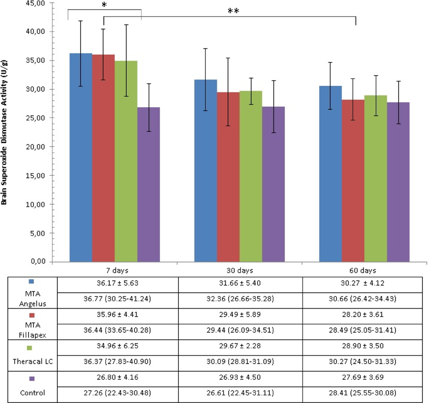
SOD activity in brain samples of rats that received different hydraulic calcium silicate cements in their dental extraction socket, as explained in the methods, and killed after 7, 30 and 60 days post operation. Data are expressed as mean ± SD in the first row and median (quartiles) in the second row. *p < 0.05 among the four groups on day 7. **p < 0.05 among MTA Fillapex applied rats on days 7, 30 and 60. p Values were determined using one-way ANOVA. SOD: superoxide dismutase; SD: standard deviation; ANOVA: analysis of variance.
GPx enzyme activity in brain samples of rats having calcium silicate cements in their alveolar socket and controls was also determined and the results are given in Figure 5. Similar to Al, TBARS, CAT and SOD results, the highest GPx measurement was obtained with MTA Angelus group on day 7. Brain GPx activity of MTA Angelus and Theracal LC groups was significantly higher compared with controls on day 7, and there was a significant difference among the study groups. As in the case of TBARS level and CAT and SOD activity measurements, GPx activities also decreased after day 7, and there was no marked difference among the study groups on days 30 and 60. The decrease in brain GPx activity throughout the study period was significant for all the studied dental cements.

GPx activity in brain samples of rats that received different hydraulic calcium silicate cements in their dental extraction socket, as explained in the methods, and killed after 7, 30 and 60 days post operation. Data are expressed as mean ± SD in the first row and median (quartiles) in the second row. *p < 0.05 among the four groups on day 7. **p < 0.05 among MTA Angelus applied rats on days 7, 30 and 60. ***p < 0.05 among MTA Fillapex applied rats on days 7, 30 and 60. ****p < 0.05 among Theracal LC applied rats on days 7, 30 and 60. All p values were determined using one-way ANOVA. GPx: glutathione peroxidase; SD: standard deviation; ANOVA: analysis of variance.
Discussion
MTA is a cementitious dental material in the form of a very fine powder used successfully for various applications in endodontics. It is a hydraulic cement, which means that it sets and is stable under water. 29 However, the oral environment is described as hostile for dental restorative materials, because freshly prepared dental cements tend to disintegrate, or washout, upon contact with fluids. MTA has been shown to be susceptible to washout; a mass loss of 5–10% was observed with MTA Angelus. 30
In vitro studies carried out using artificial saliva or other similar fluids, 12,15,22,23,31 or cell culture systems, 13,14 have shown that in addition to an initial wash out before the cement has fully set, a slower diffusion controlled release of metal ions from dental cements also occurs for days. The metals released at higher levels from MTA Angelus were chromium, arsenic and lead. 22,23 Jang et al., 31 on the other hand, observed that iron, zinc, nickel, chromium and copper, in descending order, are released mostly from MTA; however, the type of MTA was not specified in this work. 31
The material lost from the dental cement due to initial washout and the subsequent material release can cause a local or even a generalized inflammation of the gums because the gingival epithelium is permeable 32 and allows penetration of leachable components. Furthermore, metal ions released from dental cements can enter the systemic circulation, because the tissues surrounding the dental restoration are damaged during endodontic treatments. In addition, the swallowed metals can damage the intestinal mucosa and increase the permeability, in turn increasing the systemic uptake of metal ions. 33
MTA is available in several forms which differ slightly in their formulation, as well as setting times. Because long setting time can lead to increased metal ion release, in this study, we decided to use two self-setting calcium silicate dental cements, MTA Angelus and MTA Fillapex and a light-cured formulation, Theracal LC. These dental cements contain trace amounts of Al among other metals. Al content of MTA Angelus was reported to be 15,000 ppm, 10 13,400 ppm 8 and 9000 ppm. 9 On the other hand, Al content of MTA Fillapex was found to be much lower, reported as 4500 ppm 11 and 3000 ppm. 9 The level of Al in Theracal LC was found to be 6000 ppm. 9 The release of Al from these dental cements has not been studied before.
Therefore, in this study, we wanted to concentrate on Al and employed an alveolar socket implant model to observe the in vivo effects of the presence of MTA and similar dental cements on brain Al levels and subsequently on oxidative stress parameters. The used model, originally introduced by Degrood et al., 34 and later used by several other researchers, 35 –37 simulates what happens in endodontic therapy, because the dental cements within polyethylene tubes are inserted into dental extraction sockets of animals. This model was accepted to be adequate for evaluating the tissue response to tubes filled with biomaterials and can be a suitable alternative to subcutaneous implants. 35,36 We previously used the same in vivo model to initially determine whether presence of MTA-based dental cements in the alveolar socket would affect the plasma and liver Al levels 9 and erythrocyte and liver oxidative stress parameters (Demirkaya et al. unpublished results). The present report is part of these studies and was intended to focus mainly on the brain Al levels and oxidative stress parameters, as these may be of interest specifically to additional subset of readers, mainly neurologists and neuroscientists.
We detected higher levels of Al in the dental cement (MTA Angelus, MTA Fillapex and Theracal LC)–applied animals’ plasma and liver samples in our previous study, 9 and parallel to this finding, we also observed increased oxidative stress levels in erythrocyte and liver samples of these animals (Demirkaya et al. unpublished findings). In the present work, we observed significantly higher Al concentration in brain samples of MTA Angelus, MTA Fillapex and Theracal LC–applied rats when compared with controls, on day 7 after the operation. MTA Angelus group reached a transient peak on day 7, which decreased afterwards. MTA Fillapex and Theracal LC, on the other hand, reached highest Al level on day 60. This different behaviour of the dental cements might be due to the differences in setting times and the form of the cement. The setting time of MTA Angelus was observed to be 165 min, 38 which was much longer than the 15 min suggested by its manufacturer. 3 In addition, MTA Angelus has loose, sandy nature and lacks tackiness, therefore insertion and packing of MTA into canal space is difficult. 39 For MTA Fillapex, the setting time was much shorter (130 min) and it is in the form of a paste, easier to handle. 4 Setting is almost immediate for Theracal LC because it is light-cured and its placement is easy due to its formulation. 5 Therefore, the different trends of Al levels throughout the study period for these dental cements may be due to higher levels of initial washout from MTA Angelus, as opposed to higher level of element leaching in long time from MTA Fillapex and Theracal LC.
In a recent study, Şimşek et al. 40 analysed trace element levels in various organs of rats, including the brain, after implanting Micro Mega MTA, Bioaggregate and Biodentine materials into the subcutaneous tissue for 45 days. Al levels were not significantly different in the brain samples of dental material–applied rats when compared with controls. This difference might be due to the different experimental method or different biomaterials used.
Presence of Al in brain tissue induces oxidative stress. 17,41 –44 Therefore, we also measured oxidative stress parameters in rat brains in the present study. We observed significantly higher brain TBARS levels in MTA Angelus, MTA Fillapex and Theracal LC groups compared with controls on day 7, which decreased later. Peroxidation of membrane lipids leads to changes in the biological properties of the membrane, therefore is a very detrimental process. We did not measure oxidative damage to proteins and DNA in this study; however, the increase in TBARS level indicates that other biomolecules could also have been damaged in the same period. Interestingly, Eid et al. 45 have reported that exposure in vitro to hydraulic calcium silicate cements resulted in decreased ROS levels. They explained their observation by the pH elevation due to release of calcium hydroxide from these dental cements, which may decrease ROS formation.
In terms of antioxidant enzyme activities, we observed increased enzyme activities in some of the groups on day 7. In detail, CAT activity was significantly higher in MTA Angelus group, SOD activity was significantly higher in MTA Angelus and MTA Fillapex groups, and GPx activity was significantly higher for MTA Angelus and Theracal LC groups, compared with controls on day 7. These significant differences were not observed on days 30 and 60. Under oxidative stress, antioxidant enzymes such as GPx and SOD may be induced or consumed. 46 Observations have suggested that SOD level is induced by oxidative stress in the early stages of Alzheimer’s disease and is consumed in the later stage. 47 In some experimental work, Al accumulation was accompanied by a decrease in the activity of antioxidant enzymes SOD, CAT and GPx. 43 However, another study reported a decrease in SOD activity but increase in CAT and GPx activities in brain samples due to intraperitoneal Al injection. 44 We can conclude that in the present study, oxidative stress was induced and antioxidant enzymes were transiently upregulated in brain tissue of rats having the dental cements. However, there seemed to be no direct relationship between the Al levels and the oxidative stress parameters. The increase in oxidative stress biomarkers on day 7 might be due to the initial release from the dental cements of metal ions other than Al, such as chromium, which was not analysed in this study.
In conclusion, in this study, Al levels increased transiently in brains of rats having MTA Angelus, MTA Fillapex and Theracal LC in their alveolar socket. In addition, TBARS level, indicating lipid peroxidation, was significantly higher in all three dental cement application groups on day 7 and decreased afterwards. Moreover, antioxidant activities of the enzymes CAT, SOD and GPx were increased transiently in the dental cement–applied rats’ brains. Metals that leach from MTA-based dental cements can create a potential neurological threat to the organism because the brain is particularly sensitive to oxidative damage. Therefore, further studies to search for oxidative damage to DNA and proteins and neuronal damage should be done to completely understand the possible toxic effects of calcium silicate cements on the brain.
Footnotes
Acknowledgements
The authors would like to thank the Gülhane Research and Development Center Laboratory Animals Unit for their support in the housing and maintenance of rats.
Declaration of Conflicting Interests
The author(s) declared no potential conflicts of interest with respect to the research, authorship, and/or publication of this article.
Funding
The author(s) disclosed receipt of the following financial support for the research, authorship, and/or publication of this article: This study was supported by the Gülhane Military Medical Academy Research Project Fund (ARE-2013/39).