
Abstract
Fipronil, an insecticide of the phenylpyrazole class has been classified as a carcinogen by United States Environmental Protection Agency, yet very limited information is available about its genotoxic effects. Adult male and female animals were gavaged with various doses of fipronil (2.5, 12.5, and 25 mg/kg body weight (bw)) to evaluate micronucleus test (mice), chromosome aberration (CA), and comet assay (rats), respectively. Cyclophosphamide (40 mg/kg bw; intraperitoneal) was used as positive control. Another group of animals were pretreated with vitamin E orally (400 mg/kg bw) for 5 days prior to administration of fipronil (12.5 mg/kg). Fipronil exposure in both male and female mice caused significant increase in the frequency of micronuclei (MN) in polychromatic erythrocytes. Similarly, structural CAs in bone marrow cells and DNA damage in the lymphocytes was found to be significantly higher in the male and female rats exposed to fipronil as compared to their respective controls. The average degree of protection (male and female animals combined together) shown by pretreatment of vitamin E against fipronil-induced genotoxicity was 63.28%: CAs; 47.91%: MN formation; and 74.70%: DNA damage. Findings of this study demonstrate genotoxic nature of fipronil regardless of gender effect and documents protective role of vitamin E.
Introduction
Fipronil belongs to the phenylpyrazole group of pesticides and is being extensively used in the agriculture and veterinary medicine for the control of pests. Fipronil stands out from other pesticides owing to its effectiveness at low field application rate against insects that are resistant to pyrethroids, organophosphates, and carbamates. 1 Therefore, it is being used in the cockroach baits and gels, seed treatments, topical pet care products, ant baits and gels, liquid termiticides, turf and golf course products, and in the agricultural products. Fipronil noncompetitively antagonizes γ-aminobutyric acid A receptors as well as glutamate-activated chloride channels in the insect central nervous system triggering hyperexcitation and death. Its sulfone metabolites and fipronil desulfinyl (a product of photodegradation) have been reported to be more toxic to insects, mammals, fishes, and birds than the parent compound. 2 Researchers have expressed concern of potential adverse public health effects on account of widespread fipronil use in commercial and for home applications. 1,3
There is comparatively little information available about action of fipronil in vertebrates. 4 Fipronil is reported to be moderately toxic to rats and mice but highly toxic to aquatic invertebrates and fish. 4 Thyroid, liver, and kidney have been demonstrated to be the target organs (showing toxic effects) on chronic exposure of fipronil in rats. 1 In chronic toxicity studies, technical grade fipronil have been reported to cause number of toxicological effects at relatively low doses. 5 Metabolites of fipronil, especially, fipronil-desulfinyl have been reported to be 10 times more toxic to mammals than the parent compound. 1 Tingle et al. 5 have expressed important environmental concern of fipronil exposure and stated that cyclic, systemic applications of fipronil have bioaccumulative effects which in turn affect all animals along the trophic chain.
The lowest observed adverse effect level dose of fipronil (technical grade) has been documented as 1.5 ppm (0.06 mg/kg/day for male rats and 0.08 mg/kg/day for female rats); at this dose, increased incidences of seizures and alteration in thyroid hormone levels have been reported. 6 World Health Organization (WHO) has classified fipronil as a moderately toxic insecticide. Recently, a study conducted in our laboratory has shown that 28 days exposure to fipronil induces oxidative stress in the kidney, brain, and liver of mice. 7,8 In a limited but imperative study involving commercial formulation of fipronil (80% purity), fipronil has been shown to produce mutagenic effects (50 mg/kg) in the rats. 9 Celik et al. 10 have shown DNA-damaging ability of fipronil in vitro using comet assay.
The antioxidant vitamins E and C scavenge free radicals generated by normal metabolic processes as well as by environmental chemicals (pesticides) and prevent their attack on polyunsaturated fats. 11 Several researchers have shown that vitamins E and/or C reduce genotoxicity induced by variety of chemicals and drugs in experimental animals. 12 –15 Among antioxidants tested, vitamin E has been said to be the most promising vitamin in reducing genotoxic effects induced by chemicals. 12 However, there is no information available about the ameliorative effect of vitamin E against fipronil-induced genotoxicity.
Although, de Oliveira et al. 9 have described DNA-damaging potential of commercial formulation of fipronil (having 80% purity), there is no information available about genotoxic potential of technical grade fipronil. They have evaluated genotoxic effects of fipronil in one sex only, that is, female mice. Moreover, the effect of fipronil on chromosome aberrations (CAs) and the mechanism of fipronil-induced genotoxic effects are also not yet known. Based on these facts, we aimed to assess genotoxic effects of fipronil by measuring structural CAs in bone marrow cells, frequency of micronuclei (MN) in polychromatic erythrocytes (PCEs) and DNA damage in the blood lymphocytes using comet assay in both sexes of animals. Another objective of this study was to see if fipronil-induced genotoxicity has any gender-specific differences. We further aimed to evaluate protective effect of vitamin E pretreatment against fipronil-induced genotoxicity and DNA damage, since its protective effect against several other pesticides 12 –14 is well known, but not against fipronil.
Material and methods
Chemicals
Fipronil (technical grade, 98%) was generously gifted by Gharda Chemicals Ltd. (Mumbai, Maharashtra, India). Vitamin E, corn oil, Giemsa stain, potassium chloride (KCl), agarose (normal), cyclophosphamide, colchicine, and ethidium bromide were purchased from Sigma (St Louis, Missouri, USA). Fetal bovine serum (FBS) and low-melting point agarose (ultrapure agarose) were purchased from Invitrogen (UK). Sodium hydroxide, sodium chloride, and heparin were procured from Bangalore Genei (Bengaluru, Karnataka, India).
Experimental animals
Experimental animals used in this study were bred at the institutional animal house of the National Institute for Research in Reproductive Health (Mumbai, Maharashtra, India). Six to eight weeks old, healthy adult Holtzman rats (male as well as female) with an average body weight (bw) of 250 ± 25 g were used for the evaluation of CA assay and comet assay, whereas 6–8 weeks old, healthy Swiss albino mice (male and female) with an average bw of 25 ± 5 g were used for conducting micronucleus test. Male and female animals were housed in separate polypropylene cages. Animals were maintained in a room having controlled atmospheric conditions, that is, temperature: 23 ± 1°C; humidity: 55 ± 5%, and 14-h light/10-h dark cycle. Animals were provided with pelleted feed and drinking water, ad libitum. All animal experiments were approved by the Institutional Animal Ethics Committee of the institute (Project no. 17/13) as per the guidelines proposed by the Committee for the Purpose of Control and Supervision of Experiments on Animals, India.
Dosage and treatment schedule
Genotoxic effect of fipronil and possible protective effect of vitamin E was evaluated with assays which measured chromosome or DNA damage. Experimental procedures followed were as per the organization for economic cooperation and development (OECD) guidelines, that is, mammalian erythrocyte micronucleus test was conducted according to the OECD guideline 474 16 and mammalian bone marrow CA test was carried out as per the OECD guideline 475. 17 Comet assay was carried out as described by Chaubey et al. 18 No observed adverse effect level (2.5 mg/kg bw) of fipronil in rats reported by Jackson et al. 6 was the basis of dose selection and was used as the lowest dose in the present study. Other doses were in the multiple of 5 and 10 times of low dose (i.e. 12.5 and 25 mg/kg bw). Fipronil, being a photosensitive compound, optimum care was taken while preparing its doses, that is, weighing of compound in the dark and storing it in amber-colored vials. Corn oil was used as vehicle for suspending both fipronil and vitamin E. The dose of vitamin E (400 mg/kg bw) was based on earlier published reports of Aly and Donya. 13 Cyclophosphamide was used as positive control and was administered by intraperitoneal (i.p.) route. Selection of animals, that is, mice (for micronucleus test) and rats (for CA test and comet assay) was done as per the existing established experimental procedures. 19,20 Animals used for all genotoxicity experiments (both male and female) were assigned in the seven groups (Table 1).
Mammalian bone marrow CA test
The respective doses (as mentioned in Table 1) were administered to five male and five female rats in each group. At the end of the treatment, animals were killed by carbon dioxide asphyxiation. Rats were injected (i.p.) with colchicine (4 mg/kg bw) 1.5 h before killing. Both femurs were dissected out and bone marrow cells were aspirated in a 15 mL tube containing 4 mL of FBS. The resulting cell suspension was centrifuged at 800 × g/10 min. The pellet obtained was suspended in the hypotonic solution of 0.56% KCl and incubated at 37°C for 30 min followed by centrifugation at 800 × g for 5 min. The cell pellet was fixed with cold solution of glacial acetic acid/methanol, 1:3 (v/v). The pellet was centrifuged and resuspended in the same fixative for three more times to separate cells from the debris. The resulting pellet was suspended in a small amount of fixative (1 mL) and dropped (from the height of 2–3 ft so as to burst the cells) onto chilled glass slides, dried on a hot plate at 45°C for 30 s. Slides were stained with a freshly prepared Giemsa stain (10%, v/v in Sorenson buffer) for 5 min followed by washing with distilled water. Slides were scanned under 10× objective of a metaphase analyzer (Metasystem, Carl Zeiss, Germany). The selected images of metaphase chromosomes were captured using 63× oil immersion objective lens. These images were subsequently analyzed manually for CAs. About 100 metaphases per animal were evaluated (500 metaphases per sex and per group) for CAs. Percent structural CAs (SCA) were calculated by taking the average of total number of SCA observed in each group for male and female rats separately. Chromatid and chromosome-type of aberrations were noted, namely, gaps, breaks, fragments, and rings. Gaps and aneuploidy were not considered for the percent CA calculations; however, they were recorded.
Doses and treatment schedule for genotoxicity and vitamin E protective study.a
FPN: fipronil; bw: body weight; Vit E: vitamin E.
aDoses and treatment schedule was similar for rats and mice.
In vivo mammalian erythrocyte micronucleus test
In vivo mammalian erythrocyte micronucleus test was carried out in the bone marrow of Swiss albino mice (five males and five females). Mice were treated with respective doses suspended in the vehicle as mentioned in Table 1. Mice were killed and femur bones were dissected out. Bone marrow cells were aspirated from femurs in the 3 mL FBS with a syringe and 26 G needle. The resulting cell suspension was centrifuged at 800 × g for 10 min. After discarding supernatant, the pellet thus obtained was mixed in the remaining FBS (about 100 µL) to provide a random distribution of cells. The pellet was used to prepare smears on clean glass slides taking care to preserve the cell morphology so as to facilitate a satisfactory scoring. Smears were prepared as a single layer of adequately spread cells. Smears were allowed to completely air dry. Then, smears were fixed in the methanol for 5 min and stained using a 0.25% solution of May Grunwald’s stain (in methanol) for 7 min. After this, smears were stained with a diluted solution of May Grunwald’s (1:1, v/v distilled water) again for 7 min. Slides were then washed with distilled water and further stained with Giemsa (10%, v/v in Sorenson buffer) solution for 5 min. At the end, slides were washed, air dried, and observed under the light microscope (Carl Zeiss) at 100× magnification. To assess genotoxic potential, a total of 2000 PCEs and normochromatic erythrocytes (NCEs) were scored per animal for the presence of MN. Moreover, the ratio of PCEs to NCEs (PCE/NCE per mouse) was also determined to evaluate bone marrow toxicity.
Comet assay
The alkaline comet assay was performed on rat blood using method described by Chaubay et al. 18 Five male and five female adult Holtzman rats were gavaged with respective doses suspended in the vehicle as mentioned in Table 1. Blood was collected 24 h post last dose from the orbital sinus in the heparinized tubes. Twenty microliters of blood was mixed with 130 µL of pre-warmed (41°C) solution of 0.5% (w/v) low melting point (LMP) agarose in phosphate-buffered saline (PBS). The first layer of agarose was formed by dipping clean, grease-free glass slides in 0.8% w/v of normal agarose in PBS. The blood–LMP mixture (150 µL) was layered on these slides (second layer). These slides were gently covered with coverslips and held at 4°C for 15–20 min to allow agarose layer to solidify. Then, coverslips were removed gently without disturbing agarose layer. Slides were carefully immersed in a cold freshly prepared lysis buffer (2.5 M sodium chloride, 100 mM ethylenediaminetetraacetic acid, 10 mM Tris–Hydrochloride, 1% Triton X, 10% dimethyl sulfoxide, pH 10) so as to get rid of red blood cells. After holding slides in this buffer at 4°C for 2 h, slides were carefully transferred and incubated in an electrophoresis buffer (300 mM sodium hydroxide, 1 mM ethylenediaminetetraacetic acid, pH > 13) for 20 min at room temperature for unwinding of DNA. Then, electrophoresis was performed in a fresh buffer for 30 min at 4°C, 25 V, and 300 mA. In the neutralization step, slides were washed gently (three times) with a freshly prepared neutralization buffer (0.4 M Tris–hydrochloride, pH 7.5) for 5 min. At the end, slides were stained with 75 µL of 1× ethidium bromide (1 mg/mL) and were covered with coverslips. Slides were observed under the fluorescent microscope (Carl Zeiss) in the 40× objective lens with 490 nM excitation and 515 nM emission filters. Total 50 cells were randomly selected and images were captured for each animal using a comet scan software (Metasystem™, Germany) to determine the tail length (TL) of comet, tail moment (TM), tail olive moment (TMO, the product of TL and percentage of DNA in the tail), and percent DNA damage (250 cells/group/sex). The assay being photosensitive, experimental procedure was carried out in the dark to prevent light-induced DNA damage.
Calculation of protective effect of vitamin E
Protective effect of vitamin E pretreatment against fipronil-induced genotoxicity was calculated by modifying a formula given by Sankar et al.
21

Statistical analysis
Data is represented as mean ± SEM. Statistical analysis was done with the help of SPSS software. Data was analyzed statistically by the one-way analysis of variance, followed by Tukey’s post-hoc test. Results were considered statistically significant at p < 0.05.
Results
Mammalian bone marrow CA test
Quantitative analysis of the SCAs showed dose-dependent increase in the frequency of aberrations after fipronil treatment except at low dose (Figure 1). Prominent aberrations observed were breaks, fragments, gaps, and dicentric chromosomes (Figure 2). Significant increase (p < 0.05) in %SCA was observed in the male and female rats treated with medium and high doses of fipronil (FPN 12.5 and FPN 25) as compared to controls. Pretreatment with antioxidant vitamin E reduced fipronil-induced %SCA (p < 0.05) in both male (67.74%) and female rats (58.82%), respectively. A significant (p < 0.05) increase in %SCA was observed in the male as well as female rats treated with cyclophosphamide indicating sensitivity of the assay (Figure 1).

Frequency of CAs in the rat bone marrow metaphase cells after oral exposure to fipronil and vitamin E. SCA expressed as mean ± SE of n = 5 rats/sex/group. Total number of metaphase chromosomes examined was 500 per group (100 per rat) each for male and female rat. Mean values bearing different letters on error bars (a, b, and c) differ significantly between groups at p < 0.05 in Tukey’s multiple comparison post hoc test. Gaps are not considered as structural abnormalities, hence, not included in the analysis. *Percent value in the rectangle show protective effect of vitamin E pretreatment. SCA: structural chromosome aberrations; CA: chromosome aberrations.
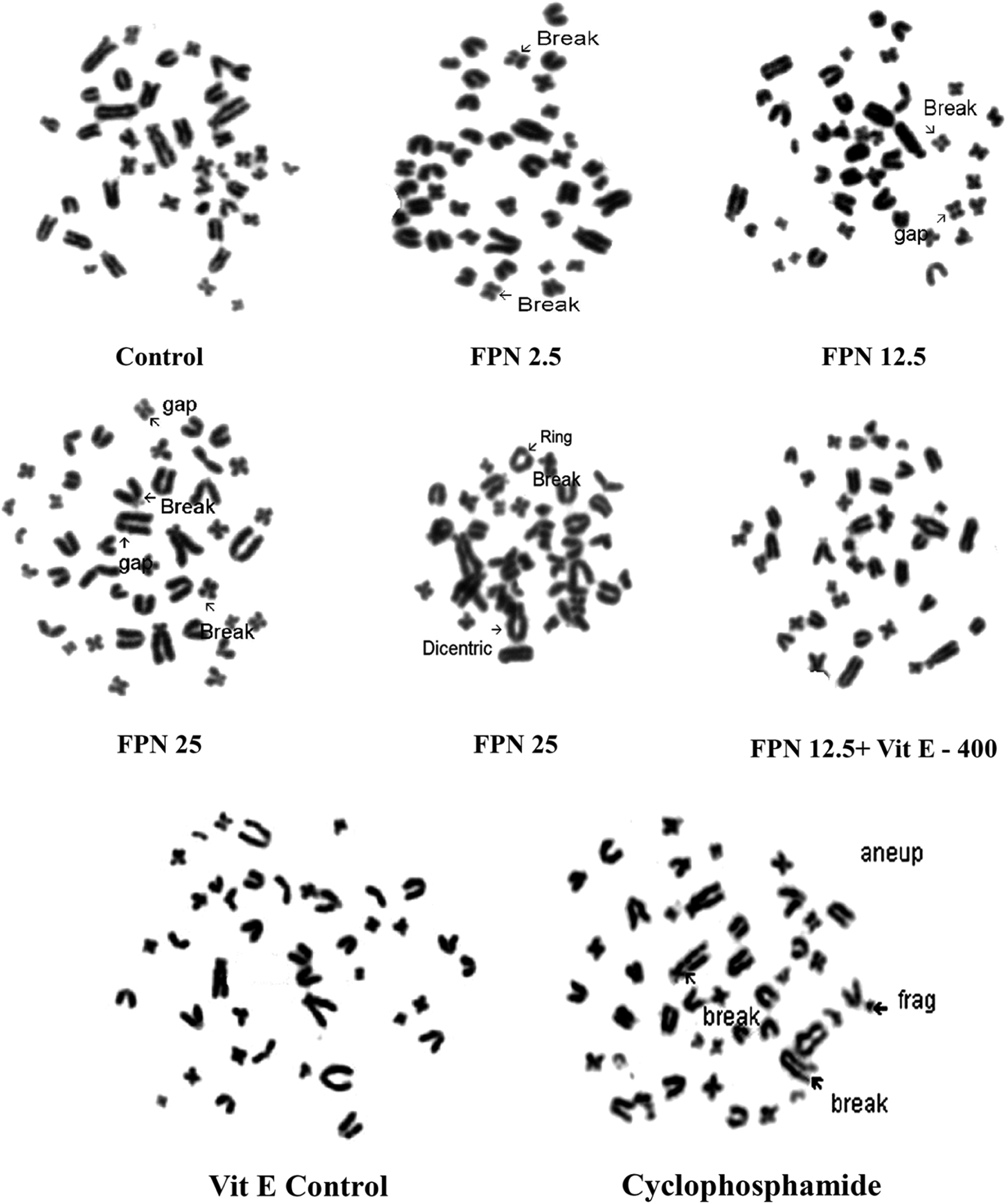
Representative images of structural CAs in rat bone marrow metaphase cells after oral treatment of fipronil and vitamin E. CA: chromosome aberration.
Mammalian erythrocyte micronucleus test
Results of the micronucleus test are given in the Table 2. Administration of medium and high doses of fipronil (i.e. FPN 12.5 and FPN 25) caused significant increase in the number (MN in polychromatic erythrocyte (MNP) + MN in normochromatic erythrocyte (MNN)) as well as frequency of micronucleated cells in both male and female mice when compared with control. Pretreatment of vitamin E for 5 days in fipronil-exposed mice (FPN 12.5) led to significant reduction (53.33% and 42.50%) in the number and frequency of MN in male and female mice, respectively. Significant increase in the frequency of micronucleated cells was observed in the both male and female mice treated with cyclophosphamide. However, there was no significant change in the PCE/NCE ratio, in both male and female mice at all doses of fipronil except in the cyclophosphamide-treated mice group (Table 2).
Frequency of micronuclei in mouse bone marrow cells after oral exposure to fipronil and vitamin E.a

FPN: fipronil; MNP: micronuclei in polychromatic erythrocyte; MNN: micronuclei in normochromatic erythrocyte; PCE: polychromatic erythrocyte; NCE: normochromatic erythrocyte.
aAverage micronuclei expressed as mean ± SE of n = 5 mice/sex/group; mean values bearing different superscript letters (b, c, d) in the same column differ significantly between groups at p < 0.05 in Tukey’s multiple comparison post hoc test.
Comet assay
A single dose of fipronil exposure over a period of 24 h in adult male and female rats caused an increase in DNA strand breaks leading to DNA migration out of nucleus into the tail of the comets in lymphocytes. Representative images of comet cells in the control and treated groups are shown in the Figure 3. Figure 4 (a, b, c and d) summarizes results of comet assay parameters, that is, DNA damage, TL, TM, and TMO in the male and female rats, respectively. Barring low dose of fipronil, TL of comets was significantly increased (p < 0.05) in lymphocyte cells of male and female rats treated with fipronil as compared to control. However, DNA damage percentage was increased in the blood lymphocytes of male and female rats at all three test doses of fipronil, albeit nonsignificantly at low dose. Prior administration of vitamin E caused significant reduction (64.38% and 64.51%) in the comet TL of male and female rats, respectively. Vitamin E had a significant (p < 0.05) protective effect (77.43% and 71.98%) against fipronil-induced DNA damage in male and female rats, respectively. In both sexes, TM (a product of fraction of DNA in the tail and TL) was significantly increased (p < 0.05) only at high dose of FPN.

Representative images of comets observed in the rat blood cells after oral treatment of fipronil and vitamin E.

(a, b, c, and d) Effect of oral exposure of fipronil and vitamin E on DNA damage, tail length, tail moment, and tail olive moment of comets observed in the blood cells of rats. Data expressed as mean ± SE, n = 5 rats/sex/group (50 cells/comets were scored per rat). Mean values bearing different error bar letters (a, b, c, etc.) differ significantly between groups at p < 0.05. Tail moment: product of fraction of DNA in the tail and tail length. *Percent value in the rectangle show protective effect of vitamin E pretreatment. SE: standard error.
Discussion
Ability of pesticides to induce genotoxic effects in directly and indirectly exposed nontarget organisms (including humans) have been demonstrated by several researchers. 12,22 Owing to its high use, fipronil and its metabolites have been reported to be leaching in the soil and water. 23,24 Le faouder et al. 25 and Liua et al. 26 have even detected fipronil residues in drinking water and in different food products such as maize, Chinese cabbage, and milk, respectively. Fipronil has also been reported to bioaccumulate in fish for about 35 days with bioconcentration factors of 321, 164, and 575 for whole fish, edible tissues, and nonedible tissues, respectively. 5 However, no reliable data is available concerning the level of fipronil exposure in humans. It is also known that fipronil promotes development of thyroid tumors in rodents by modulating cytochrome P450 through disruption of hepatic metabolism and excretion of thyroid hormones. 27 Even in humans, fipronil and its sulfone metabolite induces cytotoxicity in hepatocytes. 4 Considering these effects, a detailed study evaluating genotoxic potential of fipronil was planned so that adverse effects of tumors and hepatotoxicity could be explained better. Besides, we evaluated the protective role of vitamin E on the fipronil-induced genotoxicity. According to many authors who have studied the action of vitamins in vivo, the treatment protocols that yielded best results in terms of reduction of chromosome damage were those in which vitamin C, E, or A was administered as pretreatment or in simultaneous treatment with the clastogenic agents. As per the existing published experimental protocols, 19,20,28 we also used cyclophosphamide, a known clastogenic agent as positive control for evaluating genotoxic potential of fipronil.
In the present study, fipronil induced different types of SCA in the bone marrow cells of male and female rats. Fipronil exposure induced CAs such as dicentric chromosomes. During analysis severely damaged cells, single/double chromosome gaps, and aneuploidy were recorded but not considered for the percent CA determination, 28 since numerical aberrations may arise due to increase or decrease in chromosome number resulting into polyploidy or aneuploidy, respectively. Moreover, at second or third mitosis, these aberrations get mixed and lose identity depending upon the cell stage. Significant SCA observed at high dose show clastogenic potential of fipronil and is the first report documenting CAs induced by fipronil. In an earlier study demonstrating genotoxic potential of fipronil (commercial formulation containing only 80% active fipronil), researchers have conducted only two tests, that is, DNA damage and micronucleus formation. 9 A dose-dependent increase in the frequency of SCA observed in this study is in agreement with earlier reports evincing clastogenic potential of insecticides such as trimethyltin, 29 deltamethrin, 30 allethrin, 31 dimethoate. 32
The findings of MN analysis strongly corroborated results of CAs. These findings are in complete agreement with de Oliveira et al., 9 who reported high frequency of MN in the peripheral blood of Swiss albino mice 24 h after treatment with fipronil, albeit at high doses (25 and 50 mg/kg bw) and attributed fipronil to be mutagenic. We believe that the results of the present study clearly vindicate and supplement carcinogenicity/tumor-inducing potential of fipronil claimed by several researchers. 5,28 Erythropoietic cytotoxicity evaluation is an important component of the safety assessment. PCE counts in the peripheral blood or bone marrow are the most popular and convenient method of monitoring erythropoiesis. Reduction in the proportion of immature PCEs to mature NCEs gives us a view of mutagen-induced cytotoxicity. 33 In the present study, fipronil did not significantly alter PCE/NCE ratio indicating lack of cytotoxicity at the dose levels tested.
The intensity and the length of the comet tail are regarded as an indicator of the amount of DNA breakage. 34 Chemicals that cause damage to lysosomes and membranes of cellular system induce release of lysosomal or other DNAase into the cytoplasm of damaged cells which in turn trigger DNA double- strand breaks. In those cells that survive sub-lethal damage, such double-strand breaks could have a variety of genotoxic effects including mutation and CAs. 35 Several mutagens and carcinogens primarily affect DNA directly or modify other cellular processes associated with the integrity of the genome. These resulting structural alterations of DNA can be easily analyzed by comet assay. In the present study, important comet assay indices, that is, DNA damage and TL were significantly increased in the male as well as female rats treated with medium and high doses of fipronil as compared to control. These findings are similar to that of de Oliveira et al. who demonstrated that mice treated with high dose of fipronil (50 mg/kg) showed higher frequency of damaged nucleoids and classical comets. 9 Our findings are also in parallel with Celik et al., 10 who have shown DNA-damaging ability of fipronil with the help of comet assay in vitro. Recently (September 2014), a remarkable development in the genotoxicity testing of chemicals unfolded with OECD finally specifying a definite guideline (Guideline No. 489) for the in vivo mammalian alkaline comet assay stressing importance of this test.
It is very well known that different toxic compounds induce oxidative stress in organisms and the reduction of oxidative stress involves antioxidants/antioxidant enzymes. 36 All the parameters evaluated in determining genotoxic potential of fipronil were significantly restored by pretreatment of vitamin E. Similar to our results, vitamin E has been shown to exert protective effect against atrazine-induced DNA damage and genotoxicity. 12 Previously, numerous studies have shown the protective effect of vitamin E against genotoxic effects of several chemicals. 13,14,37,38 It has long been reported that vitamin E protects DNA from free radicals attack either by scavenging lipid peroxyl radicals, thereby terminating lipid peroxidation chain reaction that creates DNA-damaging products or by inactivating reactive oxygen species (ROS). 39 The findings of our study strongly evince involvement of similar mechanism and support free radical-mediated genotoxicity hypothesis. Thus, the probable mechanism of genotoxic effects of fipronil appears to be an increase in the free radical generation and subsequent involvement of oxidative stress. Especially, recent studies from our laboratory have shown that fipronil exposure for 28 days induces oxidative stress in the kidney and brain 7 as well as in the liver 8 of mice. Interestingly, pre-administration of vitamins E and C have also shown protective effect against oxidative stress induced by fipronil in these organs. 7,8 Rather than nucleophilic interaction with electrophilic species generated by mutagens, anti-mutagenic effect of vitamin E seen in this study could be attributed more to the inactivation of DNA free radicals appearing in DNA during its active synthesis as postulated by Aly and Donya. 13 Overall, the protective effect of vitamin E in fipronil-exposed animals was highest against the percent DNA damage (>70%) followed by CAs (60% and above), and MN formation (48%). The average degree of protection (male and female animals combined together) offered by pretreatment of vitamin E against fipronil was 63.28%: CAs; 47.91%: MN formation; and 74.70%: percentage DNA damage.
In conclusion, the results of the present study demonstrated that fipronil insecticide has clastogenic and mutagenic effects in male as well as female animals. As this study was conducted in both sexes, it is definitely more conclusive and these findings show that genotoxicity of fipronil is independent of gender effect. The mechanism of this genotoxic effects could be attributed to ROS-mediated oxidative stress as pretreatment with a known antioxidant, that is, vitamin E has shown protective effect. We strongly believe that periodic reassessment of pesticides is necessary owing to ever-changing environmental and host genetic make-up. Therefore, further detailed studies on fipronil are solicited to better characterize these adverse effects.
Footnotes
Acknowledgment
The authors are thankful to the director of Indian Veterinary Research Institute, and the director of National Institute for Research in Reproductive Health for providing necessary funds and facilities, respectively. The authors are grateful to the staff and students of the department of toxicology, National Institute for Research in Reproductive Health, namely, Mr. Pravin Salunke, Mr. Subhash Kadam, Mr. Mali, Mr. Jayant Tare, Ms. Neelam, Mr. Swapnil, Ms. Priyanka, and Ms. Paula Raijiwala for helping in the laboratory and in the animal experiments. A kind gift of technical grade fipronil from M/s. Gharda Chemicals, Mumbai, is gratefully acknowledged.
Declaration of Conflicting Interests
The author(s) declared no potential conflicts of interest with respect to the research, authorship, and/or publication of this article.
Funding
The author(s) received no financial support for the research, authorship, and/or publication of this article.