
Abstract
The use of anabolic androgenic steroids (AAS) has grown among practitioners of recreational bodybuilding, with significant contributions of designer steroids, aiming muscle hypertrophy in healthy subjects. The abusive use of AAS in general is associated with adverse effects; one of the most worrisome is cancer development. The aim of this study was to evaluate the effectiveness of the cytokinesis block micronucleus (CBMN) test in human lymphocytes in identifying risk groups for cancer development in users of AAS. Blood was collected from 15 AAS users bodybuilders (G1), 20 non-users bodybuilders (G2) and 20 non-users sedentary (G3). MN analysis was performed on a minimum of 1000 binucleated lymphocytes. The occurrence of MN was significantly higher (p < 0.05) in individuals of G1 compared to G2 and G3. The results indicate the sensitivity of CBMN in human lymphocytes in the identification of chromosomal damage in consequence of AAS.
Introduction
Anabolic androgenic steroids (AAS) are derived synthetic derivatives of testosterone that are manufactured such that their anabolic effect is maximized and their androgenic effect is minimized. 1 The anabolic activity of testosterone and its synthetic derivatives results in significant muscle mass and strength gains, 2 due to increased nitrogen retention and protein synthesis in skeletal muscle cells and diminished catabolism. 3 –5
AAS were initially developed for therapeutic purposes, but today, the greatest prevalence of AAS use is among athletes who seek improvements in their performance in sports practice and among individuals doing recreational muscle building, who are motivated by constructing a body image that satisfies them. 6,7,8 Epidemiological evidence has indicated that there is high prevalence of illicit use of these drugs in the United States, 9 Australia, 10 United Kingdom, Greece, Bulgaria and Croatia. 6
Designer steroids (DS), which are AAS designed for the purpose of enabling muscle hypertrophy among athletes and muscle-builders, have appeared on the scene to worsen this situation. These are marketed as nutritional supplements through internet stores and they have increased the incidence of AAS use because they are easy to obtain and have a more accessible price than the traditional synthetic derivatives. 11,12,13
The anabolic effect of testosterone and its derivatives is dose dependent, and significant increases in muscle size and strength only occur with doses of 300 mg per week or more, 14 that is, quantities that are much greater than clinical doses. 15 Thus, the high prevalence of use of these drugs constitutes a social and public health problem, since their abusive use is associated with adverse effects. The most frequent of these adverse effects are cardiovascular, 16 hepatic, 17,18 sexual 19 and behavioural and psychiatric. 20,21 Among the effects that have been reported, one of the most worrying effects is the development of cancer. 22 –24
Cancer results from alterations to genes involved in DNA repair mechanisms, apoptosis and cell proliferation and differentiation control. This leads to disorganized growth and loss of cell capacity to evolve to death through apoptosis. 25 Accumulation of these alterations is possible in situations of genome instability that are characteristic of the initial stages of the carcinogenic process. 26 Exposure to genotoxic agents may lead to point mutations and numerical and/or structural chromosomal alterations, which favour evolution to this state of instability. 27
Biomarkers for these changes thus take on importance in identifying groups at risk of developing cancer. Use of such markers as preventive measures may be an effective strategy for diminishing the high morbidity and mortality rates due to cancer that are observed worldwide. Among the cytogenetic tests that make use of biomarkers for genotoxicity, the micronucleus test is widely used for evaluating human exposure to potentially genotoxic agents 28 –30 and for biomonitoring groups at risk of developing cancer. 31 –34
Micronuclei are structures resulting from chromosomal fragments or entire chromosomes that were not included in the nucleus of the daughter cell during cell division. 35 They therefore reflect occurrences of aneugenic and clastogenic damage and, according to Bonassi et al., 32 they are effective markers for the risk of cancer. The micronucleus test on human lymphocytes is one of the most commonly used methods, particularly when performed with blockage of cytokinesis. 35,36 The high reliability and sensitivity of this technique have led to its constant use for genotoxic monitoring of populations, in which it has been revealed to be effective in identifying groups at risk of developing cancer. 31 –35
Several studies in which biomarkers for genotoxicity were used have shown the capacity of AAS to induce changes to genetic material, which may explain the association between cancer and use of these steroids. 29,30,37 –39
In view of the increasing frequency of AAS use and the possible association between use of these drugs and genetic damage and cancer, the present study had the aim of evaluating the use of the micronucleus test with cytokinesis block micronucleus (CBMN) on human lymphocytes as a biomarker for cancer risk among AAS users, so as to seek an effective strategy for prevention of this disease.
Material and methods
Ethical issues
This research project was approved by the Ethics Committee for Research on Human Beings of Feira de Santana State University (UEFS), under protocol no. 098/2011. The study was conducted in observance of the ethical principles of National Health Council Resolution no. 196/1996, such that all the participants received explanations regarding the justifications for and objectives of the study, and about all the procedures that would be used. Among these principles, the subjects were completely free to accept or not to accept participation in the study, and confidentiality of information was guaranteed for all individuals who agreed to participate in the study. A free and informed consent statement was signed by each participant and by the researcher.
Sampling groups
The study included 55 individuals living in the city of Feira de Santana, Bahia, Brazil, distributed into 3 distinct groups: Group I: formed by 15 male individuals who were practicing muscle building and were making use of AAS. Group II: formed by 20 male individuals who were practicing muscle building and were not making use of AAS. Group III: formed by 20 male individuals who were not doing muscle building and were not making use of AAS.
Sample characterization
A questionnaire asking about age, tobacco consumption, alcoholic drink intake and exposure to known genotoxic agents was applied to the entire sample. The AAS users were asked about: (1) the AAS used; (2) dose, frequency and duration of use. The inclusion criteria were being male and aged over 18 years old. The following were considered to be exclusion criteria: (1) tobacco consumption, (2) recent radiographic examination and (3) exposure to other agents known to be genotoxic within the last three months.
Obtaining of material for cytological analysis
Lymphocytes were obtained through collecting 5 ml of peripheral blood from the voluntary participants. This procedure was performed in the Clinical and Parasitological Analysis Laboratory (LAC) of UEFS and was done using a heparinized sterile syringe under appropriate conditions for this.
Cytological preparations: Culturing of lymphocytes from whole blood
The blood samples were transported from the LAC to the Toxicological Genetics Laboratory of UEFS, where they were processes in accordance with the protocol described by Fenech. 36
Micronucleus analysis
The micronucleus analysis was performed on a minimum of 1000 binucleated cells, under an optical microscope with a minimum magnification of 200×. This test was performed blinded in relation to the data from the questionnaire. To identify the micronuclei (Figure 1), the criteria described by Fenech 36 were used. According to this author, the cells were considered as micronuclei structures (a) measuring 1/16th to 1/3rd of the mean diameter of the main nuclei, (b) non-refractile, (c) not linked or connected to the main nuclei, (d) not overlapping the main nuclei, (e) with micronuclear boundary distinguishable from the nuclear boundary and (f) having the same staining intensity as the main nuclei.

Photomicrograph of binucleated lymphocytes with micronuclei.
Statistical analysis
To evaluate the quantitative variable of age, analysis of variance was used. The entire analysis relating to occurrences of micronuclei was performed using the conditional test for comparing proportions in situations of rare events, which is a significance test that forms an alternative to the χ2 test, along the lines of Fisher’s exact test. It is suitable for evaluating cytogenetic events when a large sample of cells is needed in order to detect occurrences of a certain chromosomal aberration. 40 In all the analyses, the significance level used was 0.05.
Results
Age
The mean age ± standard deviation for groups I, II and III was, respectively, 23.5 ± 3.2, 23.9 ± 3.1 and 22.5 ± 2.9. An evaluation on differences observed between the mean ages of the groups did not reveal any statistical significance: F = 1.246; p = 0.292.
Consumption of alcoholic drinks
In groups I, II and III, respectively, 13, 13 and 12 participants were regularly consuming alcoholic drinks. None of the individuals participating stated that they had the habit of consuming alcoholic drinks more than once a week (Table 1).
Data relating to consumption of alcoholic drinks and physical exercise practices.

Physical exercise practices
In group I, 12 participants were doing strength training five times a week and 3 of them were training only three times a week. Ten individuals in this group reported that they were regularly practicing aerobic exercises and muscle building, while the other participants in the same group stated that they were only doing muscle building. Among the individuals in group II, 10 participants were doing strength training five times a week and 10 were training three times a week. In this group, 17 participants said that they were regularly practicing aerobic exercises, while 3 said that they were only doing muscle building. In group III, all of the individuals were sedentary (Table 1).
Anabolic steroids and patterns of use
The AAS methyldrostanolone (which is a DS) was the one with greatest incidence in the group of users (75.33%). In this group, 11 individuals reported that they were consuming this drug. One individual reported using the AAS stanozolol (winstrol®). The AAS nandrolone decanoate (deca-durabolin®) and durateston® (a combination of the AAS testosterone propionate, testosterone phenylpropionate, testosterone isocaproate and testosterone decanoate) were also consumed by one individual each. Only one of the participants said that he was consuming two AAS simultaneously, which were nandrolone decanoate (deca-durabolin) and durateston. Eight users had set up cycles for their AAS use, while seven were making separate unprogrammed use. The cycle duration, total duration of use and doses used by all the AAS users are summarized in Table 2.
Data relating to AAS: duration and sum of the doses of the last cycle (DT).
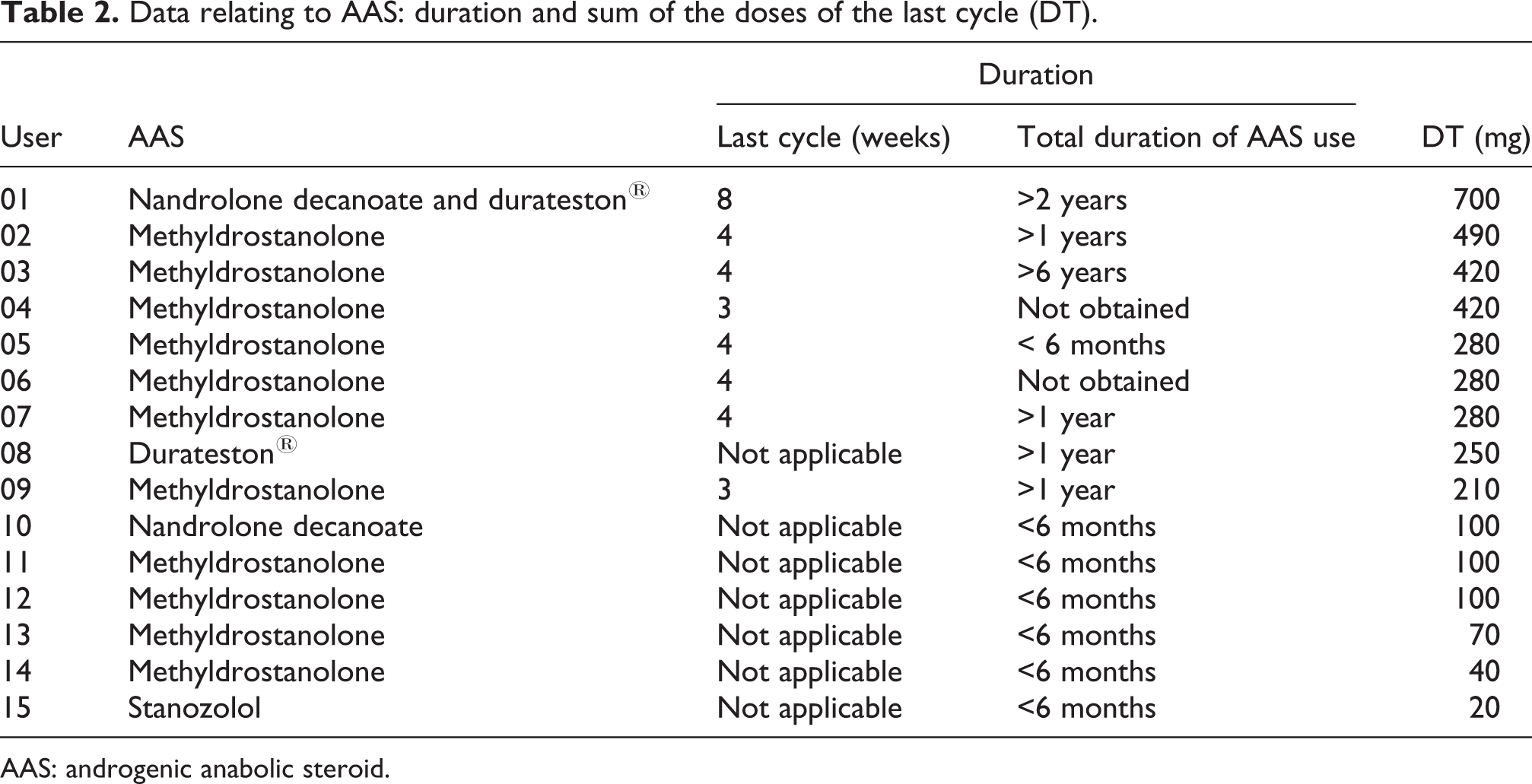
AAS: androgenic anabolic steroid.
The users (group I), except for those who were making separate and unprogrammed use, were taking methyldrostanolone at the doses recommended by the manufacturer, that is, one or two pills per day, with a maximum cycle duration of 4 weeks. Given that each pill contains 10 mg of the DS, the total doses observed among these users resulted from the different combinations possible between cycle duration (3 or 4 weeks) and the number of pills per day (one or two).
Cytogenetic analysis
The evaluation on the differences between groups I, II and III regarding occurrences of micronuclei, performed using the conditional test on proportions in situations of rare events, showed significant differences: χ2 = 6.5238, df = 2, p < 0.05. The χ2 partitions revealed that in the cells obtained from group I, the number of micronuclei was significantly greater than what was observed either in the cells from group II (χ2 = 5.2112, df = 1, p < 0.05) or the cells from group III (χ2 = 4.2231, df = 1, p < 0.05). There was no statistically significant difference in micronucleus occurrences between groups II and III (χ2 = 0.0580; DF = 1; p > 0.5). These data are presented in Table 3.
Data relating to occurrences of micronuclei in the different groups.
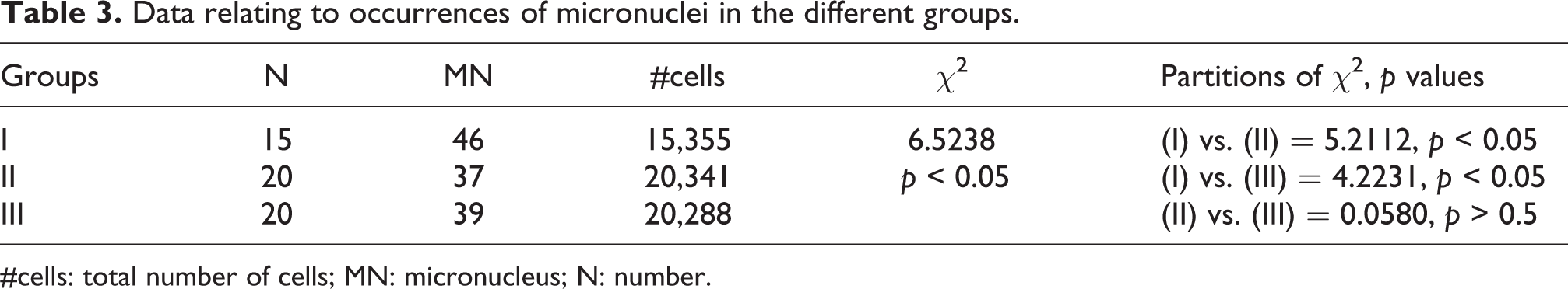
#cells: total number of cells; MN: micronucleus; N: number.
Discussion
A new group of AAS called DS is becoming very popular among young adults because they are easy to obtain and have lower prices. These factors have contributed significantly towards increasing the prevalence of AAS use in the population.12,41
Abusive use of AAS is associated with adverse effects, among which development of cancer is one of the most worrying. 22 –24 Use of primary cancer prevention measures is the most effective strategy for diminishing the high morbidity and mortality rates due to this disease, which are observed worldwide. Among such measures, use of biomarkers that denote the risk has been considered to be a valuable tool. 42 –44 One of the methods most commonly used for biomonitoring in populations exposed to genotoxic agents is CBMN, which has been shown to be effective for identifying groups at risk of developing cancer. 31 –34 However, there are no reports in the literature regarding biomonitoring of groups exposed to AAS, using this methodology. The objective of the present study was to assess the efficacy of CBMN on human lymphocytes as a biomarker for the risk of cancer among AAS users.
The results found revealed that there was greater frequency of chromosome damage, translated as micronuclei, among AAS users than among non-users. This is concordant with other studies in which the genotoxic potential of AAS was investigated. 38,39,45 –47 However, there is no consensus in the literature regarding this association, and other investigations have indicated that these drugs do not present genotoxicity. 48 –50 This divergence is diminished when only the in vivo studies conducted on humans are taken into account: although these are scarce, they mostly show increased levels of markers for genetic damage among AAS users. 29,30
It is possible that the genotoxic potential observed is influenced by the distribution and metabolism of these drugs, which cannot be adequately assessed from in vitro studies. For example, AAS such as trenbolone and nandrolone can be converted by the enzyme aromatase to the estrogen 17 beta-estradiol, in adipose, cerebral and testicular tissues. Point mutations and chromosomal damage, both in cell cultures and in vivo, have been observed in the presence of 17 beta-estradiol and its metabolites, which are also considered to be inducers of cell proliferation. 37,51 Formation of this isomer of estradiol has been one of the mechanisms proposed for explaining the genotoxicity of AAS. 29,30,47
Physical exercise practice has been correlated in some studies with increased genetic damage, and an induction mechanism based on increased production of reactive oxygen species has been proposed. 52,53 With the aim of investigating this variable, a control group composed only of sedentary individuals was included in the present study. The results obtained did not reveal any significant differences in micronucleus frequency between the individuals in group II (who practiced muscle building recreationally and mostly also performed aerobic activities) and the individuals in group III (who consisted only of sedentary individuals). These results have been corroborated by other studies. 54,55
Among the individuals in group I, the number of users of the DS methyldrostanolone, also known as methasterone, was much greater (75.3%) than the number of users of traditional AAS that have been approved as legal drugs. This large difference is due to the current popularity of DS among recreational muscle-builders, who often are unaware of the true nature of the drug. It is also likely that these users have less difficulty in stating their consumption than individuals who make use of traditional AAS. It is possible that the easy acquisition and administration of DS, together with misinformation, has contributed towards increased use of this group of AAS. In view of the high incidence of methyldrostanolone users in this study, this DS evidently made an important contribution towards the significantly higher genotoxicity observed in group I in comparison with the other groups.
Methyldrostanolone is 17 α-alkylated and, for this reason, presents a methyl group in position C-17, which makes oral administration possible. It is of interest to note that in the literature, there is a greater association between 17 α-alkylated AAS and genetic damage and cancer development than between non-alkylated AAS and these conditions. In a review of the literature on the genotoxicity of steroid hormones, Joosten et al. 37 found studies that had investigated the genotoxic potential both of 17 α-alkylated AAS (fluoxymesterone, methyltestosterone, oxymetholone and stanozolol) and of non-alkylated AAS (trenbolone and testosterone). Among all the investigations reviewed, only 17 α-alkylated AAS was capable of inducing genetic alterations, which were translated as exchanges between sister chromatids and micronuclei, both in human lymphocytes in vitro and in rodents’ bone marrow in vivo.
Analysis on the case studies in which cancer development resulting from AAS abuse has been reported reveals that, in all the studies in which the anabolic steroid administered was informed, cancer development was associated with administration of a 17 α-alkylated AAS, either separately or in association with other anabolic steroids. 22,23,24,56,57 As an example, Kosaka et al. 22 conducted a review of the literature on the development of hepatocellular carcinomas in the Japanese population resulting from prolonged therapeutic use of AAS and found that 11 cases in which it was possible to identify the ASS administered, 17 α-alkylated AAS was used in 10 of them. This use was in conjunction with non-alkylated AAS in only two of these cases.
The mechanisms involved in this promotion of genetic damage and cancer by 17 α-alkylated AAS are poorly understood. Welder et al. 58 evaluated the toxicity of 17 α-alkylated AAS and non-alkylated steroid hormones in the hepatocytes of rats. Direct hepatotoxicity was observed only in relation to the 17 α-alkylated AAS stanozolol, methyltestosterone and oxymetholone. For the AAS oxymetholone and methyltestosterone, the hepatotoxicity was also associated with diminished levels of glutathione, a tripeptide that performs a series of protective functions in cells, including against oxidative damage through formation of conjugates with free radicals. Free radicals are unstable and highly reactive, since they easily lose hydrogen atoms and form covalent bonds with cell constituents. These radicals may bind to two different DNA molecules, thus impeding their correct separation during cell division, or may bind to bases in the same DNA molecule. 29 It is possible that decreased glutathione, and consequently increased oxidative stress, is associated with more pronounced genotoxicity among 17 α-alkylated AAS, although no studies have directly investigated this relationship.
The genotoxic potential of DS has only been assessed in two studies. In these, the genotoxicity of the DS madol 39 and tetrahydrogestrinone 46 was evaluated by using the micronucleus test on V79 cell lineages. The results showed that there was a dose-dependent increase in the frequency of micronuclei in both studies, and there was a significant genotoxic response even at low concentrations of these DS.
Toxicological studies that are specific for methyldrostanolone are scarce in the literature because this drug has only been on the market for a few years: it was first identified in 2005. 12,41 Studies on the methyldrostanolone most frequently found have made reference to cases in which hepatic lesions developed and particularly cholestatic hepatic lesions. 18,59,60 These lesions occurred not only in users of this DS but also in users of 17 α-alkylated AAS in a general manner and were considered uncommon. 61 In the above-mentioned studies, these lesions developed over a short period of methyldrostanolone use, ranging from 4 to 6 weeks, which indicates the elevated hepatoxicity of this AAS, given the short exposure period and the severity of the lesion, which may lead to death. 18
Despite the limited information in the literature regarding DS, the possibility exists that much of what is known about the genotoxicity of traditional AAS that have been approved as legal drugs can be extended to DS. The similarities between their chemical structures and between their adverse effects suggest that, in many cases, the differences between these two groups of AAS are perhaps more a question of origin than of nature. Abusive use of DS can also be considered to be a reason for greater concern, given that some of them have been identified as AAS that have already been patented but which were not put on the market due to their toxicity, 62,63 or which have never been put through any toxicological evaluation by regulatory agency. 13,64 Worsening this situation, it was found in the present study that in muscle-building gyms, there was greater frequency of DS use than of other AAS, which was reflected in the final sample of users. This observation brings to light concern regarding the real prevalence of use of these drugs, which when added to the traditional AAS may mean that the prevalence rates usually reported in epidemiological studies on AAS are greatly exceeded.
The results obtained indicate the potential of AAS to induce genetic damage, particularly with regard to the DS methyldrostanolone. This was the DS most used among individuals in group 1 of this study, and it has a structure similar to that of the AAS drostanolone. Its 17 α-alkylated structure, together with the results from studies on its toxicity (including the case studies cited and the genotoxicity observed in the present study) and its great popularity, makes this an important substrate for new investigations.
The CBMN test was shown to be sensitive for detecting chromosomal damage in AAS users. However, despite the results described here, for it to be used in biomonitoring on groups at risk of developing cancer, among AAS users, studies with sample sizes of greater significance and using different AAS are needed, in order to measure the real extent of its possible application.
Footnotes
Declaration of Conflicting Interests
The author(s) declared no potential conflicts of interest with respect to the research, authorship, and/or publication of this article.
Funding
The author(s) disclosed receipt of the following financial support for the research, authorship, and/or publication of this article: This study was supported by the Feira de Santana State University (UEFS) and Foundation Support Research of the State of Bahia (FAPESB).