
Abstract
Perfluorooctanesulfonic acid (PFOS), a ubiquitous contaminant, has been used in various industrial applications. Currently few studies have documented the effects of chronic PFOS exposure on lipid metabolism, especially in aquatic organisms. The present study defined the effects of chronic exposure to low level of PFOS on lipid metabolism in F0 adult zebrafish and F1 offspring. Our findings revealed a severe fatty degeneration in the liver of F0 males treated with 0.5 μM PFOS and significant ultrastructure changes associated with substance transport or metabolism in liver and intestines (abnormal mitochondria and endoplasmic reticulum, disordered arrangement of inner microvilli within intracellular canaliculus). To address the potential trans-generational effects of PFOS exposure, the early gene expression related to lipid metabolism was measured by real-time quantitative polymerase chain reaction in F1 derived from chronically exposed parental fish. The results indicated that lepa (leptin α), kiss1 (kisspeptins), xdh (xanthine dehydrogenases), and insr (insulin receptor) were significantly upregulated in F1 while dgat1b (diacylglycerol O-acyltransferase), hb9 (motor neuron/pancreas homeobox), and Apoa1 (apolipoprotein A-I) were downregulated. These findings provided evidence that PFOS chronic exposure adversely impacts lipid metabolism in both F0 and F1 and demonstrated the validity of using zebrafish as an alternative model for PFOS chronic toxicity screening.
Introduction
Perfluorooctanesulfonic acid (PFOS) is a fully fluorinated organic compound, which has been widely used as components of fire retardants, lubricants, adhesives, paper coatings, and insecticides, and dispersed globally in various environmental media. 1,2 PFOS also results from metabolic breakdown or environmental degradation of perfluorinated compounds (PFCs) and has been one of the most prevalent PFCs detected in wildlife and humans. 3 Due to strong C–F bonds, PFOS is extremely stable to metabolic and environmental degradation and is resistant to biotransformation. Because PFOS is persistent in environment with properties of high bioaccumulation and negligible elimination, high concentrations of PFOS are frequently detected in a variety of wildlife and aquatic species. 4 For example, PFOS levels ranged from 9 to 315 ng/g (wet weight) were reported in the liver of bass from New York State (USA), and the average levels of PFOS in the fish are 8850 times higher than those found in surface water. 5 Likewise, high levels of PFOS have been detected in the liver (7760 ng/g) of plaice 6 and feral gibel carp (up to 9031 ng/g) in Belgium. 7
Weight of evidence has shown that PFOS exposure induces developmental and reproductive toxicity, immunotoxicity, endocrine disruption, and neurotoxicity in mammalian and aquatic fish models. 8,9 However, few studies have defined the effects of chronic PFOS exposure on lipid metabolism in animals, especially in aquatic organisms. Limited evidence from rat and monkey studies demonstrated pathological lesions of the liver and a decreased serum cholesterol level after subchronic or chronic PFOS exposure. 10,11 Mouse studies also showed effects on the expression of genes related to lipid metabolism, cholesterol transport, and biosynthesis following PFOS exposure. 12,13 However, very little is known about the chronic effect of PFOS on lipid metabolism in aquatic organisms. It was reported that 14 day (d) PFOS exposure (0.1–1 mg/L) significantly influenced the expression of several genes involved in energy or lipid metabolism in the liver of carp. 14 Lipid droplet accumulation was also observed in the liver of male zebrafish following 40 d exposure to PFOS (250 µg/L). 15 These studies with aquatic animals were conducted with either higher PFOS levels or used a short exposure period, and potential trans-generational effect on lipid metabolism following chronic exposure has not been investigated.
Zebrafish as an alternative in vivo model has received increased popularity in the fields of developmental biology, genetics, toxicology, reproductive studies, neurobiology, and environmental sciences. 16 Due to its high fecundity, rapid embryogenesis, short life cycle, and small size, zebrafish are easier and less expensive to maintain than mammalian models. Those traits also make zebrafish well suited for long-term exposure study in terms of labor and costs. In addition, numerous studies have documented homologies between zebrafish and human lipid metabolism. 17 Zebrafish possess many of the same gastrointestinal organs present in humans (e.g. the liver, intestine, exocrine and endocrine pancreas, and gallbladder) as well as the specialized cell types involved in lipid absorption and processing (e.g. enterocytes, hepatocytes, and acinar cells). 18 The developmental process of digestive organs and cell types are conserved between zebrafish and mammals. 19 Zebrafish express many of the genes needed to transport and metabolize lipid. 17
Despite the widespread use of PFOS in environmental media, the effects of chronic PFOS exposure on lipid metabolism have not been studied in aquatic organisms. In this study, we continuously exposed zebrafish to environmentally relevant levels of PFOS from embryonic stage to adulthood. The aim of this study is to investigate whether chronic exposure to low level of PFOS alters lipid metabolism and to explore the feasibility of using zebrafish as an alternative model for chronic toxicity screening of hazardous chemicals.
Materials and methods
Fish husbandry and embryo collection
Wild-type zebrafish (Danio rerio, AB strain) were raised under standard laboratory condition of 28°C (water temperature) with a 14:10 h dark/light photoperiod in a recirculating system according to standard zebrafish breeding protocol. 20 Water supplied to the system was filtered by reverse osmosis (pH 7.0–7.5), and Instant Ocean® salt was added to maintain the conductivity to 450–1000 μS/cm. The adult fish were fed twice daily with live artemia (Jiahong Feed Co., Tianjin, China) and dry flake diet (Zeigler, Aquatic Habitats). Zebrafish embryos were obtained from adults in tanks with a sex ratio of 1:1, and spawning was induced in the morning when the light was turned on. Embryos were collected within 1 h after spawning and rinsed in embryo medium. The fertilized embryos were staged using a stereomicroscope according to the standard method. 21 The use of zebrafish for research protocol was approved by the Institutional Animal Care and Use Committee at the Wenzhou Medical University.
Chemicals and exposure protocols
PFOS (CAS#1763-23-1, purity > 96%) was purchased from Sigma-Aldrich Chemicals (St Louis, Missouri, USA). The stock solution was prepared by dissolving PFOS in dimethylsulfoxide (DMSO). High-quality embryos at 8 h post fertilization (hpf) were divided into four groups: vehicle control (0.01% DMSO, v/v), PFOS at 0.02, 0.1, and 0.5 µM. In this study, we used static system for PFOS exposure and the dose range was chosen based on the literature 15,22 and our previous research. 2,23 The exposure paradigm used in this study would generate an internal PFOS concentration ranging from ∼7.7 to 11.1 µg/g (wet weight) in whole-body tissues of adult zebrafish in the 0.5 µM treatment group as we showed previously, 23 which are comparable to those commonly detected from various environmental fish samples (liver of plaice 7.76 µg/g; feral gibel carp 9.03 µg/g). 6,7 Briefly, embryos were first exposed to PFOS in a petri dish (100/group) for 5 d without media change, and all embryos hatched and survived in this stage. After 5 d, fish were transferred into 2 L tanks until 30 dpf. After 30 dpf, fish were raised in 10 L tanks (30/tank) until the end of experiment. Throughout the whole exposure period, 50% water was renewed with freshly prepared solutions every 5 d. Each tank was checked for fish morbidity on a daily basis and water quality was monitored on a weekly basis. Feeding was initiated at 5 dpf. Between 5 and 14 dpf, fish were fed three times daily with standard larval diet (protein 50%, fat 12%, and carbohydrate 2.5%) from Zeigler Bros (Gardners, Pennsylvania, USA), and after 14 dpf, they were fed twice daily with freshly hatched live Artemia. Equal amounts of feed were given among different groups each day. All experiments performed in this study were repeated three times with embryos derived from three different parental stocks.
Evaluation of F0 adult fish and F1 offspring
At the end of chronic exposure (180 dpf), fish from each group (n > 30) were checked for their sex, body weight, and length (measured from snout to the fork point of caudal fin). Condition factor [K = weight (g) × 100/length (cm)3] was tabulated to determine overall fitness. Breeding trials were also carried out to produce F1 offspring (F0 females were paired with F0 males from the same treatment group). Malformation and survival rate of both generations were evaluated.
Measurement of serum TC and TG in F0 generation
After 180 d exposure, 20 adult fish from each group (n = 10 per sex) were fasted for 24 h prior to blood collection. Following anesthetization in 0.01% MS-222 (tricaine methanesulfonate; Sigma-Aldrich) for 2 min, fish were dried with filter paper and the tails were cut immediately for blood collection. Serum from each fish was separated by centrifugation for 10 min at 1000g. The level of total cholesterol (TC) and triglyceride (TG) were measured immediately according to the manufacturer protocols (Applygen Technologies Inc., Beijing, China).
Histological analyses of the liver and intestine
After 180 d exposure, five fish per sex from each group were euthanized. Liver and intestine were surgically removed and immediately fixed with 4% paraformaldehyde or 2.5% glutaraldehyde for 48 h at 4°C, and then processed for hematoxylin and eosin (H&E) staining or transmission electron microscopic (TEM) examination as described previously. 24
qRT-PCR in F1 offspring
Embryos derived from F0, which have been exposed to 0.5 μM PFOS for 180 d, were allowed to develop in the absence of PFOS. At 96 hpf, three pooled samples from control and PFOS treatment group were collected (40 embryos per sample); 96 hpf was chosen based on previous studies from our group 24 and others. 22 This would allow us to compare relevant results at the same developmental stage across different studies. Total RNA from each sample was isolated using Trizol (Invitrogen, Waltham, Massachusetts, USA) and the purity of RNA was determined by 260/280 ratio using Nanodrop (Thermo Scientific, Wilmington, Delaware, USA). A sample of 0.5 μg of RNA was converted to cDNA using PrimeScript® RT reagent kit (Takara, Dalian, China) in a 10 μL volume according to the manufacturer instructions. A total of 25 ng cDNA was used for each Quantitative real-time polymerase chain reaction (qRT-PCR) using the SYBR Green PCR Master Mix (Takara, Dalian, China). The thermal cycle was as follows: 30 s at 95°C, followed by 40 cycles of 95°C for 5 s, and then 60°C for 30 s. qRT-PCR was conducted on the Mastercycler Real-plex2 Thermocycler System (Eppendorf, Westbury, New York, USA). The housekeeping gene β-actin was used as the internal reference as described 15,24 and the comparative CT (ΔΔCT) method was used for quantitative gene expression analysis. Gradient annealing temperature studies were initially completed to determine the optimal temperature for each primer set. Gel electrophoresis and melt curve analysis with Mastercycler Real-plex2 (Eppendorf) were used to confirm product specificity. All specific oligonucleotide primers (Table 1) were designed based on cDNA sequence of zebrafish and synthesized by Sunny Biotechnology (Shanghai, China).
Gene-specific primers used in quantitative real-time PCR.
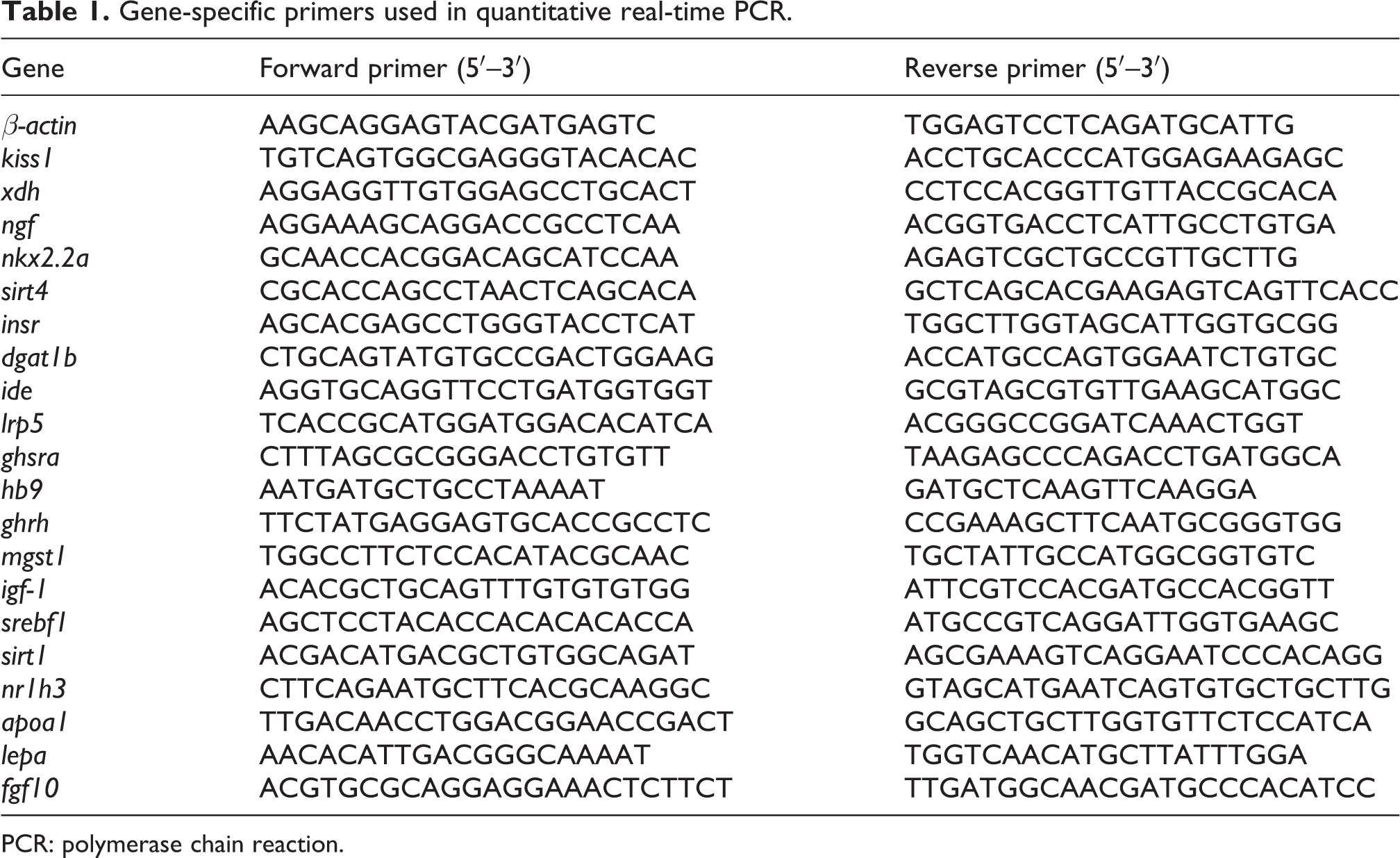
PCR: polymerase chain reaction.
Statistical analysis
The data were reported as mean ± standard error unless otherwise stated. One-way analysis of variance and Tukey’s multiple range tests were used to determine difference between groups. All statistical analysis was performed using SPSS 18.0 (SPSS, Chicago, Illinois, USA) and p < 0.05 was considered to be statistically significant.
Results
Effect of chronic PFOS exposure on the growth of F0 generation
No significant differences in F0 survival rate and malformation were observed among groups during exposure period (data not shown). After 180 d exposure, average body weight or length in the 0.5 µM group were slightly higher than controls but not statistically significant. Condition factor was increased significantly in males exposed to 0.5 µM PFOS (Table 2). Consistent with our previous study, 23 the sex ratio was also altered with a significant female dominance in the F0 0.5 µM group.
Body length, weight, and condition factor of zebrafish following exposure to PFOS for 6 months beginning 8 hpf.

PFOS: perfluorooctanesulfonic acid.
***p < 0.001 when compared with the control group of same sex; n ≥ 30 per sex for each treatment group.
Serum TC and TG levels in F0 after 180 d exposure
PFOS exposure exhibited different effects on serum lipid profiles for males and females. In females, there were no significant changes on fasting TG, but TC level was elevated in 0.5 μM group. In males, both serum TG and TC levels were significantly decreased in 0.5 μM group, and decreased levels of serum TG are also observed in 0.02 and 0.1 μM group (Figure 1).

Chronic PFOS exposure affected serum lipids in F0. Wild-type zebrafish were exposed to PFOS for 6 months starting at 8 hpf. In females, there are no significant changes on serum TG at 180 dpf, but TC level was elevated significantly at 0.5 μM. In males, significant low level of TC was observed in 0.5 μM group, while low levels of TG were noted in all PFOS treatment groups. *p < 0.05; **p < 0.01 compared with controls; n = 10 per sex per group. PFOS: perfluorooctanesulfonic acid; TG: triglyceride; TC: total cholesterol.
Histological examination of liver in F0 adult
A histological study was carried out on the livers of PFOS-treated and control F0 fish at the age of 180 d. In controls, the homogeneous parenchyma of liver are composed of network of units called hepatic lobules. The hepatic cells showed well-preserved cytoplasm and prominent nucleus (Figure 2(a) and (b)). However, liver sections from all 0.5 μM PFOS-treated fish showed hepatic cells with various grade of fatty changes comprising of tiny and large vacuoles (Figure 2(c) and (d)). The vacuolization was mainly due to accumulation of lipid droplets in the hepatocytes, which pushed nucleus to one side of cell (Figure 2(d)). The lipid droplet accumulation was observed significantly in males exposed to 0.5 μM PFOS (Figure 2(d)), but less in females, where a mild accumulation of lipid droplets was observed (Figure 2(c)). In present study, fish exposed to lower levels of PFOS (0.02–0.1 μM) did not show morphological alternations in the livers (data not shown). Macroscopically, we also noticed that the liver of controls appeared soft and sanguine when dissected at 180 d; however, the liver seemed brittle and pale in all males after 0.5 μM PFOS exposure (data not shown).

H&E staining of liver tissue from zebrafish after exposed to 0.5 μM PFOS for 180 d. Representative liver sections from vehicle control females (a) and males (b), exposed females (c) and males (d). Liver of PFOS-treated fish showed hepatic cells with various degree of lipid accumulation (c and d). The fatty degeneration was severe in males, but mild in females, where a less accumulation of lipid droplets was observed. Scale bar: 10 µm; n = 5 fish per sex from each group. H&E: hematoxylin and eosin; PFOS: perfluorooctanesulfonic acid.
TEM was used to explore the ultrastructural changes associated with altered lipid metabolism. In controls, hepatocytes with spherically shaped nuclei were arranged with distinct intercellular space (Figure 3(a)). Abundant mitochondria (Figure 3(c)) and glycogen granules (Figure 3(a)) were evident in cytoplasm. Highly developed bile canaliculus was distributed in cytoplasm with abundant microvilli neatly arranged within canaliculus (Figure 3(b)). However, in the 0.5 μM group, a great number of lipid droplets with various sizes were spread in cytoplasm (Figure 3(d) to (f)). The nuclei were irregularly shaped due to squeezing from lipid droplets accumulation in the cell (Figure 3(f)). Hepatic glycogen storage in cytoplasm was significantly reduced compared with controls. Mitochondria with fuzzy ridges were occasionally observed. The number of inner microvilli within intracellular canaliculus was decreased and the arrangement of microvilli was disordered (Figure 3(e)). Ultrastructural alterations were similar between males and females following 0.5 μM PFOS exposure, but less lipid droplets observed in females (data not shown), which is consistent with light microscope study.

Representative TEM images showing liver ultrastructure from control (a–c) and 0.5 μM PFOS-treated F0 males (d–f). In controls, hepatocytes with spherically shaped nuclei (n) were arranged with distinct intercellular space (*). Abundant mitochondria (mit) and glycogen granules were evident in the cytoplasm. Highly developed bile canaliculus (c) was distributed with abundant microvilli (mic) neatly arranged within canaliculus. However, in all PFOS-treated males, lipid droplets (lp) with various sizes were spread in cytoplasm. The nuclei (n) were irregularly shaped due to squeezing from lipid droplets accumulation. Hepatic glycogen storage in cytoplasm was significantly reduced. Mitochondria with fuzzy ridges were observed. The number of microvilli (mic) within canaliculus (c) decreased along with disordered arrangement. Ultrastructure alterations were similar between males and females following 0.5 μM PFOS exposure, but less lipid droplet observed in females; n = 5 fish per sex from each group. PFOS: perfluorooctanesulfonic acid; TEM: transmission electron microscopic.
Histological examination of small intestine in F0
Consistent with previous findings, 18 columnar-shaped absorptive enterocytes are the most numerous in F0 zebrafish intestinal epithelium. Goblet cells, as second most populous epithelial cell type, are interspersed among absorptive cells. Brush border are neatly arranged on the lumen side of columnar-shaped epithelial cells (Figure 4(a) and (b)). However, compared with controls, the number of goblet cells was higher in all males following 0.5 μM PFOS exposure for 180 d, while the number of columnar epithelial cells decreased (Figure 4(d)). There were no significant morphological changes in female intestine following PFOS exposure (Figure 4(c)).

H&E staining of small intestine from F0 after exposed to PFOS for 180 days. Representative intestine sections from control female (a) and male (b), female exposed to 0.5 μM PFOS (c) and males exposed to 0.5 μM PFOS (d). Compared with the control males, the number of goblet cells was higher in PFOS-treated males, while the number of columnar cells decreased. There were no significant morphological changes in female following PFOS exposure. Scale bar: 25 µm; n = 5 fish per sex from each group. PFOS: perfluorooctanesulfonic acid; H&E: hematoxylin and eosin.
At the level of TEM, abundant mitochondria with clear cristae were distributed in the cytoplasm of controls. The microvilli of brush borders were neatly arranged on the lumen side of epithelia cells surface. Tight junctions between cells were frequently observed (Figure 5(a) and (b)). However, upon 0.5 µM PFOS exposure, the structure and arrangement of epithelium microvilli were altered. In females, shrunk or swelling microvilli appeared alternately on the brush border (Figure 5(c)). In males, some of the epithelium microvilli disconnected and fell off to intestinal lumen (Figure 5(d)). Tight junction between cells was obscure, and cytoplasmic matrix was distributed unevenly.
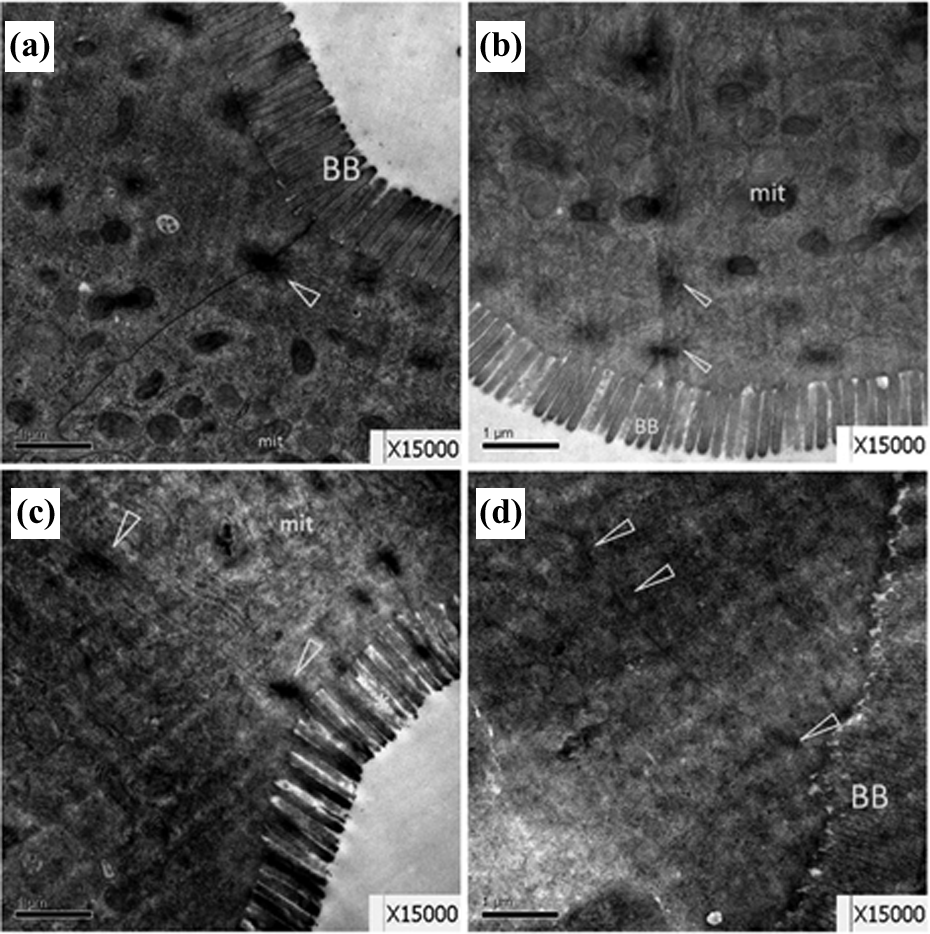
Representative TEM images showing small intestine ultrastructure from control females (a) and males (b), and 0.5 μM PFOS-exposed females (c) and males (d). In all females treated with PFOS (c), shrunk or swelling microvilli appeared alternately on the brush border (BB). In all males treated with PFOS, epithelium microvilli disconnected and fell off to intestinal lumen (d). Tight junction (▵) between cells was obscure, cytoplasmic matrix were distributed unevenly; n = 5 fish per sex from each group. TEM: transmission electron microscopic; PFOS: perfluorooctanesulfonic acid.
Effect of chronic PFOS exposure on the expression of genes related to lipid metabolism in F1 offprings
Similar to our previous study, 23 no significant difference in hatching rate at 72 hpf was observed among groups. F1 offspring derived from parental fish chronically exposed to 0.5 µM PFOS has severe deformity and low survival rate when compared with controls. Uninflated swim bladder and bent spine were the predominant malformation observed in this group. Other deformations included pericardial edema, yolk sac edema, and necrosis as we described previously 23 (see supplemental Figure 1). Without intensive and careful husbandry, fewer offspring from 0.5 µM group can reach maturity and survive to adulthood. To further investigate whether parental exposure causes potential trans-generational problems in lipid metabolism, morphologically normal F1 offspring derived from 0.5 µM PFOS-treated fish was collected at 96 hpf without further exposure. To avoid technical challenges in blood lipid and histological analysis from 96 hpf embryos, the early transcriptional changes in genes associated with lipid metabolism were measured by qRT-PCR (Figure 6). The results demonstrated that genes kiss1 (kisspeptins), xdh (xanthine dehydrogenases), nr1h3 (LXR-α or LXR-β), insr (insulin receptor α), and lepa (leptin α) were significantly upregulated compared with controls. Genes dgat1b (diacylglycerol O-acyltransferase 1), hb9 (motor neuron and pancreas homeobox 1), and apoa1 (Apoa1) were downregulated in F1. We did not see significant changes in the expression of genes including mkx2.2a (NK2 homeobox 2), sirt4 (sirtuin 4), ide (insulin-degrading enzyme), lrp5 (LDL receptor-related protein 5), ghsra (growth hormone secretagogue receptor), ghrh (growth hormone releasing hormones), mgst1 (microsomal glutathione S-transferase 1), prl (prolactin), igf1 (insulin-like growth factor 1), srebf1(sterol regulatory element binding transcription factor 1), and sirt1 (sirtuin 1) (data not shown).

The expression of genes related to lipid, glucose metabolism, and oxidative stress in F1 offspring (96 hpf) derived from chronic PFOS-exposed F0 fish. Lepa, kiss1, xdh, nr1h3, and insr were significantly upregulated compared with the vehicle controls. dgat1b, hb9, and Apoa1 were significantly downregulated in F1 (n = 3 pooled samples, 40 embryos per sample). PFOS: perfluorooctanesulfonic acid.
Discussion
The present study showed that chronic PFOS exposure caused severe liver fatty degeneration in F0 males but less severe in females. TEM examination confirmed the light microscopic findings and demonstrated the ultrastructure changes associated with substance transport and metabolism. Consistent with morphological changes in liver, similar alterations in intestine were observed. We also noticed that the expression of genes related to lipid metabolism was markedly altered in F1 larvae, indicating potential trans-generational effects.
Condition factor, as an indicator of the general “well-being” of fish, was commonly used to measure food intake and the plumpness of aquatic organisms under various conditions. 23,25 We found that the condition factor was increased significantly in males exposed to 0.5 μM PFOS compared with the controls (Table 2). This was in agreement with findings from earlier studies in which condition factor was increased in males and unchanged in females.5,25
Mammalian studies reported pathological lesions of liver in rats and monkeys after subchronic or chronic PFOS exposure. 10,11,26 Liver lesions included centrilobular hypertrophy and vacuolization and were similar in rats and monkeys. Besides pathological findings in livers, a decrease in serum TC and high-density lipoprotein (HDL) cholesterol was observed in monkeys treated with 0.75 mg/kg/d. 10 Male rats also had decreased serum TC at 14 weeks at a dose of 1.33–1.56 mg/kg/d. 11,26 In addition, several mouse studies showed the change of genes expression associated with lipid metabolism, cholesterol transport and biosynthesis process, and lipoprotein metabolism following PFOS exposure. 12,13,27 Consistent with these studies, liver sections from 0.5 μM PFOS-treated F0 fish showed various grade of fatty changes, suggesting that zebrafish may be used as an alternative model for PFOS chronic toxicity screening.
Interestingly, we noticed that PFOS chronic exposure caused different effects on the profiles of lipid metabolism between sexes. Although both sexes are affected, in general males showed more severe histological alterations. We speculate that the different extents on the alteration of lipid metabolism between sexes may be due to the sex difference in PFOS metabolism and subsequent regulation on the process of lipid synthesis and β-oxidation. The higher sensitivity displayed by males was likely related to their higher PFOS body burden. In support of this speculation, our study showed a marked difference in the body burden of PFOS between sexes with more PFOS accumulated in male fish after 5 months PFOS exposure especially at the dose of 250 μg/L (equivalent of 0.5 µM). 23 The sex difference of PFOS body burden may result mainly from higher excretion rate of females through spawning rather than higher accumulation rate in males. 23 Similar to our study, lower PFOS body burden were observed in women. Menstrual bleeding, pregnancy, and lactation were thought to be significant routes for PFOS excretion and may contribute to the lower PFOS body burden in women. 28
Evidence from animal studies indicates that increased hepatic lipid content in the absence of a strong peroxisome proliferator-activated receptor α response is a characteristic of PFOS exposure in vivo. 27,29 Lipid accumulation in livers of PFOS-treated animals may be caused by either disrupted lipid metabolism or lipid transport. Our results suggest that both possibilities may account for it as we have observed (1) macroscopic alteration of liver appearance, where the liver seemed brittle and pale in males exposed to 0.5 μM PFOS; (2) abnormal mitochondria (fuzzy ridges), reduced number of microvilli, and the disordered arrangement of microvilli within intracellular canaliculus in the hepatocytes of 0.5 μM group, suggesting a likely disturbed lipid transport due to these ultrastructure changes; and (3) reduced serum lipid level in males exposed to 0.5 μM PFOS, suggesting an impaired lipid transportation out of liver or compromised lipid biosynthetic capacity at the late stage of chronic liver disease. 30,31 The control of lipid content in liver is a dynamic process that depends on the balance among lipid synthesis, β-oxidation, and transport. Due to the limitation of present study (one time-point study design), it will be difficult to reveal the relationship between lipid transport and lipid biosynthesis capacity in this study. A well-designed time-course study with multiple endpoints in future will help address the underlying mechanisms of PFOS-induced lipid metabolism disturbance.
Distinct from previous studies, we further looked into the potential trans-generational effect of PFOS on the lipid metabolism in F1. After 6 months parental exposure (0.5 µM), F1 were collected at 96 hpf and the expression of genes associated with lipid metabolism was measured in larvae by qRT-PCR. The expression of dgat1b, which encodes for diacylglycerol acyltransferase, was decreased. This enzyme functions as a key metabolic enzyme in TG biosynthesis using diacylglycerol and fatty acyl CoA as its substrates. In addition to dgat1b, we observed a significant upregulation of nr1h3 and downregulation of apoa1 gene expression. Nr1h3 encodes liver X receptors which are highly expressed in visceral tissues and play an important role in lipid metabolism. 32 Apoa1 encodes apolipoprotein A-1, which is the major component of HDL in plasma, plays an important role in regulating the cholesterol of peripheral tissues through the reverse cholesterol transport pathway. Although the expression of a set of key genes associated with lipid metabolism was significantly changed in F1 during the early developmental stage, we did not further measure those genes at the protein level to verify qRT-PCR results because F1 derived from parental exposure had low survival rate. Further studies with detailed biochemical endpoints (e.g. TC, TG, and fatty acid level) in F1 would be useful in addressing uncertainty in the trans-generational effects of PFOS.
In this study, we also found that transcriptional changes in genes related to oxidative stress. For example, the expression of xdh (xanthine dehydrogenases/oxidase) was significantly increased in F1. This enzyme complex exists in interconvertible forms. The oxidase form generates reactive oxygen species, which plays an important role in the pathogenesis of hepatic steatosis and diabetes mellitus type I and type II. 33 Consistent with the increased expression of xdh mRNA in zebrafish, evidence from previous studies demonstrated that PFOS-induced oxidative stress in mice, rat, and zebrafish. 22,34,35 We speculate that PFOS exposure might trigger hepatocyte injury and cause lipid accumulation via xdh-mediated oxidative stress. However, due to the limitation on the initial scope of this study, we did not collect fish samples to measure xdh activity directly at the protein level. Future studies with detailed biochemical endpoints are greatly needed to test this hypothesis and elucidate specific mechanisms.
Besides genes directly related to lipid metabolism, we demonstrated a significant upregulation of lepa (leptin) and kiss1 expression in F1. The kiss1 encodes kisspeptins, which have been detected in intestine, adipose, placenta, pancreas, testes, and central nervous system. 36 Leptin receptor and kiss1 mRNA was also detected in rat islets and pancreatic β-cell line. 37,38 The inhibitory effect of leptin on insulin secretion of pancreatic β-cells has been established. 39 A recent study provided strong evidence that leptin is able to modulate kiss1 mRNA expression in the Langerhans islets and cultured CRI-D2 cell lines. 40 In addition to the changes in the expression of lepa and kiss1, we observed significant alterations in the expression of insr (insulin receptor) and hb9 (motor neuron and pancreas homeobox 1) following PFOS exposure. Hb9 (Mnx1) is required for both initial morphogenesis of pancreas and subsequent differentiation of insulin-producing islet, which is key to pancreas development and function. 41 Dysfunction of insulin receptor and components of the downstream signaling cascade would cause insulin resistance that eventually leads to disrupted glucose metabolism. Taken together, these results indicate that PFOS chronic exposure may affect pancreatic development and function to control insulin synthesis and secretion, and therefore modulate glucose metabolism as well. In the present study, although the expression of a set of key genes associated with lipid or glucose metabolism was significantly changed in F1 during the early stage, the inference is hard to make on the logic and temporal relationship between these genes since we only observed the transcriptional changes at one time-point (96 hpf). These interesting results would build foundation and provide insights for a future detailed mechanistic study.
In summary, we utilize environmental relevant exposure paradigm and demonstrate that PFOS chronic exposure alters lipid metabolism in both F0 and F1 generations. These results provide strong evidence on the validity of using zebrafish as an alternative model for chronic toxicity screening. However, little is known on the process and mechanisms by which PFOS disrupts the complex signaling network associated with lipid metabolism in aquatic organisms, and future research is greatly needed in this area.
Footnotes
Acknowledgement
We thank Zhouxi Fang for technical help with TEM.
Declaration of Conflicting Interests
The author(s) declared no potential conflicts of interest with respect to the research, authorship, and/or publication of this article.
Funding
The author(s) disclosed receipt of the following financial support for the research, authorship, and/or publication of this article: This work was supported by funding from the National Natural Science Foundation of China (no. 21277104) and the Key Project of Zhejiang Provincial Natural Science Foundation (LZ13B070001).
References
Supplementary Material
Please find the following supplemental material available below.
For Open Access articles published under a Creative Commons License, all supplemental material carries the same license as the article it is associated with.
For non-Open Access articles published, all supplemental material carries a non-exclusive license, and permission requests for re-use of supplemental material or any part of supplemental material shall be sent directly to the copyright owner as specified in the copyright notice associated with the article.