
Abstract
Hepatorenal toxicities are an important side effect of anthracycline antibiotics. The objective of this study was to determine whether sesamin (Ses) protects against acute doxorubicin (DOX)-induced hepatorenal toxicities. Rats received daily treatment with either 0.5% carboxymethylcellulose (10 mL/kg) or Ses (10, 20 and 40 mg/kg) orally for 10 days, followed by an intravenous injection at day 8 of either saline (10 mL/kg) or DOX (20 mg/kg). Hepatorenal toxicity was assessed by measuring the levels of serum creatinine (Cre), blood urea nitrogen (BUN), aspartate aminotransferase (AST), alanine aminotransferase (ALT) and alkaline phosphatase (ALP). The protein expression of 4-hydroxynonenal (4-HNE) in hepatorenal tissues was evaluated using immunohistochemistry. The malondialdehyde (MDA) content and antioxidant activity in the kidney and liver tissues were also measured. The results suggest that pretreatment with Ses ameliorated DOX-induced liver and kidney injury by lowering the serum ALT, AST, ALP, Cre and BUN levels (p < 0.05 or p < 0.01), and the histological damage to the liver and kidney tissues induced by DOX compared to control were also significantly attenuated by Ses. Furthermore, Ses significantly decreased the DOX-induced increase of MDA and 4-HNE and increased the activity of CAT, SOD and GPX compared to the DOX-treated rats (p < 0.05 or p < 0.01), whereas the change of DOX + Ses (10 mg/kg) group is not significant compared to the DOX-treated group (p > 0.05). These findings indicate that Ses elicits a typical protective effect against DOX-induced acute hepatorenal toxicity via the suppression of oxidative stress.
Introduction
Doxorubicin (DOX) is a potent cancer chemotherapeutic agent with efficacy against a broad range of malignancies. 1 However, its clinical use is severely restricted by dose-dependent toxicity in various tissues, including the heart, liver and kidneys. 2 Experimental studies in animals showed that DOX causes renal toxicity and produces progressive glomerular injuries. 3 –5 DOX-induced hepatotoxicity was also reported. 6,7 These adverse effects of DOX restrict its clinical use.
The exact mechanism of DOX-induced toxicity remains unclear. Several researchers proposed that DOX-induced toxicity is most likely mediated by the formation of an iron–anthracycline complex that generates reactive oxygen species (ROS), which in turn initiate free radical-mediated chain reactions resulting in the conversion of membrane unsaturated fatty acid into lipid peroxidase. 8 This view is supported by the observation that antioxidants prevent DOX-induced toxicity in both experimental animals and humans. 9,10
Sesame seeds (Sesamum indicum L.) are widely used as dietary supplements, and their oil has been used in human diets for thousands of years and is believed to provide health benefits. Sesamin (Ses), one of the major lignans in sesame seeds, has received a great deal of interest. Several studies showed that Ses has antioxidative, anti-inflammatory, anti-apoptosis, antihypertensive and anti-carcinogenic properties. 11 –15 Recent studies showed that Ses exerts beneficial effects on heart, liver and kidney injuries induced by a variety of pathological conditions. 14,16,17 Reports from our laboratory demonstrated that Ses ameliorates DOX-induced cardiotoxicity. 18 We hypothesize that Ses inhibits the excessive generation of oxidative stress, thus providing an overall protective role against liver and kidney toxicities induced by DOX administration in rats.
Materials and methods
Chemicals
Ses was purchased from Inner Mongolia Kailu Irrigation Pharmaceutical Co., Ltd. (Inner Mongolia, China). DOX was purchased from Sigma Chemical Co. (St Louis, Missouri, USA). Superoxide dismutase (SOD), malondialdehyde (MDA), catalase (CAT) and glutathione peroxidase (GPX) assay kits were obtained from Jiancheng Bioengineering Institute (Nanjing, China). Alanine aminotransferase (ALT), aspartate aminotransferase (AST), alkaline phosphatase (ALP), blood urea nitrogen (BUN) and creatinine (Cre) diagnostic kits were obtained from Sysmex Corporation (Japan). An antibody against 4-hydroxy-2-nonenal (4-HNE) was purchased from Abcam Co. (Cambridge, Massachusetts, USA).
Animals and treatments
Adult male Sprague–Dawley rats (250 ± 20 g, n = 50) were provided by the Experimental Animal Center of Hebei Medical University (Shijiazhuang, China). All animals were treated in accordance with the Guide for Care and Use of Laboratory Animals published by the US National Institutes of Health. The animals were housed under standard laboratory conditions (12-h light:12-h dark and 24 ± 3°C). Food and water were provided ad libitum. The rats were randomly divided into five groups as follows: control, DOX, and DOX + Ses (10, 20 and 40 mg/kg). The control group received carboxymethylcellulose (CMC; 0.5%) orally for 10 consecutive days. The DOX group received CMC orally for 10 consecutive days and a single dose of DOX (20 mg/kg, intraperitoneally (i.p.)) on day 8. Ses was administered intragastrically once a day for 10 consecutive days, and a single dose of DOX (20 mg/kg, i.p.) was administered on day 8. The dosing volume was 1 mL/100 g body weight. The mortality, general condition and body weight of the animals were observed during the entire experiment. Then, on day 10, 30 min after the last Ses was administered, all rats were anaesthetized. The abdomen of each rat was opened, and the blood samples were collected from the abdominal aorta. Next, the liver and kidney were rapidly removed and washed with ice-cold saline, part of which was stored in liquid nitrogen and part of which was fixed in 4% paraformaldehyde.
Assessment of survival and general toxicity
The general condition and mortality of the experimental rats were recorded daily during the entire experimental period. At the end of the experiment, all rats were anaesthetized, and the abdomen of each rat was opened. The fluid that accumulated in the abdominal cavity was collected with a syringe and scored according to a graded scale of 0 to +++ (0: non; +: mild; ++: moderate; and +++: severe). 19
Estimation of biochemical parameters
The blood was centrifuged at 3000g for 15 min to separate the serum and was stored at −80°C for biochemical analyses. The levels of ALT, AST, ALP, BUN and Cre in the serum were determined as sensitive indicators of hepatorenal damage, according to the manufacturer’s protocol for the detection kit, with a CHEMIX-180 automatic biochemistry analyser (Sysmex, Japan).
Histopathological analysis
Some of the liver and kidney tissues obtained from each animal were fixed in 4% paraformaldehyde, dehydrated in ascending grades of alcohol and embedded in paraffin. Four-micrometre thick sections were obtained and stained with haematoxylin and eosin (H&E), and histological examination was conducted using a light microscope (Olympus BX-50 Olympus Corporation, Tokyo, Japan).
Immunohistochemistry
The protein expression of 4-HNE in the liver and kidney were characterized by immunohistochemistry. The tissue sections were dewaxed and then incubated with 1× target retrieval solution in a microwave oven for 15 min at 98°C for antigen retrieval, followed by treatment with 3% hydrogen peroxide for 15 min at room temperature and 5% bovine serum albumin for 30 min. These sections were incubated with a primary antibody against 4-HNE (1:500) overnight at 4°C. The sections were then washed with phosphate-buffered saline and incubated with horseradish peroxidase-conjugated secondary antibodies for 2 h at room temperature. To develop the colour, sections were treated with peroxidase substrate 3,3′-diaminobenzidine and counterstained with haematoxylin. Six sections from each group were evaluated in a blinded manner.
Determination of MDA content and antioxidant enzyme activities
The liver and kidney tissues were rinsed and homogenized in the following buffer (in mM): Tris-hydrochloric acid, 10; sodium chloride, 137; Na2EDTA, 1; dithiothreitol, 0.5; and sucrose, 250, at pH 7.4 using a homogenizer (T 18 basic Ultra-Turrax®; Mandel Scientific Company Inc., Guelph, Canada). The homogenate was centrifuged at 1000g for 15 min at 4°C. The supernatants were removed, and the total protein concentration was measured using a protein assay kit. The supernatants were used for biochemical assays. The levels of SOD, CAT, GPX and MDA in the tissue homogenate were determined using a commercially available assay kit according to the manufacturer’s instructions.
Statistical analyses
All values are presented as the means ± SD. Statistical analysis was performed using one-way analysis of variance and Dunnett’s test (SPSS for Windows v11.0). Differences of p < 0.05 were considered statistically significant.
Results
Effects of Ses on the survival and general toxicity in DOX-induced rat acute injury
The general condition and mortality of the experimental rats were recorded daily during the entire experimental period. We observed that two rats died in the DOX-treated group. However, no mortality was observed in any other groups. Moreover, reduced appetite, decreased activity and progressive physical exhaustion were found in the DOX-treated group (data not shown). These animals also showed scruffy fur and developed a light yellow tinge compared to the control and Ses + DOX-treated group. We also observed that rats in the DOX alone group developed ascites, as determined by a grossly distended abdomen and later confirmed during necropsy. At necropsy, the most prominent gross pathological change in the rats treated with DOX was excessive amounts of pericardial, pleural and peritoneal fluids. The effusion intensity score was severe in 100% of the DOX-treated group compared with the control group (p < 0.01). However, treatment with Ses significantly decreased the amounts of pericardial, pleural and peritoneal fluids. Compared with the DOX-treated group, the effusion intensity score improved to different degrees in the Ses (10, 20 and 40 mg/kg)-treated groups (p < 0.01; Table 1).
The influence of Ses on doxorubicin-induced abdominal, pleural and pericardial effusion intensity score in surviving rats.a
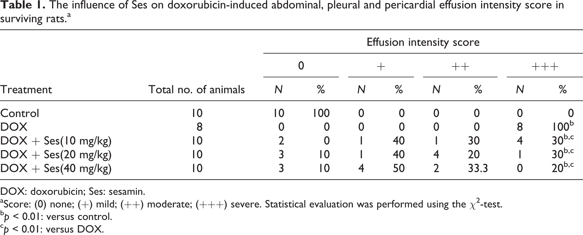
DOX: doxorubicin; Ses: sesamin.
aScore: (0) none; (+) mild; (++) moderate; (+++) severe. Statistical evaluation was performed using the χ2-test.
bp < 0.01: versus control.
cp < 0.01: versus DOX.
Effects of DOX and Ses treatment on serum enzymes
The serum levels of ALT, AST, ALP, BUN and Cre are widely used clinically as parameters for the diagnosis of hepatorenal diseases. As shown in Tables 2 and 3, DOX alone induced significant increases in serum ALT, AST, ALP, BUN and Cre levels compared with the control group (p < 0.01), and these increases were effectively attenuated by Ses (10, 20, and 40 mg/kg) treatment to different degrees (p < 0.05 or p < 0.01). However, none of the assessed parameters in the Ses (10, 20 and 40 mg/kg) treatment groups returned to the control group levels. These results suggest that Ses effectively protects liver and kidney function against DOX-induced hepatorenal toxicity.
Effect of Ses on the levels of serum ALT (U/L), AST (U/L) and ALP (U/L) in serum of male rats after DOX administration.a

AST: aspartate aminotransferase; ALT: alanine aminotransferase; ALP: alkaline phosphatase; DOX: doxorubicin; Ses: sesamin.
aMean ± SD; n = 8.
bp < 0.01: versus control.
cp < 0.05: versus DOX.
dp < 0.01: versus DOX.
ep < 0.05: versus control.
Effect of Ses on the levels of BUN (mmol/L) and Cre (umol/L) in serum of male rats after DOX administration.a

BUN: blood urea nitrogen; DOX: doxorubicin; Cre: creatinine; Ses: sesamin.
aMean ± SD; n = 8.
bp < 0.01: versus control;
cp < 0.05: versus DOX.
dp < 0.01: versus DOX.
Effects of DOX and Ses treatment on liver and kidney histology
The hepatorenal toxicity induced by DOX in rats was further assessed using haematoxylin and eosin-stained sections. The liver sections from the control group showed a regular cell distribution and normal integrated structures of the liver lobules (Figure 1(a)). However, administration of DOX caused clear histopathological changes in the rat liver. The trabecular structure of the liver is blurred with loss of definition of liver plates, nuclei are contracted, pyknotic with condensed chromatin and focal or single-cell necrosis (Figure 1(b)). By contrast, the severe hepatic injury induced by DOX was significantly decreased by Ses treatment (Figure 1(c) to (e)). Representative examples of the histological appearance of the rat kidney are shown in Figure 2. The microscopic examination of the kidney in the control group revealed normal histological parameters (Figure 2(a)). However, the kidney tissues from the DOX-treated group showed widespread structural abnormalities, with significant oedema and vacuolation, hydropic degeneration, desquamation and necrosis atrophy observed in the epithelial cells of the proximal and distal tubules. There were also changes in the glomerulus, such as shrinkage and widening of the capsular space (Figure 2(b)). When Ses was administered to the DOX-treated rats, except for the low-dose group (10 mg/kg; Figure 2(c)), the kidney had a normal appearance upon histological examination with reversal of DOX-induced renal damage (Figure 2(d) and (e)).

Effects of Ses on histopathological changes in DOX-treated hepatic tissue. (a) Section in control rat liver showing the normal morphology, CV; (b) liver of rat exposed to a single dose of DOX (20 mg/kg, i.p. on day 8) showing the trabecular structure of the liver is blurred and loss of definition of liver plates, nuclei are pyknotic with condensed chromatin () and focal or single-cell necrosis( ); (c) DOX-treated hepatic tissue with Ses (10 mg/kg); (d), DOX-treated hepatic tissue with Ses (20 mg/kg); and (e) DOX-treated hepatic tissue with Ses (40 mg/kg). Ses treatment markedly attenuated the DOX-induced liver tissue injury and restored more or less the same histopathological picture observed with the control group. Bar = 50μm. DOX: doxorubicin; Ses: sesamin; CV: central vein.
Effects of Ses on histopathological changes in DOX-treated renal tissue. (a) Section in control rat kidney showing the normal morphology; (b) kidney of rat exposed to a single dose of DOX (20 mg/kg, i.p. on day 8) showing significant oedema and vacuolation, hydropic degeneration () and necrosis atrophy (*) observed in the epithelial cells of the proximal and distal tubules. The glomerulus is atrophic and the capsular space is extensive (); (c) DOX-treated renal tissue with Ses (10 mg/kg); (d) DOX-treated renal tissue with Ses (20 mg/kg); (e) DOX-treated renal tissue with Ses (40 mg/kg). Ses treatment, except the low-dose group (10 mg/kg), markedly attenuated the DOX-induced kidney tissue injury and restored more or less the same histopathological picture observed with the control group. Bar = 50μm. DOX: doxorubicin; Ses: sesamin.
Ses ameliorates DOX-induced oxidative stress
Table 4 indicates that the content of MDA was significantly increased in the livers and kidneys (p < 0.01) of rats treated with DOX compared with that of the control group. Ses (20 and 40 mg/kg) treatment obviously reversed the DOX-induced changes with the content of MDA in rat liver and kidney tissue homogenate (p < 0.05 or p < 0.01) compared to the DOX-treated group. Whereas the change of DOX + Ses (10 mg/kg) group is not significant compared with the DOX-treated group (p > 0.05). Immunohistochemical staining was used to examine the protein expression of 4-HNE as an index of lipid peroxidation (Figures 3 and 4). Figure 3(f) indicates that the protein expression of 4-HNE (0.77 ± 0.07) was significantly increased in the livers of rats treated with DOX compared with that of the control group (0.24 ± 0.02; p < 0.01), which was significantly prevented by Ses (20 and 40 mg/kg) treatment. Analogously, in rat kidney tissue, Ses (20 and 40 mg/kg) obviously decreased the protein expression of 4-HNE (0.42 ± 0.02 and 0.38 ± 0.01, respectively) compared with that of the DOX-treated group (0.54 ± 0.02; p < 0.01, Figure 4(f)).
Effect of Ses on the levels of MDA in liver and kidney tissues of male rats after DOX administration.a

MDA: malondialdehyde; DOX: doxorubicin; Ses: sesamin.
aMean ± SD; n = 8.
bp < 0.01: versus control.
cp < 0.05: versus control.
dp < 0.01: versus DOX.
ep < 0.05: versus DOX.
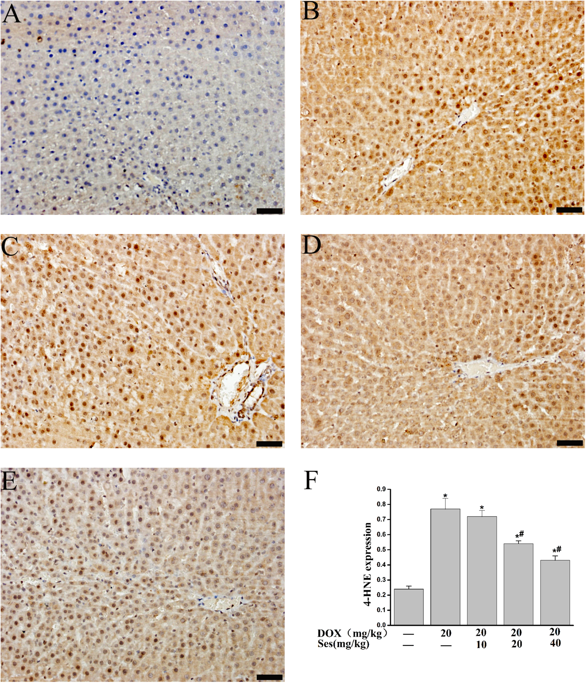
Ses protection from DOX-induced oxidative stress in hepatic tissue. Immunohistochemical staining, followed by a semi-quantitative analysis of positive staining were performed to measure oxidative damage by 4-HNE. The protein expression of 4-HNE was significantly increased in liver tissues exposed to a single dose of DOX (20 mg/kg, i.p. on day 8), which was normalized by treatment with Ses (20, 40 mg/kg). Data are presented as means ± SD (n = 6). *p < 0.01: versus control; #p < 0.01: versus DOX. Bar = 50 μm. DOX: doxorubicin; Ses: sesamin; 4-HNE: 4-hydroxynonenal.

Ses protection from DOX-induced oxidative stress in renal tissue. Immunohistochemical staining, followed by a semi-quantitative analysis of positive staining were performed to measure oxidative damage by 4-HNE. Data are presented as means ± SD (n = 6). *p < 0.01: versus control; #p < 0.01: versus DOX. Bar = 50 μm. DOX: doxorubicin; Ses: sesamin; 4-HNE: 4-hydroxynonenal.
Effects of DOX and Ses on antioxidative enzymes
The measured data of the SOD, CAT and GPX in the liver and kidney tissues are summarized in Figure 5. DOX treatment led to a significant depletion in the activity of SOD, CAT and GPX in the liver and kidney tissue homogenate compared with that of the control group (p < 0.01). Whereas Ses (20 and 40 mg/kg) treatment obviously reversed the reduced activity of SOD, CAT and GPX induced by DOX in rat liver tissue homogenate (p < 0.05 or p < 0.01) compared with the DOX-treated group.

Effects of Ses on the activity of SOD, CAT, GPx in the hepatic and renal tissue of DOX-induced acute injury rats. Values are expressed as means ± SD (n = 8). *p < 0.05 and ** p < 0.01: versus control; #p < 0.05 and ##p < 0.01: versus DOX. DOX: doxorubicin; Ses: sesamin; SOD: superoxide dismutase; CAT: catalase; GPx: glutathione peroxidase.
Discussion
The liver and kidney have many vital functions such as detoxification, protein synthesis, production of biochemicals necessary for digestion and excretion or retention of various substances according to specific body needs. However, the liver and kidney are the most common target organs for chemically induced injuries. 20 –22 Many chemotherapy drugs have hepatorenal toxicity. 23,24 In the current study, 20 mg/kg DOX was injected into rats, and serum markers that indicate hepatic and renal damage were determined 2 days after administration. Serum BUN and Cre levels as well as ALT, AST and ALP activities increased in DOX-treated rats. Our histopathological findings also confirmed the presence of hepatic and renal injury in DOX-treated rats. These results are consistent with previous studies. 4,6
DOX treatment induces oxidative stress as well as apoptotic and necrotic changes in organs. 25 Lipid peroxidation refers to the oxidative degradation of lipids. It is the process by which free radicals steal electrons from the lipids in cell membranes, resulting in cell damage. The end products of lipid peroxidation are reactive aldehydes, such as MDA and 4-HNE. In the current study, significant increases in MDA and 4-HNE levels were found in the examined organs of DOX-treated rats. The results indicate that there is a strong relationship between oxidative stress and DOX-related hepatorenal toxicities.
Because DOX has significant antitumour activity, novel methods to reduce or prevent detrimental side effects are expected to increase its effectiveness in anticancer therapy. Recently, attention has focused on natural products as sources to develop safe and potent agents for preventing DOX-induced toxicity. 26 –28 Ses is one of the most abundant ligands in sesame seeds. 29 It exhibits multiple biological functions, such as inhibition of inflammation, 30 carcinogenesis and oxidative stress. 31,32 In addition, Ses attenuates hypertension, serum and hepatic cholesterol and serum triglycerides. 33 –35 Furthermore, Ses also exhibited protective effects in various models of hepatorenal injuries. 14,16 Our results demonstrate that Ses protects the liver and kidney functions by lowering serum ALT, AST, ALP, Cre and BUN levels. Furthermore, the protective effect of Ses against DOX was also evaluated histopathologically in the liver and kidney. The histological damage induced by DOX was also significantly decreased by Ses pretreatment. These findings indicate that Ses is a potential protective agent against DOX injury. Current data also show that Ses significantly decreased the DOX-induced increase of MDA and 4-HNE and increased the activity of CAT, SOD and GPX compared to the DOX administered rats. The pretreatment of rats with Ses significantly alleviated the oxidative stress induced by DOX. Therefore, Ses protects against DOX-induced hepatic and renal injury partially due to its antioxidative properties. However, the exact mechanism of Ses is unclear. The remaining question is how the Ses increase multiple antioxidants. One possibility is that Ses increases these antioxidants by targeting activation of their common transcription factors in the upstream. Recent studies demonstrated that Ses mediates antioxidative and anti-inflammatory effects through the activation of sirtuin 1 (SIRT1) or inhibition of nuclear factor κB. 18,36 Forkhead box O3a (FOXO3a) is one of the key transcription factors in the antioxidant defence system, on the other hand, FOXO3a is regulated by sirt1. FOXO3a is one member of the FOXO family proteins which have been implicated in the regulation of oxidative stress and several other diverse physiologic processes including stress resistance, cell differentiation, cell cycle arrest, metabolism and apoptosis. It has been demonstrated that FOXO3a reduces ROS by the transcriptional activation of superoxide dismutase 2 and CAT. 37,38 Further studies are needed to identify the protective effect of Ses administration and the exact mechanism of Ses on DOX-induced liver and kidney injury.
In conclusion, these results suggest that Ses treatment is useful for decreasing oxidative stress and injury in the liver and kidneys of DOX-treated rats. This suggests that Ses may have a role in the attenuation and prevention of serious complications from DOX. The combination of Ses with DOX is a novel strategy that may protect against DOX-induced hepatorenal toxicity in clinical practice.
Footnotes
Declaration of Conflicting Interests
The author(s) declared no potential conflicts of interest with respect to the research, authorship, and/or publication of this article.
Funding
The author(s) disclosed receipt of the following financial support for the research, authorship, and/or publication of this article: This work was supported by the fund from the National Natural Science Fund of China (81101465, 81202401, 81473292), the Natural Science Foundation of Hebei Province of China (H2013206292), the Foundation of High Educational Institute of Hebei (ZD2013057), 2014 Project of College Students’ Innovation and Entrepreneurship Training Program Project in Hebei Province (201410089015, 201410089048).